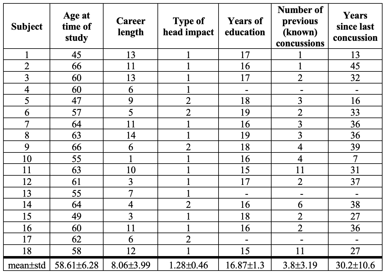
0457
Abnormal spontaneous brain fluctuations present in retired football players1KITE, Toronto Rehabilitation Institute, Toronto, ON, Canada, 2School of Biomedical Engineering, McMaster University, Hamilton, ON, Canada, 3Imaging Research Centre, St. Joseph's Healthcare Hamilton, Hamilton, ON, Canada, 4Electrical and Computer Engineering, McMaster University, Hamilton, ON, Canada, 5Radiology, McMaster University, Hamilton, ON, Canada
Synopsis
Keywords: Traumatic brain injury, fMRI (resting state), ALFF
Novel methods are required to understand the scale of potential brain changes in collision sport athletes. with many former athletes having developed neurocognitive deficits or neurological disorders. This study used resting state functional MRI (rsfMRI) to examine if retired professional football players (n=18) had functional brain abnormalities based on a personalized amplitude of low-frequency fluctuations (ALFF) and Z-scoring approach. Brain injuries were identified if the ALFF Z-score exceeded 3 standard deviations from the healthy control mean. Thirty regions were abnormal in more than half of the retired athletes. Cerebellar and central, sub-cortical brain regions were most often seriously abnormal.Introduction
Sport-related concussions are a concern and known risk for those currently playing collision sports1. However, in recent years, the effects of these injuries in retired athletes have been more extensively studied. For example, in former professional athletes, cognitive, emotional, and physical deficits have been observed2. Whether these sequelae are directly tied to a functional neuropathology is less well-researched. Brain health following sport-related concussions has been extensively researched using resting state functional magnetic resonance imaging (rsfMRI)3. However, in former elite athletes, most research has utilized network connectivity analyses, often demonstrating alterations to the default mode, visual, and dorsolateral frontal networks4,5. An alternative approach to functional connectivity analysis is to examine low frequency oscillations (0.008-0.09 Hz), present in rsfMRI acquisitions6. The amplitude of low-frequency fluctuations (ALFF) and fractional ALFF (fALFF) can be used to determine spontaneous neural activity and frequency composition of the BOLD signal in each voxel7,8. The purpose of this study was to (i) examine if retired Canadian Football League (rCFL) players with a history of concussions and repetitive sub-concussive head impacts have any functional abnormalities present years after retirement from professional sport and their last (known) concussion, and (ii) to identify brain regions that most commonly show disturbances in spontaneous brain activity years after competitive play. It was hypothesized that, using a personalized analysis approach, the rCFL subjects would show decreased ALFF and fALFF in some brain regions relative to healthy controls.Methods
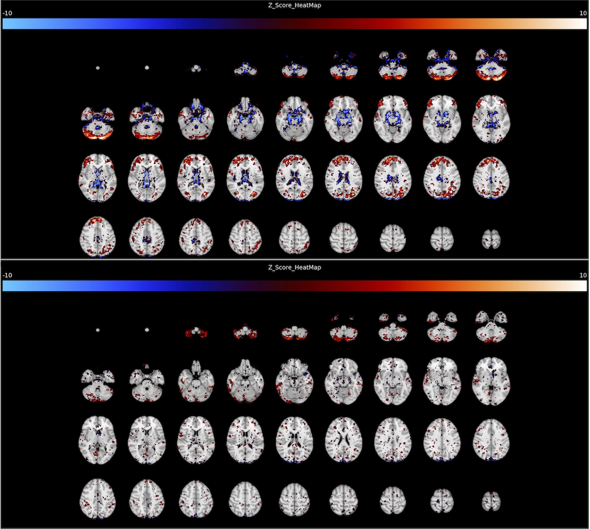
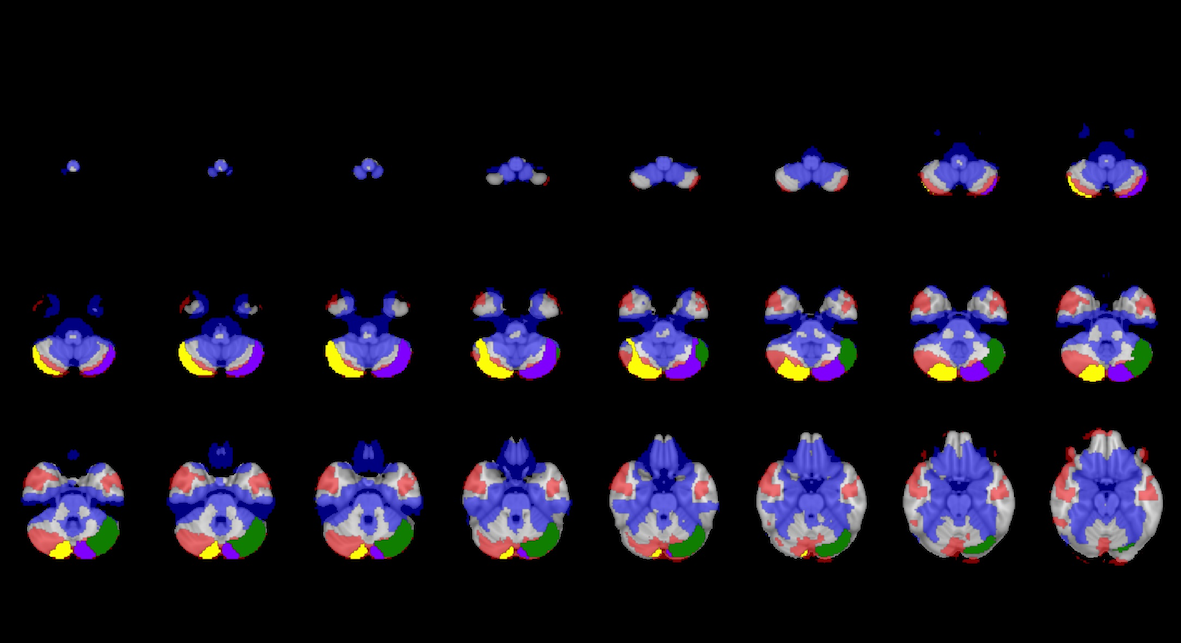
Eighteen rCFL players (male, aged 58.78±6.10) were scanned using a 3T GE MR750, had demographic information recorded (Figure 1), and completed a battery of clinical tests (Immediate Post-Concussion Assessment Tool (ImPACT), Beck’s Depression Inventory II (BDI-II), and the Short-Form 36 (SF-36)) to measure cognitive, emotional, and physical health. Healthy age and sex-matched control data (n=62, male, aged 58.81±5.69) were downloaded from the OASIS-3 study9. Preprocessing was performed using CONN10; ALFF metrics were computed and subsequently, voxel-wise and regional Z-scores were computed using MATLAB (vR2020b). 142 brain regions-of-interest (ROIs) were examined from the cortical, sub-cortical (Harvard-Oxford)11 and cerebellar (Probabilistic FNIRT) brain atlases12. Brain ROIs were considered abnormal if the Z-score exceeded 3 standard deviations from the healthy control mean. The 3 standard deviation threshold was deemed appropriate because values exceeding that limit fall outside 99.7% of the normal data and are thus fairly considerable outliers. Through correlational analyses, associations between participant demographics, clinical data, and the total number of their abnormal (+3≤Z-score≤-3) ALFF and fALFF brain ROIs was computed. A Bonferroni correction was applied to correct for family-wise errors in the correlation tests. Therefore, for demographic and concussion history correlations (i.e., 6 measures in total), the p-value threshold of significance was p<0.008 (i.e., 0.05/6= 0.008), and the p-value threshold of significance was p<0.003 (i.e., 0.05/15= 0.003) for the clinical test correlations (i.e., 15 clinical test metrics considered).Results
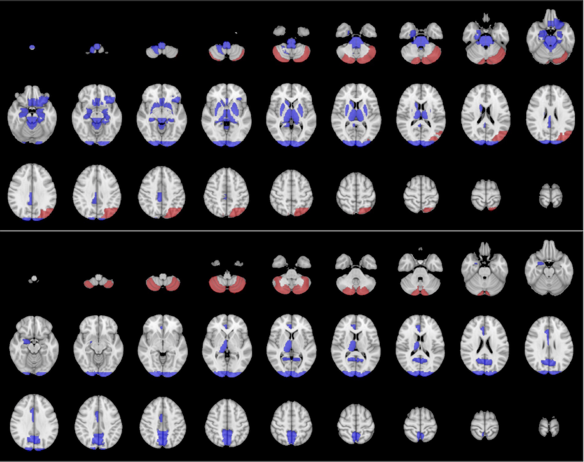
The Z-scoring analysis showed 30 ROIs to be significantly abnormal in more than 50% of the rCFL subjects using ALFF, and 13 ROIs in more than 25% of rCFL subjects were abnormal based on fALFF (Figure 2). Abnormal ROIs included the right amygdala, right thalamus, bilateral occipital pole, and bilateral cerebellum crus II (Figures 3,4). The total number of positive fALFF Z-score outliers significantly correlated with longer career length (r= 0.7, p= 0.0012). Based on Z-scores of the six most commonly damaged ROIs, the right amygdala was significantly correlated with the number of previous concussions (NPC) (r= 0.639, p= 0.0078) based on ALFF data. Also, although not significant, ALFF results from the NPC correlated with the left occipital pole (r= 0.518, p= 0.0398), right occipital pole (r= 0.586, p= 0.0171), and thalamus (r= 0.538, p= 0.0314). Additionally, the right thalamus was correlated with chronic pain (r= -0.494, p= 0.0372), and the left cerebellum crus II was correlated with impulse control (r= -0.501, p= 0.0343), emotional well-being (r= 0.509, p= 0.0311) and pain (r= 0.473, p= 0.0473). Moderately strong but not significant correlations from the fALFF data included the left occipital pole with NPC (r= 0.549, p= 0.0276), the right cerebellar crus II and physical functioning (r= -0.471, p= 0.0487), and the right thalamus with motor speed (r= -0.488, p= 0.0399) and impulse control (r= 0.481, p= 0.0435).Discussion
Spontaneous brain activity, a measure of brain health, was compromised in retired athletes in comparison to matched controls. Each subject exhibited a unique profile of abnormalities, but our analyses showed that cerebellar and central sub-cortical brain regions were most frequently and seriously abnormal. Furthermore, moderate correlations were found between the most frequently abnormal ROIs and clinically testable neurobehavioural metrics.Conclusions
With an objective and personalized brain assessment technique, clinicians can more effectively connect post-concussion symptoms with focal brain injuries, and through that understanding, managed tailored treatment plans to improve brain recovery. Further research should be performed on improving the clinical feasibility of rsfMRI and ALFF analysis for concussion patients, and on comparing the ALFF method to other rsfMRI analysis techniques.Acknowledgements
No acknowledgement found.References
1. Zuckerman SL, Kerr ZY, Yengo-Kahn A, et al. Epidemiology of sports-related concussion in NCAA athletes from 2009-2010 to 2013-2014: incidence, recurrence, and mechanisms. Am J Sports Med 2015;43(11):2654–2662.
2. Terpstra AR, Vasquez BP, Colella B, et al. Comprehensive neuropsychiatric and cognitive characterization of former professional football players: implications for neurorehabilitation. Front Neurol 2019;10:71.
3. Zhu DC, Covassin T, Nogle S, et al. A potential biomarker in sports-related concussion: brain functional connectivity alteration of the Default-Mode Network measured with longitudinal resting-state fMRI over thirty days. J Neurotrauma 2015;32(5):327-341.
4. Hampshire A, MacDonald A, Owen AM. Hypoconnectivity and hyperfrontality in retired American football players. Sci Rep 2013;3(1):2972.
5. Plourde V, Rohr CS, Virani S, et al. Default mode network functional connectivity after multiple concussions in children and adolescents. Arch Clin Neuropsychol 2020;35(3):302-311.
6. Lowe MJ, Dzemidzic M, Lurito JT, et al. Correlations in low-frequency BOLD fluctuations reflect cortico-cortical connections. NeuroImage 2000;12(5):582-587.
7. Zang YF, He Y, Zhu CZ, et al. Altered baseline brain activity in children with ADHD revealed by resting-state functional MRI. Brain Dev 2007;29(2):83–91.
8. Zou QH, Zhu CZ, Yang Y, et al. An improved approach to detection of amplitude of low-frequency fluctuation (ALFF) for resting-state fMRI: fractional ALFF. J Neurosci Meth 2008;172(1):137-141.
9. LaMontagne PJ, Benzinger TLS, Morris JC, et al. OASIS-3: longitudinal neuroimaging, clinical, and cognitive dataset for normal aging and Alzheimer Disease. MedRxiv. 2019.
10. Whitfield-Gabrieli S, Nieto-Castanon A. Conn: A functional connectivity toolbox for correlated and anticorrelated brain networks. Brain Connect 2012;2(3):125-141.
11. Desikan RS, Ségonne F, Fischl B, et al. An automated labeling system for subdividing the human cerebral cortex on MRI scans into gyral based regions of interest. Neuroimage 2006;31(3):968-980.
12. Diedrichsen J, Balster JH, Cussans E, et al. A probabilistic MR atlas of the human cerebellum. Neuroimage 2009;46(1):39-46.
Figures