0442
Identifying pathological differentiation of cervical squamous cell carcinoma with APTw and IVIM
Zhonghong Xin1, Junqiang Lei1, Jianhong Peng2, Xiande Lu2, Jiang Nan2, Yaping Zhang2, Xiaohui Wang2, Jun Zhu2, and Jianxiu Lian3
1Radiology, the First Hospital of Lanzhou University, Lanzhou, China, 2the First Hospital of Lanzhou University, Lanzhou, China, 3Philips Healthcare, Beijing, China
1Radiology, the First Hospital of Lanzhou University, Lanzhou, China, 2the First Hospital of Lanzhou University, Lanzhou, China, 3Philips Healthcare, Beijing, China
Synopsis
Keywords: Uterus, Diffusion/other diffusion imaging techniques, amide proton transfer weighted imaging,cervical cancer,squamous cell carcinoma of the cervix
The pathological differentiation of cervical squamous cell carcinoma (CSCC) determines the therapy method and prognosis. APTw, DWI and IVIM sequences were performed for predicting pathological differentiation. 27 patients with well-moderately differentiation, 13 patients with poorly differentiation and 15 healthy volunteers were enrolled. APT SI, ADC, D*, D and f values were calculated for comparing among different groups. Results showed parameters except D* differed significantly between CSCC and normal. There were statistically significant differences in AUC of APT SI, D and f between well-moderately group and poorly differentiated group. APTw and IVIM can be used to identify pathological differentiation of CSCC.Introduction
Cervical cancer (CC) is the fourth leading cause of cancer death in women [1], in which squamous cell carcinoma of the cervix (CSCC) is the most ordinary pathological subtype. The pathological differentiation of CSCC is essential for therapy and prognosis. The biopsy is the golden standard for identifying pathological differentiation with the disadvantages of invasive, susceptible to factors such as differences in operator experience, tumor heterogeneity and size. Amide proton transfer weighted (APTw) and intravoxel incoherent motion(IVIM) models can non-invasively detect the microstructural characteristics of tumor tissue, and reflect cellular metabolism and pathophysiological information, which have been used in the preoperative diagnosis and pathological grading of malignant tumor. APT signal intensity (APT SI) is the main parameter of APTw, which reflect changes of water signal intensity after exchange of amide and water proton[2]. IVIM is an extension of DWI-MRI that can accurately reflect cell density and microcirculation status of tissues by distinguishing water molecule movement and microcirculation perfusion, and effectively evaluate pathological type and grading of CC [3,4]. The parameters derived from IVIM include D, D* and f values. The study indicated that IVIM parameters as independent potential biomarkers could evaluate the pathological characteristics of CC[4]. The objective of this study is to compare APTw and IVIM with traditional DWI model to explore the value of diagnosing the degree of differentiation of CSCC.Methods
40 patients with CSCC and 15 healthy volunteers were enrolled. All participants were performed using 3.0T MR scanner (Ingenia 3.0T CX, Philips Healthcare). APTw was scanned using 3D Multishot TSE. ADC values are calculated based on the image of 2 b values (0, 800s/mm2). IVIM were scanned by using 12 b values (0,20,100,150,200,300, 400,500,600,800,1000 and 1200 s/mm2). Post-processing was performed by using ISP software (Intellispace Portal; Version 10.1; Philips Healthcare, Best, the Netherlands) and MITK Diffusion software (sDMAS-2018.07-314-g1780784feb, German Cancer Research Center). The largest cross-section of primary lesions was used to measure parameters of APTw, DWI and IVIM by two experienced radiologists. The ROIs were delineated including the solid parts of tumor and avoiding large blood vessels, degeneration, cystic changes, hemorrhage and artifacts. Quantitative analysis was performed to APT SI, ADC, D, D* and f. The parameters between the groups were compared with independent t-tests. Diagnostic performance was evaluated with a ROC analysis. The statistical analyses were performed using SPSS 26.0 and MedCalc 20.0.Results
40 patients (median 53 years; range 31-77 years) and 15 healthy volunteers (median 31 years; range 22-57 years) were included in this study. Patients were divided into a well-moderately differentiated group(n=27) and a poorly differentiated(n=13). Parameters except D* (P=0.573) differed significantly between CSCC and normal cervix (P< 0.001). Significant differences were found in APT SI and D between well-moderately differentiated and poorly differentiated group (APT SI: 2.94±0.13%, 3.08±0.18%, P=0.005; D: 0.70±0.16, 0.53±0.22, P=0.009). Parameters except D* (P=0.558) arrived the higher diagnostic performance between CSCC and normal cervix (P <0.001) (Table1 and Figure1). Comparing well-moderately and poorly differentiated CSCC, AUC of APT SI, D and f were 0.751, 0.732 and 0.689; sensitivity of them were 69.23%, 53.85%, 69.23% and specificity of them were 74.07%,100%,66.67%, respectively (Table2 and Figure2). The differences were statistically significant (P<0.05).Discussion
The APT SI of CSCC was higher than normal cervical in this study, which was consistent with tumors of other organs in previous researches[3,5]. A significant difference was found in APT SI between well-moderately differentiated and poorly differentiated CSCC (2.94±0.13%, 3.08±0.18%). It was suggested that APT SI can preliminarily evaluate the pathological differentiation of CSCC. Compared with well-moderately differentiated group, the poorly had higher degree of malignancy, higher cell density, more pronounced nuclear atypia and micronecrosis [6]. All of these may lead to an increase in the amount of free proteins and peptides in tissue, which leads to an increase in APT SI. This study showed that D* was difficult to evaluate the differentiation degree of CSCC, which may be due to the abundant blood supply and high cell density of malignant tumors, the former increases blood flow and the latter decreases blood flow velocity, resulting in no significant change in D* value. The Brownian motion of water molecules is closely related to the microenvironment of tissues. Therefore, the ADC and D of CSCC was significantly lower than normal cervix. The D value of the poorly differentiated group was lower than well-moderately differentiated group, which may be due to the faster proliferation and higher density of cells, which significantly hindered the diffusion of water molecules. In this study, D showed better performance than ADC. It indicated that D value may have a better ability to evaluate the pathological differentiation. Our research showed the f value in CSCC was significantly lower than normal cervix, but the difference was not statistically significant between well-moderately and poorly differentiated groups. The higher f value indicates higher proportion of microcirculation, richer blood supply and more active proliferation.Conclusion
APT and IVIM can be used to diagnose CSCC and provide more accurate quantitative information for clinical diagnosis and treatment. Compared with IVIM, APT has higher diagnostic performance in distinguishing the degree of differentiation of CSCC.Acknowledgements
We would like to appreciate Jianxiu Lian and Ke Jiang for their technical support.References
1. Sung, H., et al., Global Cancer Statistics 2020: GLOBOCAN Estimates of Incidence and Mortality Worldwide for 36 Cancers in 185 Countries. CA: A Cancer Journal for Clinicians, 2021. 71(3): p. 209-249. 2. van Zijl, P.C.M. and N.N. Yadav, Chemical exchange saturation transfer (CEST): What is in a name and what isn't? Magnetic Resonance in Medicine, 2011. 65(4): p. 927-948. 3. Li, B., et al., The utility of APT and IVIM in the diagnosis and differentiation of squamous cell carcinoma of the cervix: A pilot study. Magnetic Resonance Imaging, 2019. 63: p. 105-113. 4. Du, S., et al., Relationship between 18F-FDG PET metabolic parameters and MRI intravoxel incoherent motion (IVIM) histogram parameters and their correlations with clinicopathological features of cervical cancer: evidence from integrated PET/MRI. Clinical Radiology, 2019. 74(3): p. 178-186. 5. Meng, N., et al., Application of the amide proton transfer-weighted imaging and diffusion kurtosis imaging in the study of cervical cancer. European Radiology, 2020. 30(10): p. 5758-5767 Meng, N., et al., Evaluation of amide proton transfer-weighted imaging for endometrial carcinoma histological features: a comparative study with diffusion kurtosis imaging. European Radiology, 2021. 31(11): p. 8388-8398.Figures
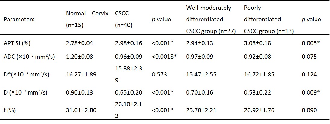
Table 1. *Statistically significant.
CSCC, cervical squamous cell carcinoma; APT SI, signal intensity of amide proton transfer; ADC, apparent diffusion coefficient; D, pure molecular diffusion; D*, pseudo-diffusion coefficient; f, perfusion fraction.
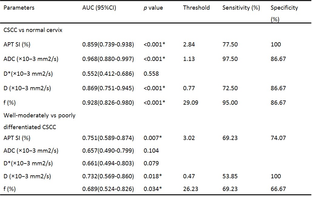
Table 2. Diagnostic performance of APT
SI, ADC, D, D*, and f value in identifying CSCC and normal cervix,
well-moderately vs poorly differentiated CSCC.
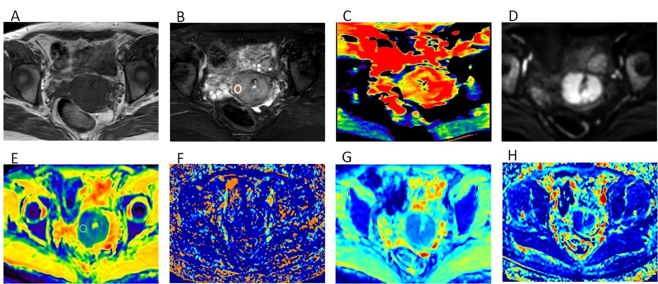
Figure 1. FA 37-year-old woman with moderately differentiated CSCC
The lesion of the cervix showed slightly
hypo-intense signal on T1WI (A),
hyper-intense signal on T2WI(B) and DWI(D). The APT SI was 2.97% (C). ADC
map was generated using b values of 0 and 800 s/mm2, the
diffusion of the lesion was limited(E). D*, D and f maps were obtained from the
IVIM (F-H). The ADC, D*, D and f values of the moderately differentiated CSCC
were 0.95×10⁻³mm2/s,16.51×10−3mm2/s, 0.74 ×10⁻³mm2/s
and 26.47%, respectively.
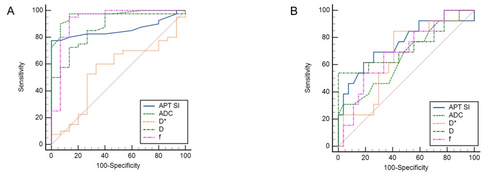
Figure 2. (A)ROC curve of the APT SI, ADC, D, D*, and f in
identifying CSCC and normal cervix;
(B) ROC curve of the APT SI,
ADC, D, D*, and f in identifying well-moderately and poorly differentiated
CSCC.
DOI: https://doi.org/10.58530/2023/0442