0440
Accelerated multi-shot diffusion MRI using deep learning denoising1Department of Medical Physics, Memorial Sloan Kettering Cancer Center, New York, NY, United States, 2Department of Radiology, Memorial Sloan Kettering Cancer Center, New York, NY, United States
Synopsis
Keywords: Pelvis, Diffusion/other diffusion imaging techniques, image reconstruction
Multi-shot EPI is commonly used to compensate for geometric distortions and increase spatial resolution in body diffusion MRI, with a price tag of longer scan times. This work presents an alternative technique to k-space undersampling to accelerate the acquisition, which is based on reducing the number of repetitions at high b-value and denoising the resulting images using a convolutional neural network. The proposed deep learning denoising technique is demonstrated to accelerate the acquisition of multi-shot diffusion MRI acquisition of patients with rectal cancer and reduce the scan time beyond the duration of a single-shot diffusion MRI acquisition.
Introduction:
Body diffusion MRI presents significant challenges such as increased susceptibility to geometric distortions and lower SNR [1]. The latter requires to performs several repetitions at high b-value for signal averaging at the expense of increasing scan time. Multi-shot EPI can reduce geometric distortions and increase spatial resolution compared to conventional single-shot EPI [2]. However, scan time increases linearly with the number of shots. k-space undersampling can be employed to accelerate the acquisition, but acceleration rates are limited by the low baseline SNR of high b-value acquisitions. This work presents an alternative technique to k-space acceleration by reducing the number of acquisitions in the high b-value acquisition and performing denoising using a convolutional neural network. The proposed deep learning denoising technique is demonstrated to accelerate multi-shot diffusion acquisition on patients with rectal cancer.Methods:
Denoising network: A residual convolutional neural network trained using single-shot diffusion data was developed (Figure. 1). The inputs to the network are the noisy high b-value image (reduced number of repetitions) and the low b-value image, which serves as a reference as described previously [3]. The output is the denoised high b-value image. The architecture of the network included 21 layers of 3x3x64 convolution filters, batch normalization and nonlinear Rectified Linear Unit (RELU). Images were divided into 60x60 patches with 20 pixels stride to increase number of training datasets (each 2D image resulted into 188 patches). The network was trained using single-shot diffusion MRI data previously acquired on 110 patients with rectal cancer using different 3T scanners (GE Healthcare). High b-value data were acquired with NEX=16 (number of repetitions) and reconstructed into images corresponding to NEX=1 (first repetition only) and NEX=16 (all repetitions). A total of 868372 patches corresponding to 4619 slices were employed for training.Loss function: Training was performed using a loss function that combines the mean squared error (L2) and the absolute value error (L1) to improve edge preservation:
$$ L = \sum_n {\left\Vert \hat{\phi}_n - y_n \right \Vert _2 }^2 +\lambda \sum_n {\left \Vert \hat{\phi}_n - y_n \right \Vert_1},$$
where $$$ \hat{\phi}_n = x_{n,1}-\psi\left(\vec{x}_n ;\vec{\Theta}\right) $$$ is the output of the network, $$$\vec{x}_n$$$ is the input, $$$y_n$$$ is the reference for training and $$$\lambda$$$ is the parameter that weights the contribution of the L1 term respect to the L2 term (set to 0.25).
Application to multi-shot diffusion in patients with rectal cancer: Multi-shot diffusion acquisition was performed on 11 patients with rectal cancer using two 3T scanners (GE Healthcare Premier and Architect). Data were acquired with 2 shots and included $$$b=0$$$ with NEX (number of repetitions) = 2 and b-800 with NEX=16. Raw data were read in Matlab using the Orchestra software (GE Healthcare) and multi-shot reconstruction was performed using a custom version of the MUSE algorithm [4] for the following configurations: LB ($$$b=0$$$ data), HB1 ($$$b=800$$$ with 1 repetition), HB2 ($$$b=800$$$ with 2 repetitions), HB4 ($$$b=800$$$ with 4 repetitions) and HB16 ($$$b=800$$$ with 16 repetitions). HB1, HB2 and HB4, which correspond to 16-fold, 8-fold and 4-fold acceleration of the original high b-value acquisition respectively, were denoised using the proposed deep learning technique The Apparent Diffusion Coefficient (ADC) was also computed.
Image quality evaluation: Blinded qualitative evaluation of image quality was performed by an expert body radiologist using the scale indicated in Table 1. HB2, HB4 and HB16 images were presented in a random order to the radiologist.
Results:
Figures 2 and 3 show the deep learning denoising results in two different representative patients respectively. They depict axial images through the pelvis, and zoomed view to the rectum, demonstrating optimal demarcation of the rectal wall layers and surrounding structures with good image quality on all views provided, NEX=4 slightly better than NEX=2 for Figure 2 and similar quality of NEX=4 and NEX=2 for Figure 3. Denoising results are very close to the reference HB16 despite the high acceleration. HB4 presents the highest performance among all accelerations, which is confirmed by the scoring results in Table 2. The score for HB4 is even higher than the reference HB16, which is due in part to the shorter acquisition time in HB4 that reduces the effect of gas-related motion in the rectum. The 4-fold acceleration in multi-shot HB4 would enable to improve image quality and even reduce the scan time of the conventional single-shot acquisition by a factor of 2.Conclusion:
This work demonstrated the application of deep learning denoising to accelerate the acquisition of multi-shot diffusion MRI data in the rectum. An acceleration factor of 4 with respect to the original multi-shot acquisition was feasible with even better image quality. The 4-fold acceleration in a 2-shot acquisition translates to a 2-fold acceleration compared to conventional single-shot diffusion MRI. The acceleration technique proposed in this work would increase the performance of multi-shot diffusion and enable the use of a higher number of shots for further increase in image quality without additional scan timeAcknowledgements
NIH grant R01‐CA244532 and a seed grant from the Department of Radiology at Memorial Sloan Kettering Cancer CenterReferences
1) Barth, B.K., Cornelius, A., Nanz, D., Eberli, D. and Donati, O.F., 2015. Diffusion-weighted imaging of the prostate: image quality and geometric distortion of readout-segmented versus selective-excitation accelerated acquisitions. Investigative radiology, 50(11), pp.785-791.
2) Chen, X., Zhang, Y., Cao, Y., Sun, R., Huang, P., Xu, Y., Wang, W., Feng, Q., Xiao, J., Yi, J. and Li, Y., 2018. A feasible study on using multiplexed sensitivity-encoding to reduce geometric distortion in diffusion-weighted echo planar imaging. Magnetic resonance imaging, 54, pp.153-159.
3) Kaye, E.A., Aherne, E.A., Duzgol, C., Häggström, I., Kobler, E., Mazaheri, Y., Fung, M.M., Zhang, Z., Otazo, R., Vargas, H.A. and Akin, O., 2020. Accelerating prostate diffusion-weighted MRI using a guided denoising convolutional neural network: retrospective feasibility study. Radiology: Artificial Intelligence, 2(5).
4) Chen, N.K., Guidon, A., Chang, H.C. and Song, A.W., 2013. A robust multi-shot scan strategy for high-resolution diffusion weighted MRI enabled by multiplexed sensitivity-encoding (MUSE). Neuroimage, 72, pp.41-47.
5) Zhang, K., Zuo, W., Chen, Y., Meng, D. and Zhang, L., 2017. Beyond a gaussian denoiser: Residual learning of deep cnn for image denoising. IEEE transactions on image processing, 26(7), pp.3142-3155.
Figures
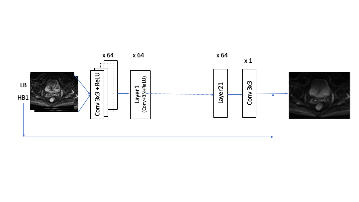
Figure. 1: Schematic of the R-CNN network, where the inputs are the noisy high b-value image (HB) and the low b-value image (LB), and the output is the denoised high b-value image. [3, 5]
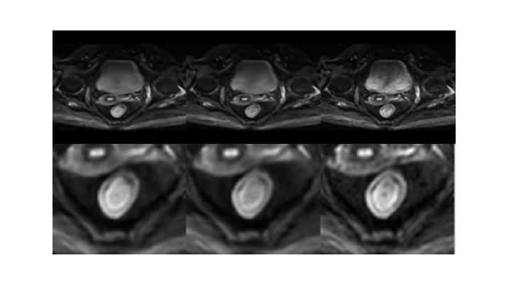
Figure. 2: Comparison of denoising results: NEX=2 (left), NEX=4 (center), and reference with NEX=16 (right) for a representative patient with rectal cancer. The bottom row shows zoomed images on the rectal region.
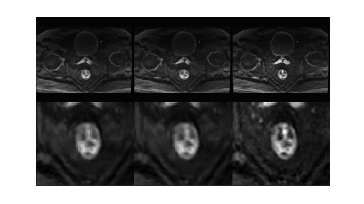
Figure 3: Comparison of denoising results: NEX=2 (left), NEX=4 (center), and reference with NEX=16 (right) for a different patient with rectal cancer. The bottom row shows zoomed images on the rectal region.
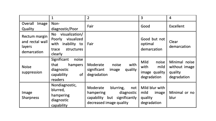
Table. 1: Scoring criteria employed by the radiologists to assess image quality
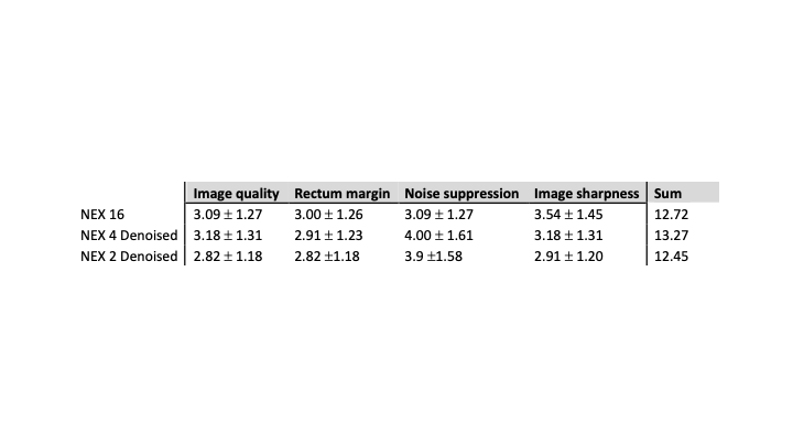
Table. 2: Image quality scoring results for the high b-value images with NEX=16, NEX=4 and NEX=2. Denoised images with NEX=4 (4-fold acceleration with respect to the reference) presented even higher image quality scores than the reference, which is due in part to the shorter acquisition that reduces the effect of gas-related motion in the rectum