0439
Radiomics of Multiparametric MRI in Tumor Grading of Endometrial Cancer1Department of Diagnostic Radiology, The University of Hong Kong, Hong Kong, Hong Kong, 2GE Healthcare, Taipei, Taiwan
Synopsis
Keywords: Uterus, Cancer, Endometrial Cancer; Tumor Grade
Random forest models were constructed to predict tumor grade (grade 1-2 vs. grade 3) of endometrial cancer based on radiomics features extracted from quantitative T1, T2, proton density maps generated by synthetic MRI and apparent diffusion coefficient maps generated by diffusion-weighted imaging. The classification model based on features extracted from all the quantitative maps achieved the highest area under the curve of 0.804 compared to models constructed based on single quantitative map.
INTRODUCTION
Tumor grade is an important prognostic factor of endometrial cancer (EC) 1. Previous studies have explored the correlation between tumor grade and mean apparent diffusion coefficient (ADC) of EC but results were inconclusive 2,3. This could be in part due to tumor heterogeneity, in which radiomics offer further assessment. Radiomics based on multiparametric MRI was able to predict lymphovascular space invasion and stratify preoperative risk of EC 4,5. Motivated by these studies, we aim to distinguish low-grade (grade 1-2) and high-grade (grade 3) EC using radiomics analysis on quantitative T1, T2 and proton density (PD) maps generated by synthetic MRI and ADC maps generated by DWI.METHODS
This prospective study selected 109 patients with EC (mean age: 58 years), including 86 patients with tumor grade 1-2 (G1-2), and 23 patients with grade 3 (G3). Each patient underwent synthetic MRI and diffusion-weighted imaging (DWI) (b values: 0, 400, 800) before surgery on a 1.5T scanner (SIGNA Explorer, GE Healthcare, USA) with a 16-channel coil. Tumors were delineated by a radiologist with >10 years’ experience in pelvic MRI on synthetic T2WI and DWI on b=800. The volume-of-interests (VOIs) on synthetic T2WI and DWI were then mapped to T1/T2/PD maps generated from synthetic MRI and ADC maps from DWI (Figure 1). 960 radiomics features were extracted from the VOIs on each quantitative map. A 5-fold cross validation was implemented on the whole dataset. All radiomics features from single quantitative map or all maps were selected by Mann-Whitney U test with p-value<0.05 and least absolute shrinkage and selection operator (LASSO) regression, and then built a random forest model. To minimal the influence of imbalanced subgroups, balanced weights were used to build models on two classes, with each class weighted inversely proportional to the corresponding frequency. In order to provide comprehensive estimation of diagnostic performance based on the small sample size, the micro-averaged receiver operating characteristic (ROC) curve and the area under the ROC curve (AUC) were used, combining the predictions from 5-fold cross validation 6. The workflow of our study was shown in Figure 2.RESULTS
The micro-averaged ROC curves were shown in Figure 3. The random forest models constructed based on radiomics features from T1 map, T2 map, PD map, ADC map and all maps achieved micro-averaged AUCs of 0.602, 0.717, 0.746, 0.644 and 0.804, respectively, in determining different tumor grades of EC.DISCUSSION AND CONCLUSION
In this study we built random forest models to predict low-grade (G1-2) or high-grade (G3) EC using radiomics features extracted from each quantitative map and all maps separately. The classification model based on features from all maps achieved highest AUC of 0.804, showing high performance in predicting tumor grading of EC. This may provide useful preoperative information on the histopathologic characteristics of EC that would contribute to treatment planning and disease stratification.Acknowledgements
Authors thank Dr. Philip P.C. Ip for his helps on histopathologic assessmentReferences
- Amant F, Moerman P, Neven P, et al. Endometrial cancer. The Lancet. 2005;366(9484):491-505.
- Tanaka T, Terai Y, Fujiwara S, et al. Preoperative diffusion-weighted magnetic resonance imaging and intraoperative frozen sections for predicting the tumor grade in endometrioid endometrial cancer. Oncotarget. 2018;9(93):36575-36584.
- Bonatti M, Pedrinolla B, Cybulski AJ, et al. Prediction of histological grade of endometrial cancer by means of MRI. Eur J Radiol. 2018;103:44-50.
- Lefebvre TL, Ueno Y, Dohan A, et al. Development and Validation of Multiparametric MRI–based Radiomics Models for Preoperative Risk Stratification of Endometrial Cancer. Radiology. 2022:212873.
- Luo Y, Mei D, Gong J, et al. Multiparametric MRI-Based Radiomics Nomogram for Predicting Lymphovascular Space Invasion in Endometrial Carcinoma. J Magn Reson Imaging. 2020;52(4):1257-1262.
- Tsoumakas G, Katakis I, Vlahavas I. Mining Multi-label Data. Boston, MA: Springer US; 2010. p. 667-685.
Figures
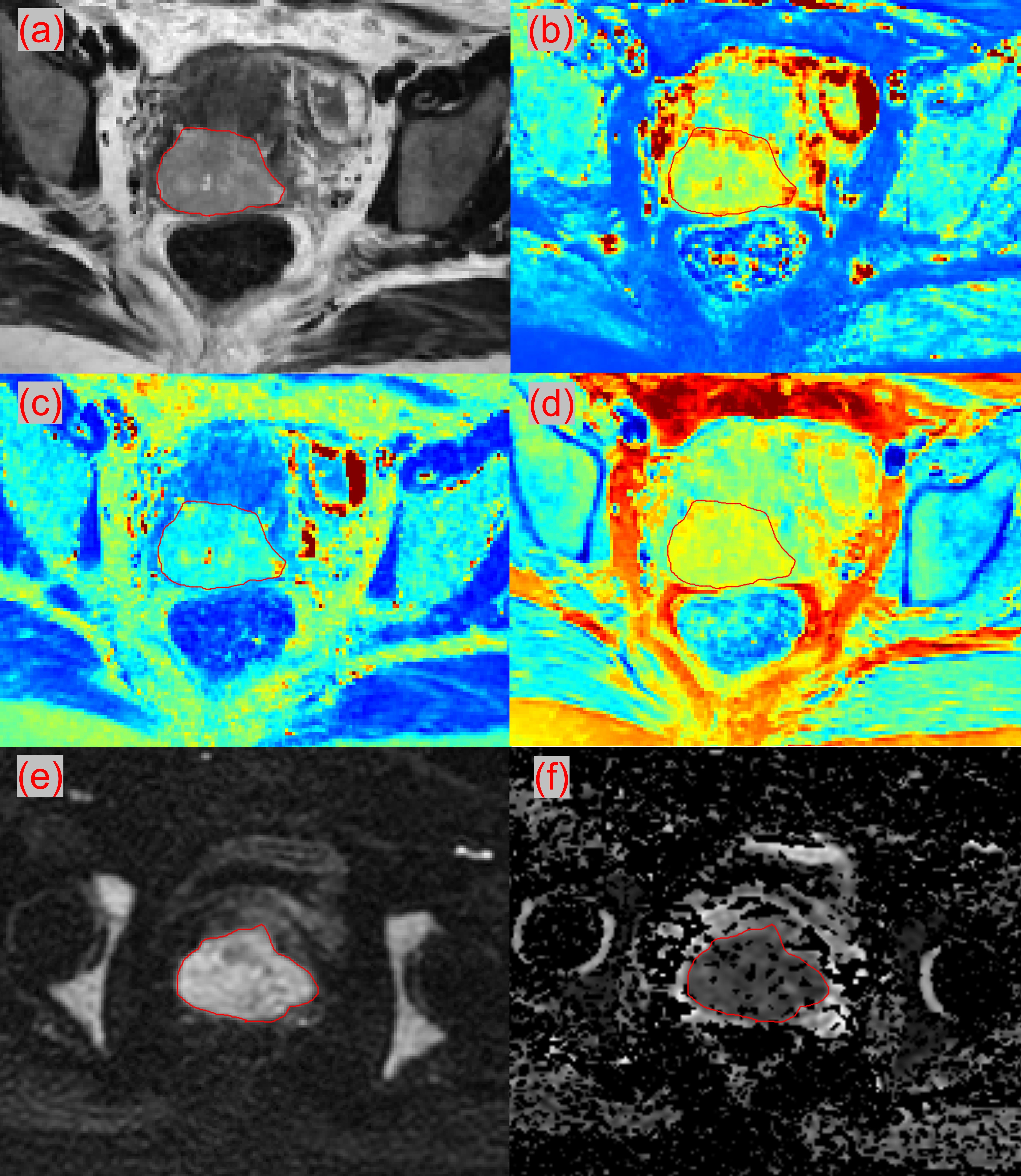
Figure 1. Example of (a) synthetic T2W image, (b) T1 map, (c) T2 map, (d) PD map, (e) DWI with b=800 and (f) ADC map.
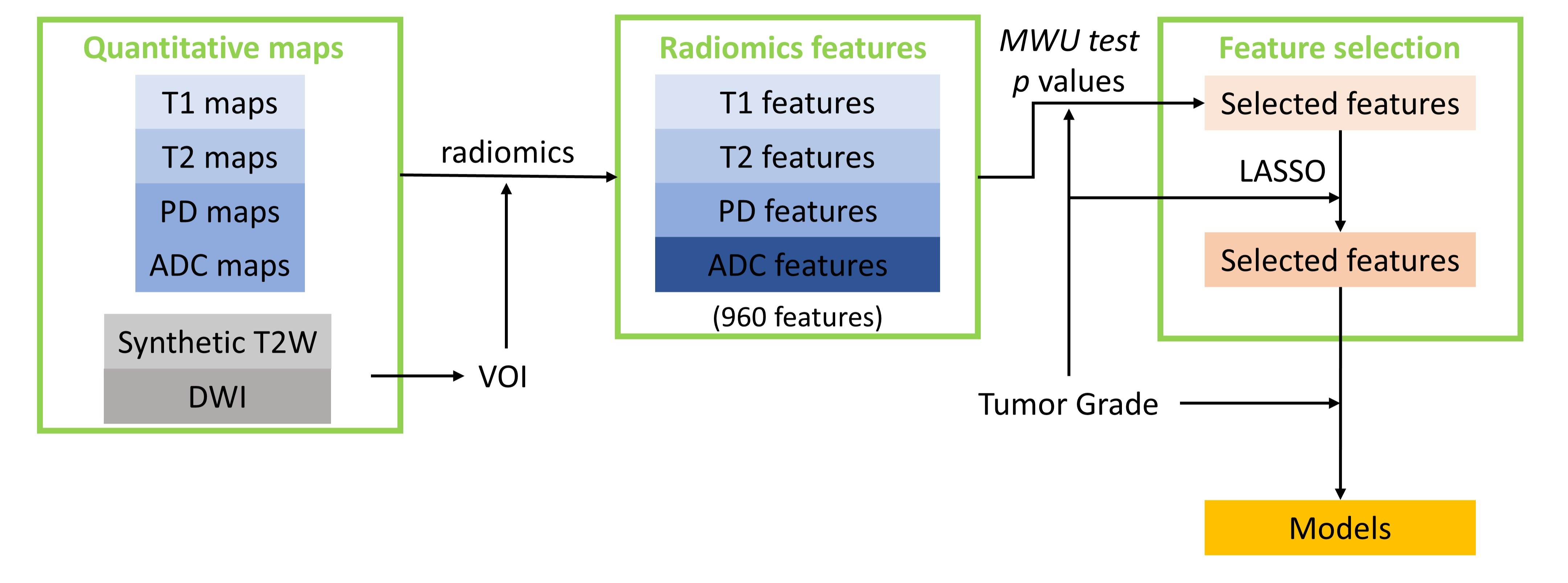
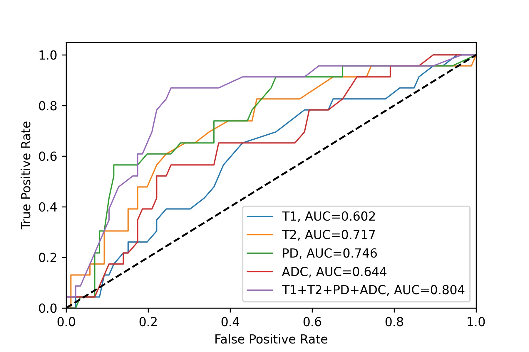
Figure 3. The micro-averaged ROC curves and corresponding AUCs for radiomics features extracted from T1 map, T2 map, PD map, ADC map and all maps.