0438
APT weighted imaging combined with IVIM to evaluate Her-2 gene expression in endometrial cancer1Department of Radiology,, The First Affiliated Hospital of Dalian Medical University, Dalian, China, China, 2Department of Radiology, The First Affiliated Hospital of Dalian Medical University, Dalian, China, China, 3Clinical & Technical Support, Philips Healthcare, Beijing, China, China, 4Clinical & Technical Support, Philips Healthcare, Shanghai, China, China
Synopsis
Keywords: Pelvis, Quantitative Imaging
it is necessary to find a method for non-invasive monitoring of Her-2 status. MRI with characteristics of multi-direction, multi-parameter, multi-function, high soft-tissue resolution, and non-invasiveness, has become the preferred method for evaluation of uterine lesions.Introduction and Purpose
We aimed to investigate the value of amide proton transfer weighted imaging(APTw) and intravoxel incoherent motion(IVIM) in evaluating the expression of human epidermal growth factor receptor-2(Her-2) gene in endometrial cancer (EC)Materials and Methods
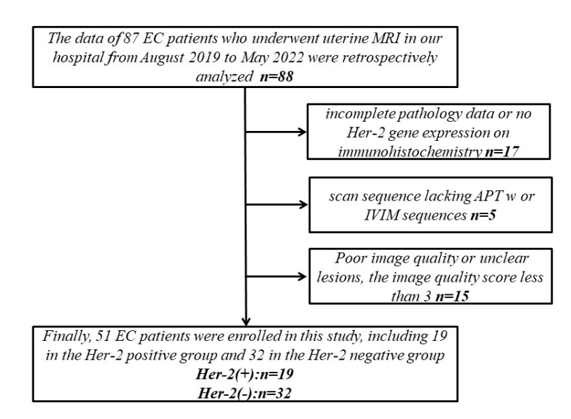
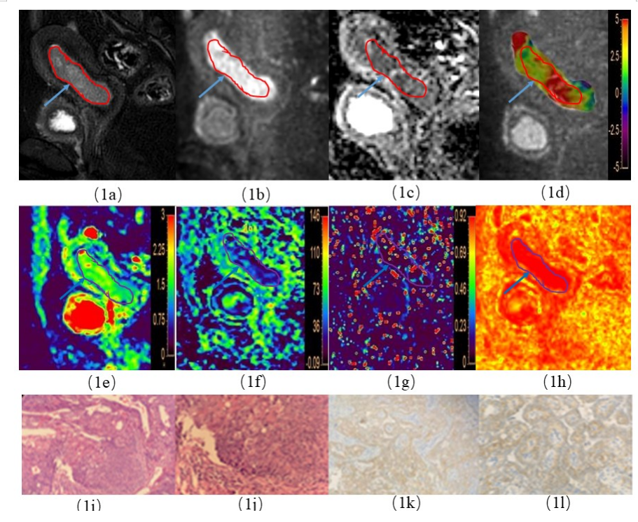
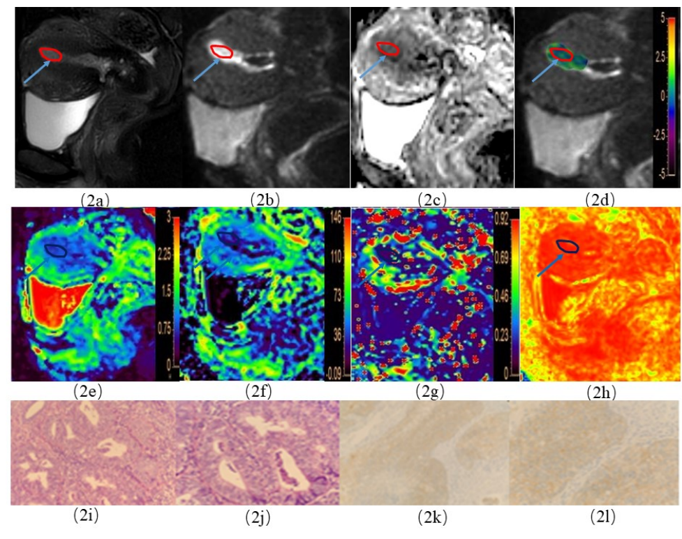
The clinical and imaging data of 51 EC patients who were pathologically confirmed in our hospital from June 2019 to May 2022 were retrospectively analyzed, and they were divided into Her-2 positive (19 cases) and Her-2 negative groups (32 cases). MR examination included APTw and IVIM imaging. The quantitative maps of APT values and IVIM parameters including the low apparent diffusion coefficient (D), fast apparent diffusion coefficient (D*), and perfusion fraction (f) were obtained. The ROC curve was used to evaluate the performance of parameters for discrimination of EC Her-2 gene expressions. Logistic regression was used to assess the association between the Her-2 positive EC and the risk factors. The Delong test was used to compare the differences among the AUCs. The correlation between parameters by APTw and IVIM methods was analyzed by Spearman correlation analysis.Results
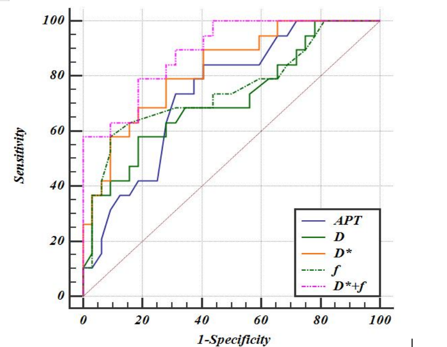
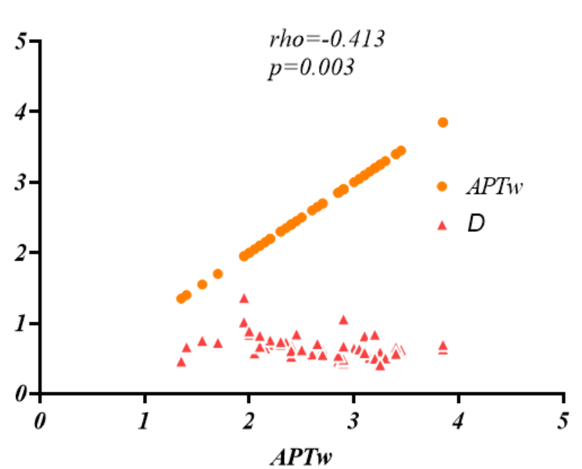
The APT value, D* value, and f value of EC in the Her-2 positive group were higher than those in Her-2 negative group, while the D value of EC in the Her-2 positive group was lower than that of the Her-2 negative group, and the difference were statistically significant (P<0.05). Multivariate logistic regression analysis showed that D* and f were independent risk factors for predicting EC Her-2 gene expression. There was a certain correlation between the APT value and D value (r=-0.413, P<0.05). And the ROC curves indicated that the combination of D* and f values could distinguish the Her-2 gene expression of EC with the highest diagnostic efficacy (AUC=0.890), even without significant difference to those by APT (AUC=0.740), D (AUC=0.714) or D* (AUC=0.822) value, separately.Discussion and Conclusion
The human epidermal growth factor receptor-2 (Her-2) gene is a proto-oncogene located on the long arm of chromosome 17, encoding a transmembrane tyrosine kinase receptor, which is overexpressed in the patients of breast cancer, gastric cancer, ovarian cancer, endometrial cancer (EC) and other malignant tumors. The overexpression of Her-2 can cause abnormal cell proliferation thus leading to malignant transformation, and it is related to tumor angiogenesis and tumor metastasis [1-3]. A previous study has shown that the expression Her-2 gene in EC was related to tumor differentiation degree, deep muscle invasion (DMI), and lymph node metastasis (LNM), and affects clinical treatment decisions [4].At present, the expression of Her-2 is generally detected by taking biopsy specimens for immunohistochemistry (IHC) and fluorescence in situ hybridization (FISH). Due to the spatial and temporal heterogeneity in the expression of Her-2, the status of Her-2 may also change during the treatment of the lesion, and thus repeated detection of the Her-2 gene expression is required after tumor recurrence and/or metastasis, and before the second-line Her-2 targeted therapy. However, it is not feasible in clinical practice to reassess Her-2 status through repeated tissue biopsies. Therefore, it is necessary to find a method for non-invasive monitoring of Her-2 status.
We found that APT value, D* value, and f value were higher in the Her-2 positive group than in the Her-2 negative group, meanwhile, the D* value and f value are independent risk factors for predicting Her-2 expression in EC. The combination of D* and f values showed a high diagnostic efficacy in differentiation of Her-2 gene expression in EC, while without significant difference to those by APT, D, D* separately. No significant correlation was found between APTw and IVIM parameters (D* and f) except D.
Acknowledgements
No.References
[1]. Diver EJ, Foster R, Rueda BR, Growdon WB: The Therapeutic Challenge of Targeting HER2 in Endometrial Cancer. Oncologist 2015, 20(9):1058-1068.2.
[2]. Buchynska LG, Brieieva O, Iurchenko NP: Assessment of HER-2/neu, с-MYC and CCNE1 gene copy number variations and protein expression in endometrial carcinomas. Experimental Oncology 2019, 41.3. [3]. Laterza MM, Ciaramella V, Facchini BA, Franzese E, Liguori C, De Falco S, Coppola P, Pompella L, Tirino G, Berretta M et al: Enhanced Antitumor Effect of Trastuzumab and Duligotuzumab or Ipatasertib Combination in HER-2 Positive Gastric Cancer Cells. Cancers (Basel) 2021, 13(10).4.
[4]. Morrison C, Zanagnolo V, Ramirez N, Cohn D, Kelbick N, Copeland L, Maxwell L, Fowler J: HER2 Is an Independent Prognostic Factor in Endometrial Cancer: Association With Outcome in a Large Cohort of Surgically Staged Patients. Journal of Clinical Oncology - J CLIN ONCOL 2006, 24:2376-2385.
Figures