0414
Functional Abnormalities of Cerebellum in Vascular Cognitive Impairment1Department of Radiology, Zhongnan Hospital of Wuhan University, Wuhan, China, 2GE Healthcare, MR Research China, Beijing, Beijing, China, 3Zhongnan Hospital of Wuhan University, Wuhan, China
Synopsis
Keywords: Dementia, Brain Connectivity, vascular cognitive impairment
The potential prevalence of vascular cognitive impairment (VCI) is high; early diagnosis and treatment is essential for disease prognosis. In this study, compared to healthy controls (HCs), there were differences of functional connectivity (FC) between the cerebellum and cerebral regions of VCI patients, mainly involving default mode network (DMN), sensorimotor network (SMN) and frontoparietal network (FPN). Furthermore, FC between multiple sub-regions in the cerebellum reduced in patients with VCI compared to HCs. These findings may expand our understanding of the neural mechanisms of VCI in the perspective of neurovascular coupling.Objective
VCI is the second leading cause of dementia, despite those well-known pathological sources, the neural basis of VCI remains incomplete understood[3]. Recent neuroimaging studies have extensively described the atlas partitioning of the cerebellar cortex and its functional mappings related to cognitive function in cortical and subcortical brain regions. In recent years, the neuroimaging community has produced many novel techniques on the fine dissection mapping of the cerebellum[4], and the involvement of cerebellar-brain functional connectivity(FC) has produced numerous novel findings[5]. These results have contributed to the study of the cerebellum's involvement in the physiology and development of normal brain function, which also advanced the understanding of brain disorders, including cognitive disorders. However, mappings of alterations in VCI remains largely unexplored. This study aimed to perform functional FC analysis using high-resolution dissection mapping of the cerebellum, including the intra-cerebellar FC and cerebellar-cerebral FC, to investigate the potential associations between abnormal cerebellar connectivity and changes in cognitive function.Materials and Methods
MRI data were collected on a 3.0T MR scanner (Signa 750w, GE Healthcare, USA). 72 VCI patients, including 38 small vessel mild cognitive impairment (SVMCI) and 34 post-stroke mild cognitive impairment (PSMCI), and 43 demographically matched HCs were included in this study. This study dissected the cerebellum using Diedrichsen 2009 Probabilistic Atlas of the Cerebellum[6]. The atlas was open for download and available to all researchers. The atlas divided the entire cerebellum into 34 sub-regions, to characterize FC alterations in cerebellar sub-regions with other brain regions of VCI patients, this study performed seed-to-voxel FC in 34 regions of the cerebellum to obtain whole-brain FC correlation coefficients at the voxel level. Changes of FC between sub-regions in the cerebellum and from each cerebellar sub-region to the cerebral seed points in VCI patients were calculated, and the association of these changes with cognitive function was examined.Results
Compared to HCs, a total of 11 cerebellar sub-regions in VCI patients had significant differences of FC in the cortical and sub-basal ganglia cortical brain regions, mainly involved in the DMN, SMN, and FPN brain regions. In the intra-cerebellar FC analysis, 45 or 8% of the total number of cerebellar connections had significant inter-group differences of mainly reduced FC respectively in SVMCI and PSMCI patients. Both SVMCI and PSMCI subjects showed significant decreases in FC of cerebellar subregions to the cerebral cortex and sub-basal ganglia cortex (P < 0.05, FWE correction). Significant positive correlations were found between FC and MoCA scores in both 38 subregions of cerebellum-cerebral FC and 34 subregions of intra-cerebellum FC.Discussion and conclusion
Our results were consistent with recent studies of changes in cerebellum structure and function as VCI progresses[7]. This study examined FC changes between the cerebellum's cognitive sub-regions and the whole brain in VCI patients with HCs. First, after adjusting for age, sex, and education, the brain regions with altered FC were mainly distributed in the DMN, FPN and SMN. The present study showed significant changes in FC between the cerebellum and cerebral cognitive regions in the SVMCI and PSMCI groups. However, the pathophysiology of how the cerebellum causes dystonia is unclear. Further, the dysfunction of the SMN in AD and aMCI patients has been confirmed in other studies[8], which suggests reduced cerebellar subregions' connectivity to the SMN and diminished FC of internal cerebellar sub-regions in VCI patients may be predictive markers of VCI. Therefore, reduced connectivity between the cerebellum and thalamus in VCI patients may indicate an impairment in the key relay point of the cerebello-thalamo-cortical pathway and affect the connectivity of the cerebellum to cerebral cognitive networks (including the DMN and limbic networks), thereby affecting cognitive function. The finding of prominent intra-cerebellar and cerebellar-cerebral FC abnormalities in VCI patients adds to the evidence for a possible role of the cerebellum in cognitive processes. Patterns in FC changes in VCI patients and their correlations with neuropsychological measures may be a pathophysiological foundation for cognitive impairment, which provides new information to explore the involvement of the cerebellum in VCI that may aid the early diagnosis of VCI.Acknowledgements
The author(s) declared no potential conflicts of interest with respect to the research, authorship, and/or publication of this article.References
[1]Stoodley C J. The cerebellum and cognition: evidence from functional imaging studies[J]. Cerebellum, 2012,11(2):352-365.
[2] Stoodley C J, Valera E M, Schmahmann J D. An fMRI study of intra-individual functional topography in the human cerebellum[J]. Behav Neurol, 2010,23(1-2):65-79.
[3] Hautzel H, Mottaghy F M, Specht K, et al. Evidence of a modality-dependent role of the cerebellum in working memory? An fMRI study comparing verbal and abstract n-back tasks[J]. Neuroimage, 2009,47(4):2073-2082.
[4] Blackwood N, Ffytche D, Simmons A, et al. The cerebellum and decision making under uncertainty[J]. Brain Res Cogn Brain Res, 2004,20(1):46-53.
[5] Ren Y, Guo L, Guo C C. A connectivity-based parcellation improved functional representation of the human cerebellum[J]. Sci Rep, 2019,9(1):9115.
[6] Fu Z, Zhao M, Wang X, et al. Altered neuroanatomical asymmetries of subcortical structures in subjective cognitive decline, amnestic mild cognitive impairment, and Alzheimer’s disease[J]. Journal of Alzheimer's Disease, 2021,79(3):1121-1132.
[7] Jacobs H I, Hopkins D A, Mayrhofer H C, et al. The cerebellum in Alzheimer’s disease: evaluating its role in cognitive decline[J]. Brain, 2018,141(1):37-47.
[8] Cai C, Huang C, Yang C, et al. Altered patterns of functional connectivity and causal connectivity in salience subnetwork of subjective cognitive decline and amnestic mild cognitive impairment[J]. Frontiers in neuroscience, 2020,14:288.
Figures
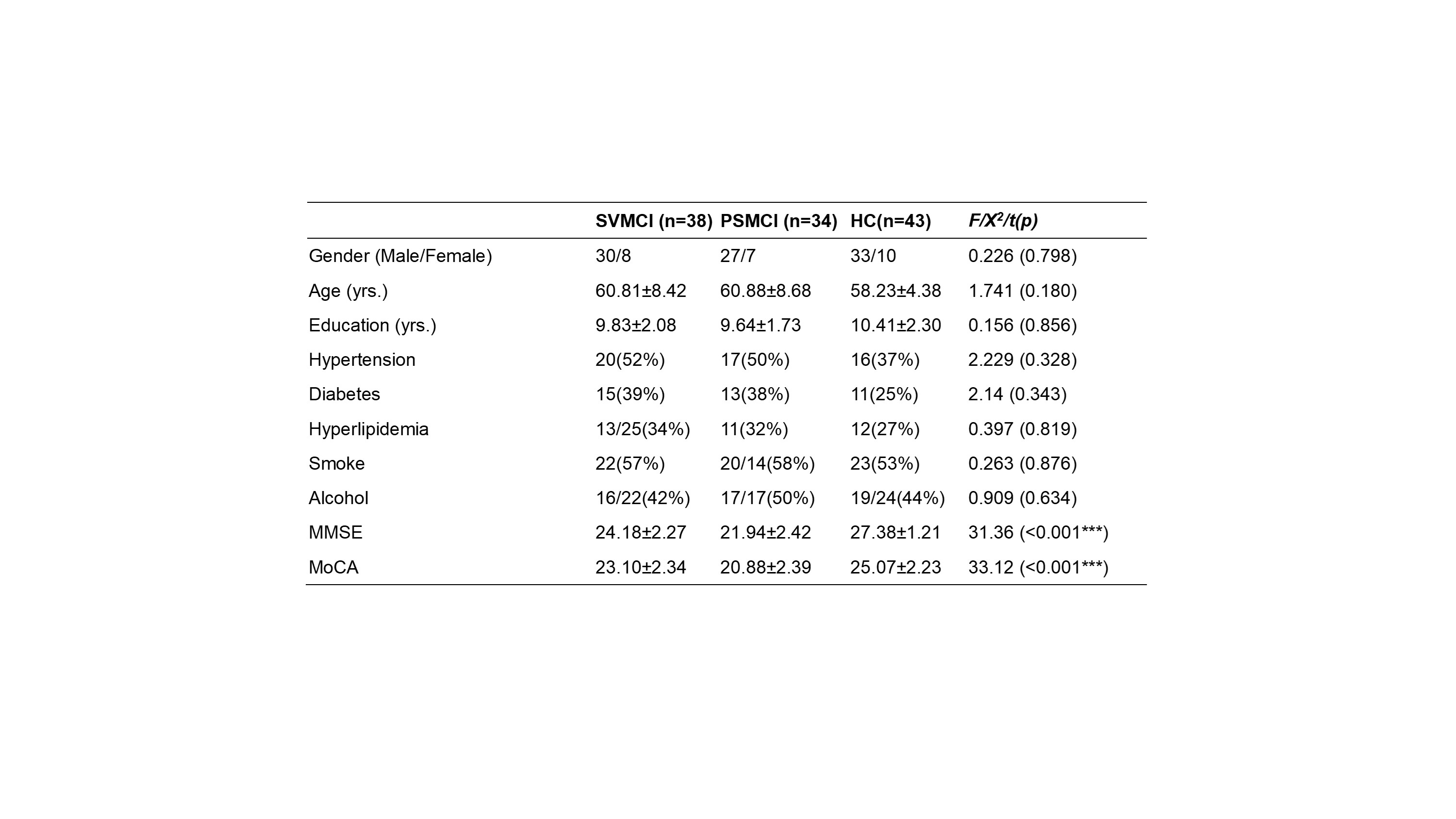
FIGURE 1 Demographics of 122 subjects recruited in this study
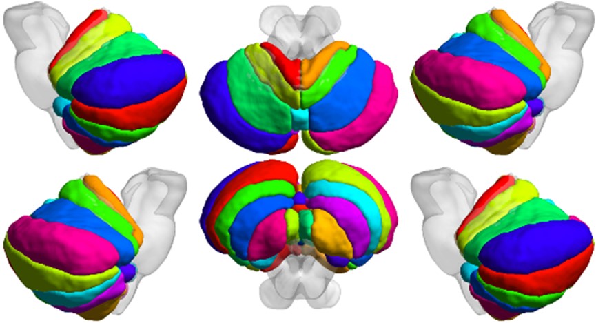
FIGURE 2 Diedrichsen 2009 cerebellar probability atlas
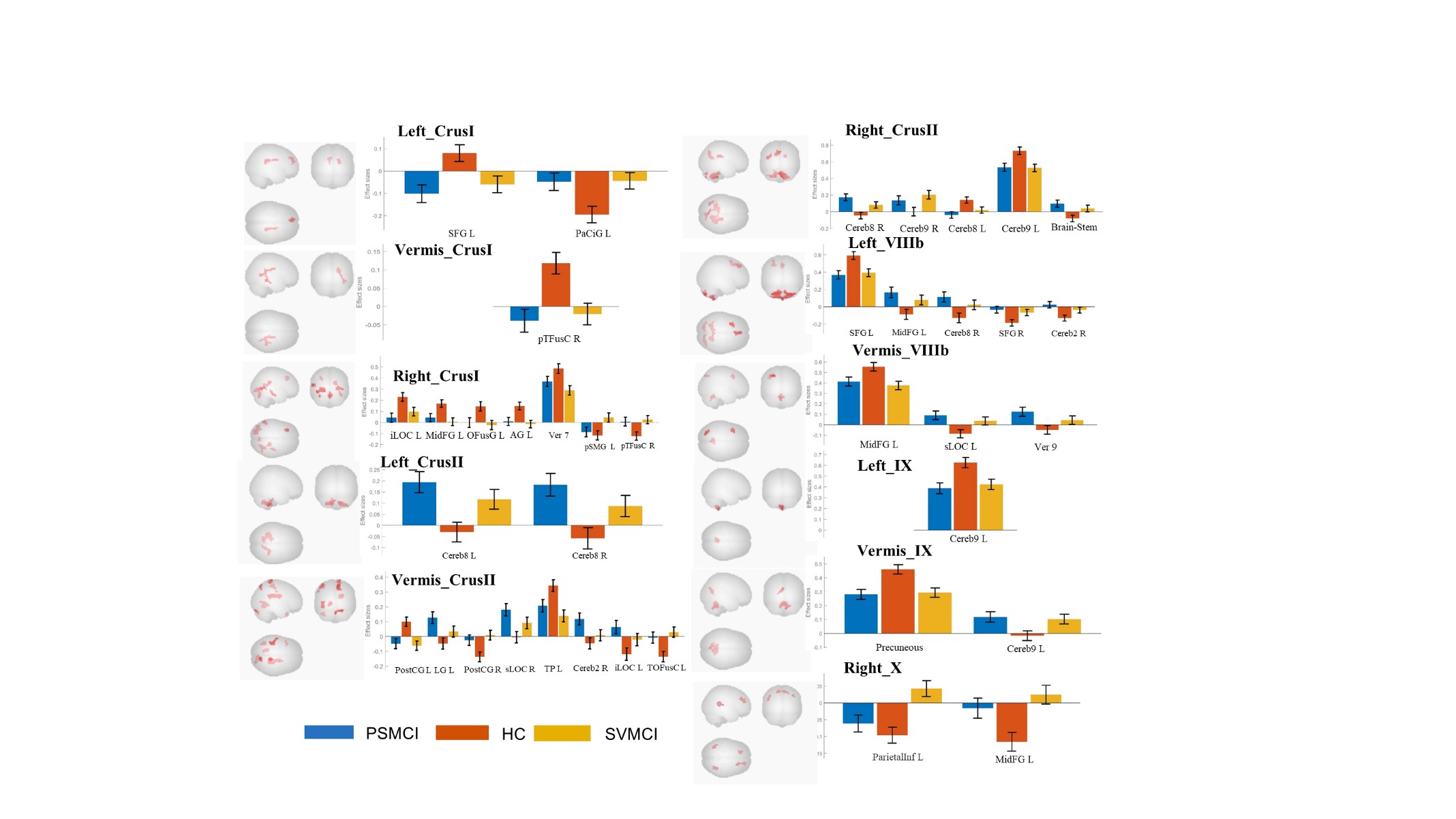
FIGURE 3 Differences in FC of cerebellar subregions to the cerebral cortex and sub-basal ganglia cortex. SVMCI, small vessel mild cognitive impairment; PSMCI, post-stroke mild cognitive impairment; HCs, healthy controls
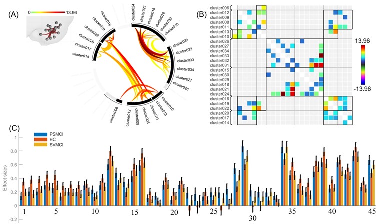
FIGURE 4 (A) A connectome map of the cerebellum dissected into 34 regions; (B) A matrix map of the cerebellum dissected into 34 regions; (C) Bar graph of 45 groups of connections with significant differences within the cerebellum, accounting for 8% of the total number of connections, which demonstrates the differences in strength and directionality of connections between the three groups.SVMCI, small vessel mild cognitive impairment; PSMCI, post-stroke mild cognitive impairment; HC, healthy controls
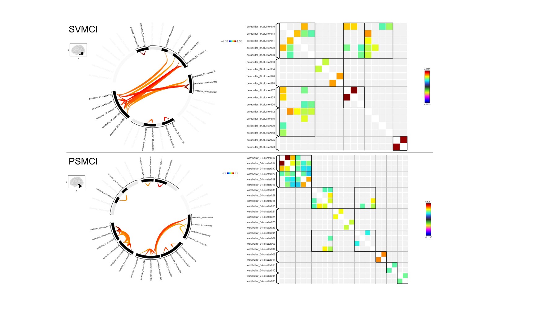
FIGURE 5 | Correlation between the functional connectivity (FC) and cognitive function scores in the SVMCI and PSMCI green patients. Significant correlation between FC changes and cognitive function scales include MOCA in 34 cerebellum sub-regions (Bonferroni corrected, p < 0.05). Age, gender, and years of education were used to control variables of the results; SVMCI, small vessel mild cognitive impairment; PSMCI, post-stroke mild cognitive impairment; HC, healthy controls; MOCA, Montreal Cognitive Assessment