0382
Deep Learning Reconstruction for Combined 8-fold Accelerated Parallel Imaging and Simultaneous Multislice Acquisition1Siemens Healthineers, Princeton, NJ, United States, 2Siemens Healthcare GmbH, Erlangen, Germany, 3Department of Radiology, NYU Grossman School of Medicine, New York, NY, United States
Synopsis
Keywords: Machine Learning/Artificial Intelligence, MSK
Combining parallel imaging (PI) and simultaneous multislice (SMS) acceleration realized a clinical 4-fold accelerated 2D TSE MRI of the knee. However, 8-fold acceleration with conventional reconstruction methods suffers from significant image quality degradation. We propose a complete DL approach for combined slice separation and k-space-to-image reconstruction of SMS-PI-accelerated knee MRI with tunable denoising strength and super-resolution image enhancement. The proposed methods enable artifact-free image reconstruction of 8-fold accelerated 2D TSE MR images in multiple planes and with multiple image contrasts. Clinical evaluations suggest equivalence of image quality and detection rates of 8-fold S2P4 DL reconstructions compared to the reference standard.Introduction
Despite achievements in accelerating MR imaging, there is still a high demand for faster imaging at high image quality since the demand for MRI is continuously growing. The combination of parallel imaging (PI) and simultaneous multislice (SMS) acceleration [1] realized clinical 4-fold accelerated (S2P2; 2-fold SMS×2-fold PI) 2D TSE MRI of the knee and other peripheral joints [2]. However, 6-fold S2P3 and 8-fold S2P4 acceleration with conventional reconstruction methods suffer from a significant image quality degradation, including a large drop in the signal-to-noise ratio (SNR) and the presence of inter-slice leakage and aliasing artifacts [3]. With the increasing success of deep learning (DL) accelerated MRI reconstruction models, a dedicated approach incorporating the SMS imaging model into a DL framework is highly desirable to enable an end-to-end framework for optimal reconstructions tailored for this use case [4]. We propose a complete DL approach for combined slice separation and k-space-to-image reconstruction of SMS-PI-accelerated knee MRI with tunable denoising strength and super-resolution image enhancement.Methods
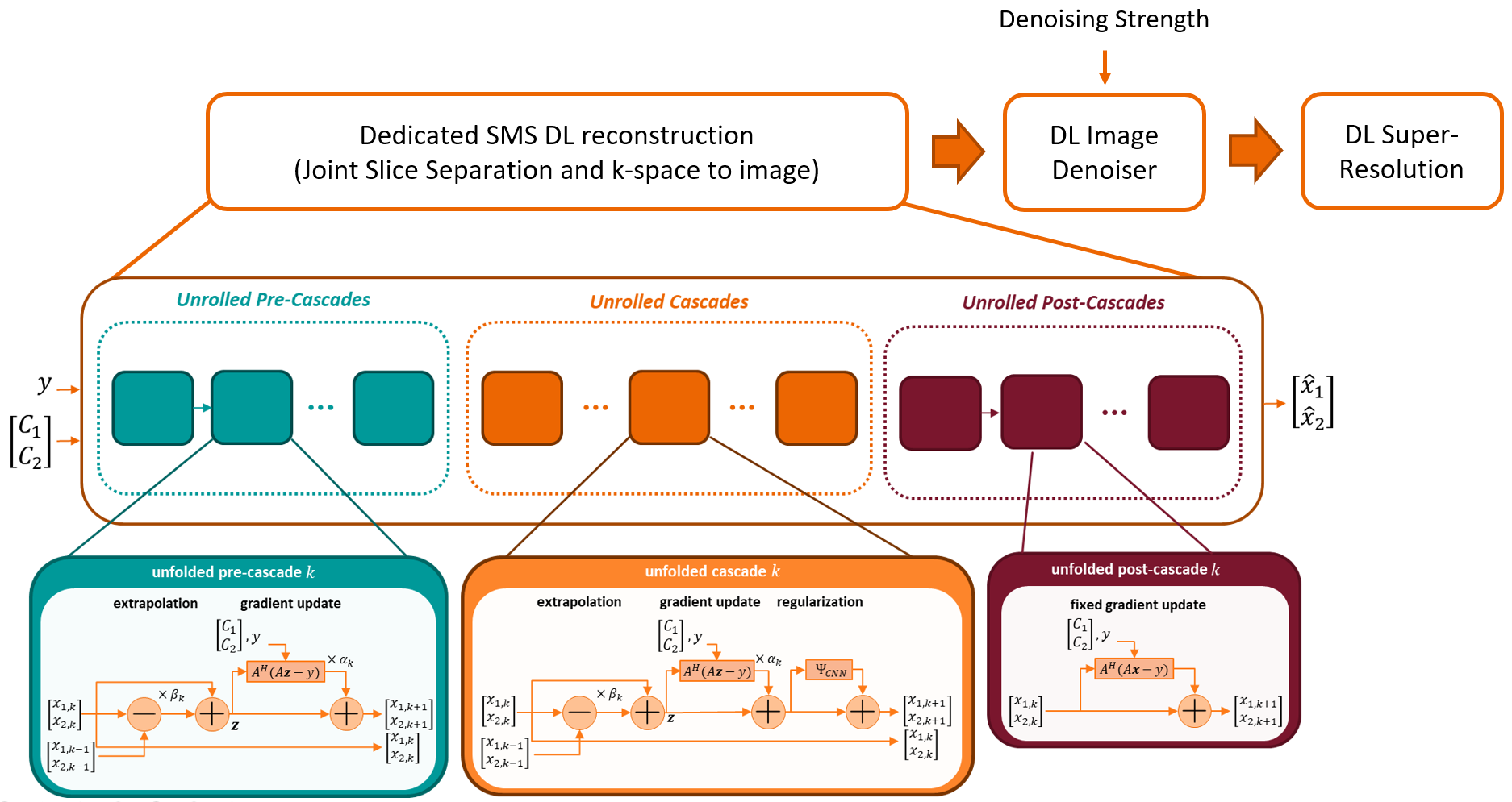
We propose a DL SENSE-based SMS-PI-accelerated reconstruction with sensitivity maps estimated from a separate reference scan [5]. The DL network unrolls a proximal gradient algorithm with an extrapolation mechanism based on Nesterov momentum and a convolutional neural network (CNN) regularizer [6]. Current reconstruction approaches to SMS-PI-accelerated data are performed in two separate stages, first separating the data of aliased slices before applying a PI-base reconstruction. On the other hand, the proposed reconstruction network accepts SMS-accelerated data as input. It outputs the images of all acquired slices, handling both SMS and PI accelerations in a single step.The forward and backward linear operators used in gradient steps also model the SMS acquisition using a SENSE formulation [7]. While the backward operator can be implemented with the adjoint of the multislice coil sensitivities and asymptotically separates the slices, direct separation can be achieved with a slice SENSE inversion, i.e., by using the pseudo-inverse of the C×S coil sensitivities at every voxel where C is the number of coils and S the number of slices. In the SMS protocol, acquiring one of the slices with a phase modulation improves the coil array’s conditioning; such phase modulation can translate into a circular shift in the MRI images. The circular shift is inverted after reconstruction. To avoid boundary artifacts from the shifts, the regularizer CNN can be made equivariant to circular shifts by performing periodic padding of the feature maps at every convolutional layer.
A DL denoising module is plugged at the end of the unrolled network to generate output with an adjustable denoising strength. The denoised image is obtained through a convex combination of the unrolled network's output and a denoising network's output. Finally, the sharpness of the MR images is enhanced using a DL single image super-resolution network to upscale the input spatial resolution with the risk of hallucination minimized through hard data consistency constraints.
Materials and Experimental Setting
Experiments were performed on 1.5T and 3T data acquired using a S2P2 5-minute 2D TSE knee protocol (118,794 2D slices for training, 13,342 2D slices for testing). Retrospective down-sampling was applied to S2P2 data with an equispaced sampling mask to obtain corresponding S2P4 data. Ground truth was obtained from S2P2 using standard GRAPPA reconstruction. The dedicated DL SMS reconstruction model was trained using combined complex-L1 loss and Wasserstein GAN adversarial loss. The image denoiser and super-resolution networks were trained using complex-L1 loss. The proposed research application was compared against corresponding non-DL SENSE-based reconstruction [5].Clinical Evaluation
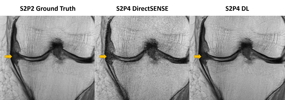
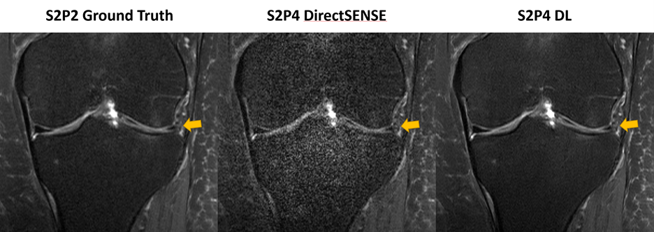
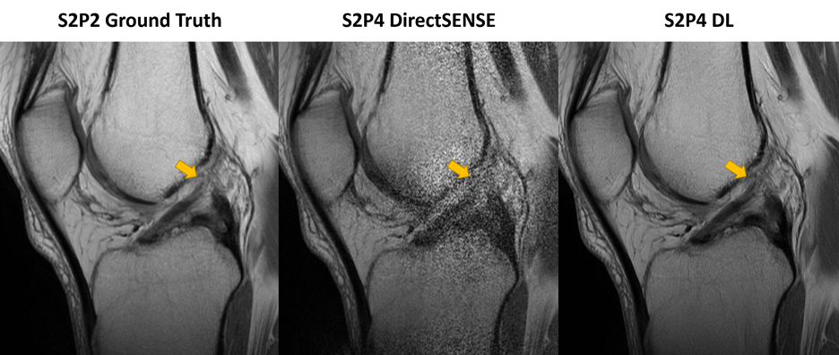
25 participants with painful knee conditions prospectively underwent S2P2 5-minute 2D TSE knee protocol at 3T. The corresponding S2P2 ground truth, S2P4 DL, and S2P4 non-DL SENSE datasets were randomized and independently evaluated by a musculoskeletal radiologist. Outcome variables included image quality using 5-point Likert scales (1=worse, 5=best) and the detection of meniscal, tendinous, ligamentous, and osteocartilaginous injuries. Statistical analyses included difference, kappa-based inter-method agreements, and interchangeability tests. P-values of less than 0.05 were considered statistically significant.Results
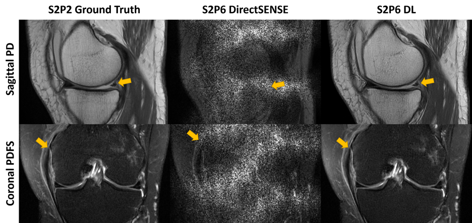
Quantitative evaluation: Compared to the ground truth, the proposed dedicated DL SMS reconstruction model (1.5T: PSNR=38.468 dB, SSIM=0.938; 3T; PSNR=39.123 dB, SSIM=0.943) significantly outperforms the conventional SENSE-based approach (1.5T: PSNR=17.904 dB, SSIM=0.653; 3T: PSNR=17.845 dB, SSIM=0.612).Clinical Evaluation: The image quality was similar (p=0.58) between S2P4 DL (4[3-5]) and S2P2 ground truth (4[3-5]), and significantly better (p<0.001) than S2P4 non-DL SENSE (2[1-3]) (Figure 2). The detection frequencies of abnormalities were statistically similar (p=0.23-0.88) between S2P4 DL and S2P2 ground truth and significantly lower on S2P4 non-DL SENSE MR images (Figure 3). S2P4 DL and S2P2 ground truth high inter-method agreements (κ=0.73-0.98) were statistically interchangeable for detecting meniscal, tendinous, ligamentous, and osteocartilaginous injuries (Figure 4). DL image reconstruction did not omit any findings and did not add artificial structures. Perspective: early results suggest the feasibility of DL reconstruction of 12-fold S2P6 acceleration (Figure 5).
Conclusion
The proposed DL SMS reconstruction model enables the artifact-free image reconstruction of 8-fold S2P4 accelerated 2D TSE MR images in multiple planes and with multiple image contrasts. Clinical evaluation suggests equivalence of image quality and detection rates compared to an S2P2 reference standard and superiority to an S2P4 non-DL SENSE reference standard. Early results suggest the feasibility of 12-fold S2P6 DL acceleration.Acknowledgements
References
[1] Fritz J, Fritz B, Zhang J, Thawait GK, Joshi DH, Pan L, Wang D. Simultaneous Multislice Accelerated Turbo Spin Echo Magnetic Resonance Imaging: Comparison and Combination With In-Plane Parallel Imaging Acceleration for High-Resolution Magnetic Resonance Imaging of the Knee. Invest Radiol. 2017 Sep;52(9):529-537. Doi: 10.1097/RLI.0000000000000376. PMID: 28430716.
[2] Del Grande F, Rashidi A, Luna R, Delcogliano M, Stern SE, Dalili D, Fritz J. Five-Minute Five-Sequence Knee MRI Using Combined Simultaneous Multislice and Parallel Imaging Acceleration: Comparison with 10-Minute Parallel Imaging Knee MRI. Radiology. 2021 Jun;299(3):635-646. doi: 10.1148/radiol.2021203655. Epub 2021 Apr 6. PMID: 33825510.
[3] Fritz J, Guggenberger R, Del Grande F. Rapid Musculoskeletal MRI in 2021: Clinical Application of Advanced Accelerated Techniques. AJR Am J Roentgenol. 2021 Mar;216(3):718-733. doi: 10.2214/AJR.20.22902. Epub 2021 Feb 3. PMID: 33534618.
[4] Lin, Dana J. MD; Walter, Sven S. MD; Fritz, Jan MD. Artificial Intelligence–Driven Ultra-Fast Superresolution MRI: 10-Fold Accelerated Musculoskeletal Turbo Spin Echo MRI Within Reach. Investigative Radiology: November 2, 2022 - Volume - Issue - 10.1097/RLI.0000000000000928 doi: 10.1097/RLI.0000000000000928
[5] Uecker M, Lai P, Murphy MJ, Virtue P, Elad M, Pauly JM, Vasanawala SS, Lustig M. ESPIRiT—an eigenvalue approach to autocalibrating parallel MRI: where SENSE meets GRAPPA. Magnetic resonance in medicine. 2014 Mar;71(3):990-1001.
[6] Herrmann J, Koerzdoerfer G, Nickel D, Mostapha M, Nadar M, Gassenmaier S, Kuestner T, Othman AE. Feasibility and implementation of a deep learning MR reconstruction for TSE sequences in musculoskeletal imaging. Diagnostics. 2021 Aug 16;11(8):1484.
[7] Zahneisen B, Ernst T, Poser BA. SENSE and simultaneous multislice imaging. Magnetic resonance in medicine. 2015 Nov;74(5):1356-62.
Figures