0381
Rapid 3D SPirAl Respiratory and Cardiac Self-gated (SPARCS) Cine Imaging with a DEep learning-based rapid Spiral Image REconstruction (DESIRE)1Stanford Univeristy, Stanford, CA, United States, 2Beijing University of Posts and Telecommunications, Beijing, China, 3Department of Radiology and Biomedical Imaging, University of California, San Francisco, San Francisco, CA, United States
Synopsis
Keywords: Machine Learning/Artificial Intelligence, Cardiovascular
We developed a rapid 3D self-gated cardiac cine technique, using a variable-density undersampled randomized stack of spiral gradient echo sequence to perform cine evaluation of the whole left ventricle. Our proposed slice-by-slice deep learning-based imaging reconstruction technique for self-gated free-breathing 3D stack of spiral cardiac cine imaging can produce cine images with high temporal (40 ms) and spatial resolution (1.25x1.25x8 mm) within a 20s acquisition time with <1s deep learning inference time.Introduction
Breath-held segmented cine imaging is the gold standard for analyzing cardiac function, but it uses ECG gating and requires 10-12 breath-holds to cover the left ventricle. Our group has developed a continuous-acquisition respiratory and cardiac self-gated cine sequence for free-breathing cardiac function acquisition using 2D stack spiral acquisition1. However, the acquisition still takes 2-3 min to cover the left ventricle, and it would benefit from the SNR efficiency of 3D imaging. Thus, we develop a rapid 3D self-gated cine, using randomized stack of spiral gradient echo sequence to perform 3D cine evaluation of the left ventricle2. Iterative compressed sensing image reconstruction of 3D spiral imaging is time-consuming and not clinically feasible. In this project, we develop high-resolution 3D cardiac cine imaging using rapid spiral acquisitions and deep learning-based imaging reconstruction to enable rapid online reconstruction for cardiac cine imaging feasible.Methods
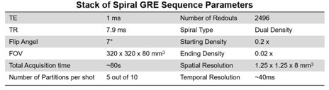
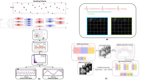
Data Acquisition: 11 patients were imaged on a 3T SIEMENS scanner (MAGNETOM Skyra; Siemens Healthineers, Erlangen, Germany). Imaging occurred 3 minutes after the injection of 0.15 mmol/kg of Clariscan or Gadovist. Stack of spiral gradient echo parameters are show in Table 1. Our stack-of-spiral sampling strategy consisted of a variable kz-density stack of dual density spirals rotated by the golden angle. The kz-t sampling pattern is shown on the top panel of Figure 1(A). We acquired 5 partitions out of 10 total partitions. The first partition (shown in rectangles and black spirals) of each measurement is set to kz=0 and used as a self-gating navigator. The other sampled kz partition in each measurement are randomly picked from a Bates distribution3:$$P(t,n) = -P_c+P_t\lbrace{\frac{n}{2(n-1)!}\sum\limits_{k=0}^{n}(-1)^k\tbinom{n}{k}(nt-k)^{n-1}sgn(nt-k)}\rbrace$$
where $$$P(t,n)$$$ is desired sampling partition index, $$$P_t$$$ is fully-sampled partition number, $$$P_c$$$ is central partition index, n is density factor, n is larger for more centralized distribution. To distribute the spirals across the partition we made sure that no kz partitions were repeated in each group, and we insured that there were equal numbers of spirals collected in each half of the kz plane.
Image Reconstruction for Reference Images: Reference images were reconstructed with a 3D iterative L1-SENSE–based reconstruction technique for 3D SPARCS imaging. The reconstruction pipeline for the reference images is shown in Figure 1. The automatic cardiac self-gating pipeline is shown in Figure 1(A): Self-gating cardiac signals were determined using the center point of the central partition each group of spiral partitions for all receiver coils. Principal component analysis was performed to separate the cardiac and respiratory components followed by frequency spectrum analysis to extract filtered cardiac and respiratory signals. Figure1(B) shows the auto coil selection over whole 3D volume. Figure 1(C) shows the retrospective cardiac binning followed by rigid motion correction in k space and non-Cartesian spiral L1-SENSE reconstruction4,5,6. The objective function was solved using non-linear conjugate gradient algorithm with 30 iterations.
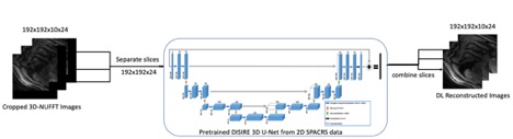
Proposed Image Reconstruction Network: Figure 2 shows the proposed 3D DESIRE U-Net image reconstruction framework. Following binning and motion correction, the 3D SPARCS data is reconstructed using a 3D NUFFT followed by optimal coil combination. The real and imaginary parts of the 2D images from each Z-location were concatenated into two channels and fed into the complex-convolutional 3D DESIRE-UNET that was previously trained for 2D SPARCS data7.
Results
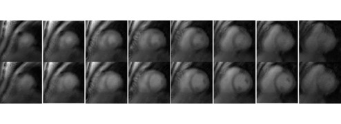
Figure 3 shows the DL reconstruction results cine images for different retrospective under sampled rates. 2x corresponds 40s acquisition 3x corresponds 30s acquisition and 4x corresponds 20s acquisition Figure 4 shows the end diastole and end systole images reconstructed by our proposed reconstruction techniques for whole heart coverage. The best trade-off between data acquisition and reconstruction quality was for the 30-second data acquisition. The proposed network had an inference time of <1 seconds as compared to 90 minutes for the 3D-L1 SENSE reconstruction.Conclusion and Discussion
Our proposed deep learning-based imaging reconstruction technique for self-gated free-breathing 3D stack of spiral cardiac cine imaging can produce cine images with high temporal and spatial resolution with a 20s acquisition with a deep-learning inference time of <1s. We anticipate that image quality could be further improved using the pre-trained DESIRE network followed by transfer-learning with fully sampled 3D SPARCS cine data. We will also investigate a 4D U-Net model for a 3D+time reconstruction.Acknowledgements
No acknowledgement found.References
1. Zhou R, Yang Y, Mathew RC, et al. Free-breathing cine imaging with motion-corrected reconstruction at 3T using SPiral Acquisition with Respiratory correction and Cardiac Self-gating (SPARCS). Magn Reson Med. 2019;82(2):706-720.
2. Wang X, Wang J, Zhou R, Salerno M.Rapid Free-breathing 3D SPirAl Respiratory and Cardiac Self-gated (SPARCS) Cine Acquisition Using an Undersampled Stack-of-Spirals. In Proceedings of the ISMRM 30th Annual Scientific Sessions, London, England, UK, 2022
3. K. Buchanan, T. Adeyemi, C. Flores-Molina, S. Wheeland and D. Overturf, "Sidelobe behavior and bandwidth characteristics of distributed antenna arrays," 2018 United States National Committee of URSI National Radio Science Meeting (USNC-URSI NRSM), 2018, pp. 1-2.
4. Otazo R, Kim D, Axel L, Sodickson DK. Combination of compressed sensing and parallel imaging for highly accelerated first-pass cardiac perfusion MRI. Magnetic Resonance in Medicine 2010;64:767–776.
5. Feng L, Grimm R, Block KT, et al. Golden-angle radial sparse parallel MRI: Combination of compressed sensing, parallel imaging, and golden-angle radial sampling for fast and flexible dynamic volumetric MRI. Magnetic Resonance in Medicine 2014;72:707–717.
6. Knoll F, Schwarzl A, Diwoky C, Sodickson DK. gpuNUFFT – An Open Source GPU Library for 3D Regridding with Direct Matlab Interface Proc Intl Soc Magn Reson Med. 22 (2014). p 4297
7. Wang J, Weller DS, Kramer CM, Salerno M. DEep learning-based rapid Spiral Image REconstruction (DESIRE) for high-resolution spiral first-pass myocardial perfusion imaging. NMR Biomed. 2022;35:e4661.
Figures