0335
3-Dimensional DANTE-prepared sequence with non-rigid motion-correction for high-resolution dark-blood aortic imaging1Biomedical Engineering, King's College London, London, United Kingdom, 2MR Research Collaborations, Siemens Healthcare Limited, Camberley, United Kingdom, 3Institute for Biological and Medical Engineering, Pontificia Universidad Católica de Chile, Santiago, Chile, 4Millennium Institute for Intelligent Healthcare Engineering, Santiago, Chile, 5School of Engineering, Pontificia Universidad Católica de Chile, Santiago, Chile
Synopsis
Keywords: Vessels, New Signal Preparation Schemes, Pulse Sequence Design
Dark-blood imaging is an important tool for vascular imaging in cardiovascular disease. A novel free-breathing water/fat DANTE-prepared sequence is introduced for 3D dark-blood imaging of the thoracic and abdominal aorta. The framework integrates image navigation to enable translational and non-rigid motion correction resulting in a predictable scan time along with dual-echo Dixon gradient-echo readout for robust fat suppression. Results from healthy subjects and patients demonstrate the feasibility of the sequence for high-resolution, aortic imaging in ~7-minute acquisition time.Introduction
Dark-blood cardiovascular MR imaging has been applied for the assessment of the aorta and the aortic vessel wall along with the detection of atherosclerosis, thrombosis or hemorrhage. Conventional dark-blood techniques leverage the motion sensitivity of flowing protons to null the blood signal from flowing blood. These are, however, confined to 2D acquisitions and require breath-holds for respiratory motion correction, impeding patient comfort, as well as providing incomplete vessel coverage. Several 3D imaging methods, which use preparation pulses and/or variable flip angle readouts have been proposed in the literature for dark-blood imaging with improved spatial resolution and spatial coverage1,2,3,4. For comprehensive imaging of the thoracic and abdominal aorta, techniques that are insensitive to spin velocity such as the delay alternating with nutation for tailored excitation (DANTE) are promising, as they can overcome limitations related to inhomogenous blood suppression due to the complex flow patterns especially in the thoracic aorta5,6. In this study we introduced a research sequence for dark-blood imaging of the thoracic and abdominal aorta, which employed a DANTE preparation pulse to achieve blood signal suppression. Our framework incorporated a dual-echo Dixon gradient-echo readout for robust fat suppression across the thorax and the abdomen and integrated an image navigator (iNAV) for respiratory and non-rigid motion correction along with low rank patch-based denoising (HD-PROST) to enable a short and predictable scan of ~7 minutes.Methods
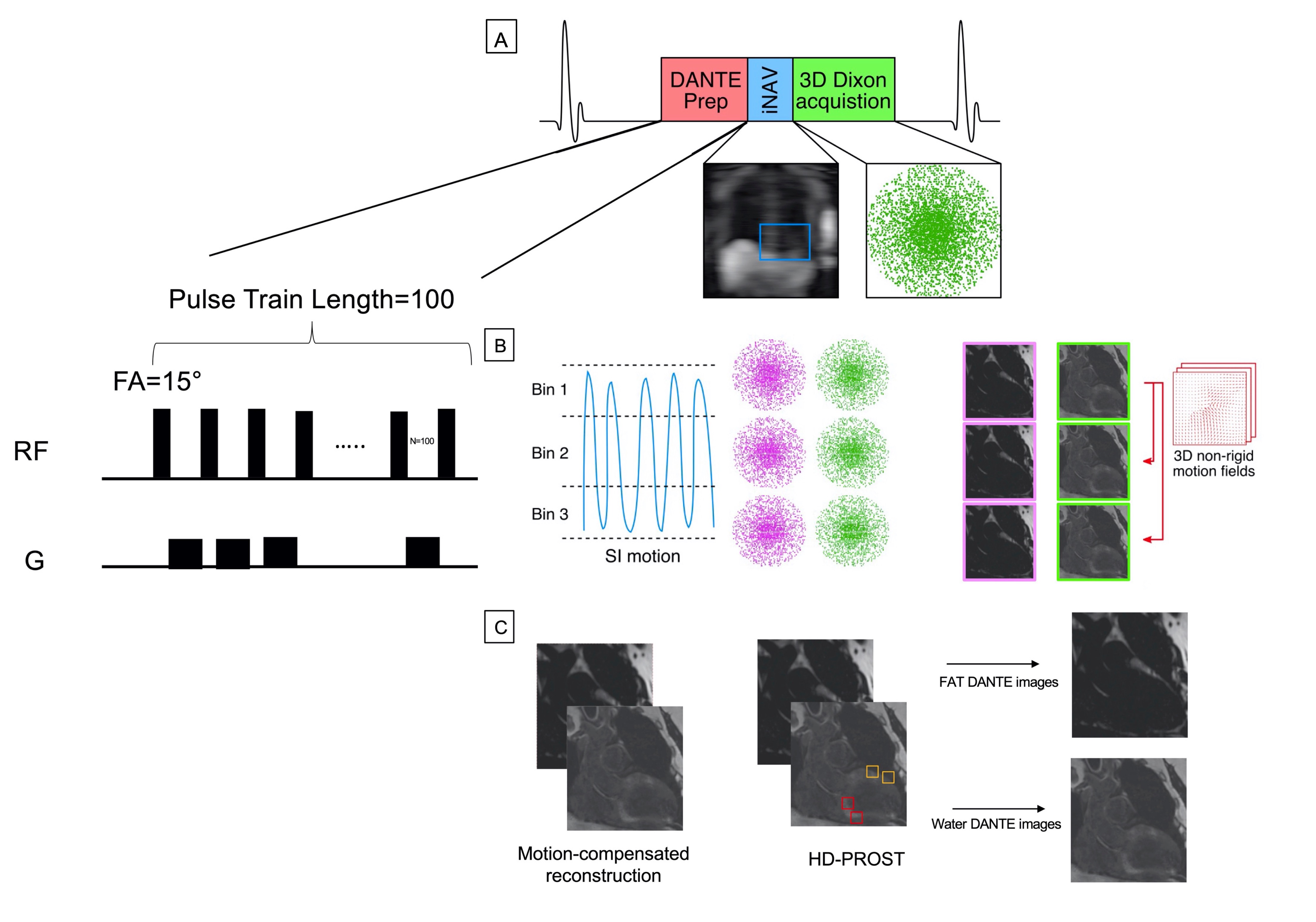
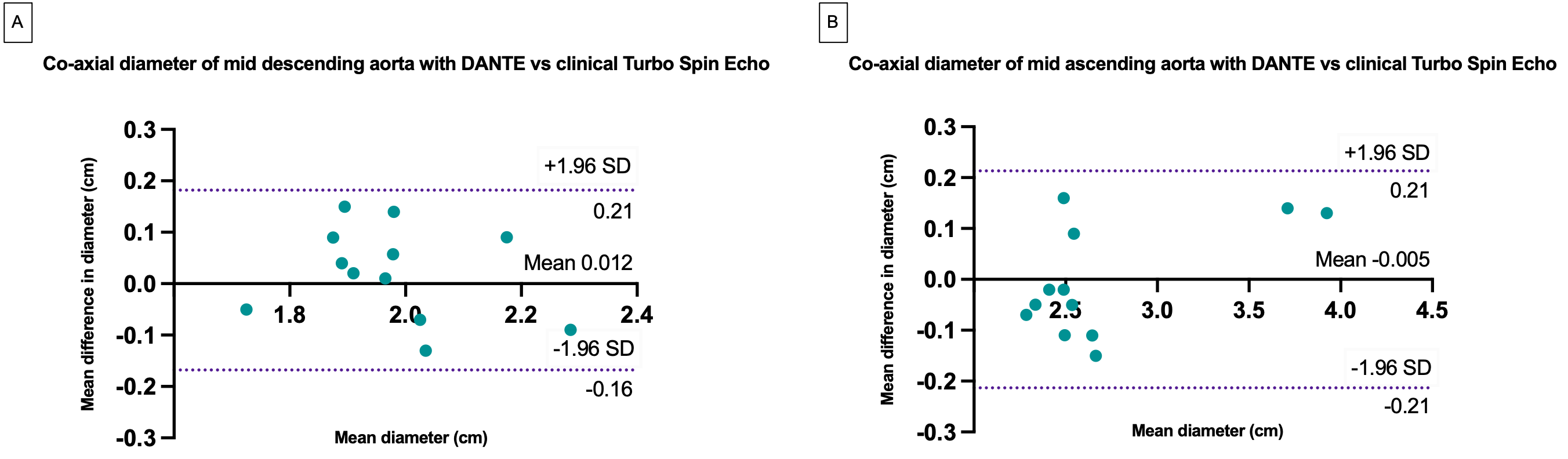
The proposed DANTE-prepared imaging sequence consists of an ECG-triggered dual-echo 3D DANTE-prepared module and a spoiled gradient echo read-out method (Fig 1). 3D dual-echo data are acquired with an undersampled variable-density golden-step Cartesian trajectory with spiral profile order sampling7. Coronal dual-echo 2D iNAVs using 7 low flip-angle excitation pulses precede the 3D acquisition and are used to estimate superior-inferior (SI) and right-left (RL) motion by tracking a template around the aorta and the base of the heart. The SI motion is used to group the acquired data into equally populated respiratory bins, and binned 3D data is corrected for rigid SI and RL respiratory motion to the center of the corresponding bin. Respiratory-resolved bin images are then reconstructed with iterative SENSE and subsequently used to estimate the non-rigid bin-to-bin respiratory motion fields, using the end expiratory bin as reference position8. These non-rigid deformation fields are incorporated into a generalized matrix formulation for motion-compensated MR reconstruction with patch-based low-rank denoising to finally obtain two co-registered motion-compensated water and fat DANTE datasets. Five healthy subjects and two patients with thoracic aortic disease who underwent a clinically indicated MRI scan (6 male, age 33±5 years old) were scanned on a 1.5T system (MAGNETOM Aera, Siemens Healthcare, Erlangen, Germany) with the proposed water/fat DANTE sequence and the clinical dark-blood 2D TSE sequence (field of view=410x280, slice thickness 6mm, TE/TR=45/800ms, bandwidth=850Hz/px, flip angle: 180°, Echo train length=11). Data for the water/fat DANTE sequence were acquired with the following parameters: coronal orientation, 1.4mm3 isotropic resolution, field of view=400x300x88-125mm3, flip angle=15°, TE1/TE2/TR=2.38/4.76/6.83ms, bandwidth=495Hz/px, subject-specific mid-diastolic acquisition window (100-120ms), 3-fold undersampling. The DANTE preparation pulse used a flip angle =15° and pulse train length =100. Scan time was recorded. Co-axial vascular dimensions at the level of the mid-ascending and mid-descending aorta were obtained with both methods and compared by the same blinded reader using Bland-Altman analysis.Results
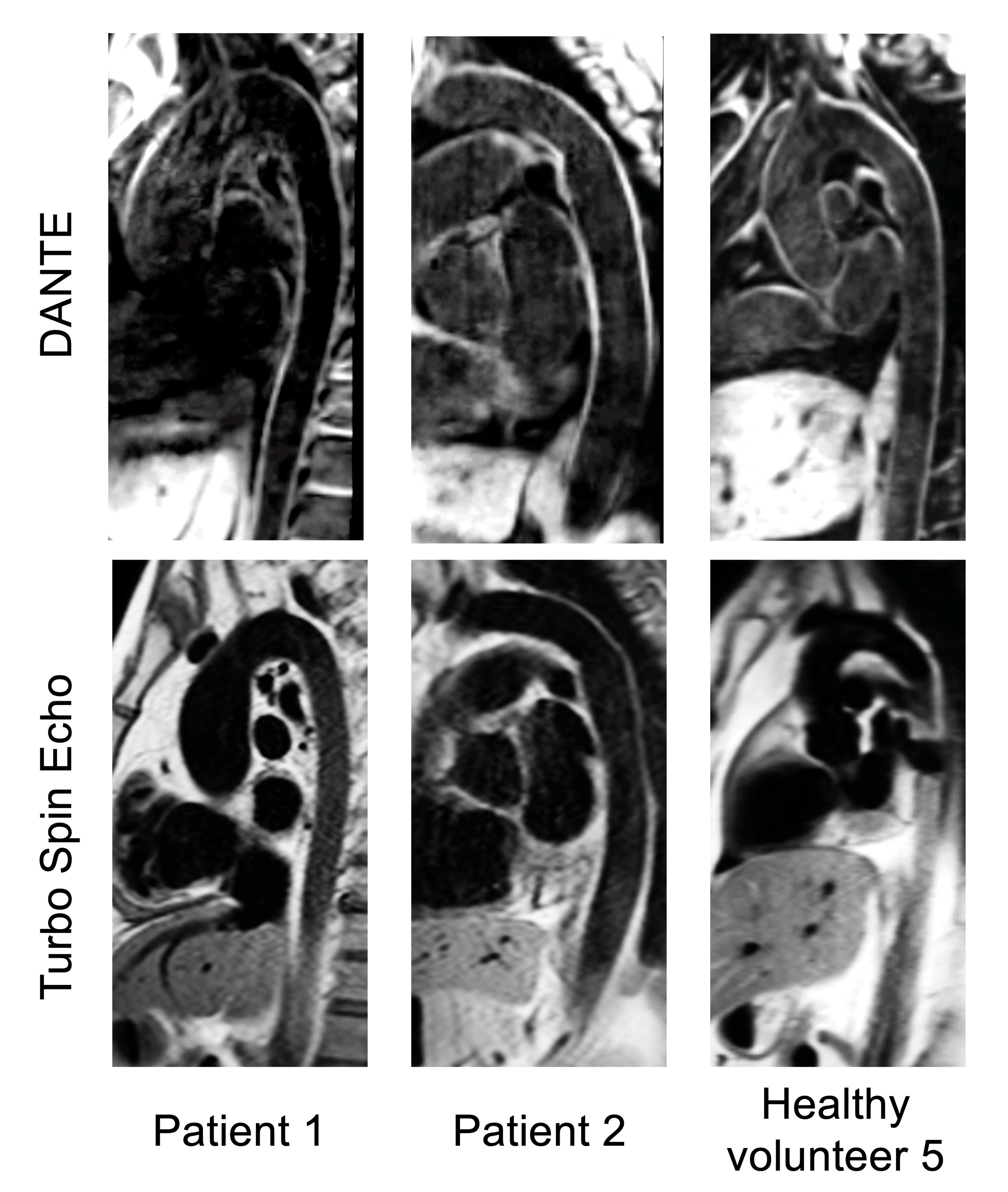
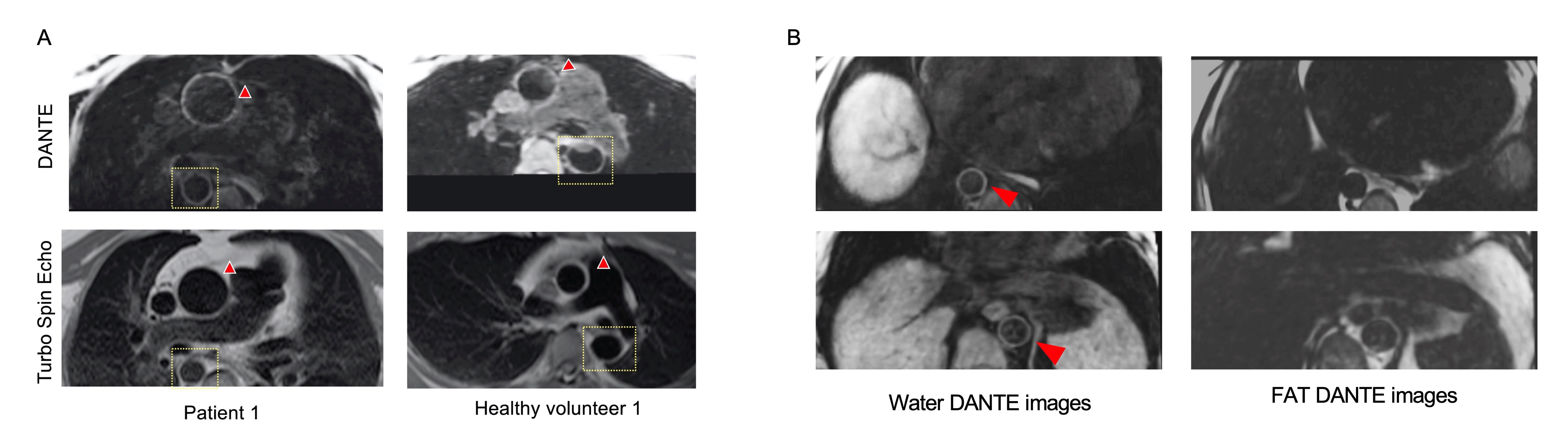
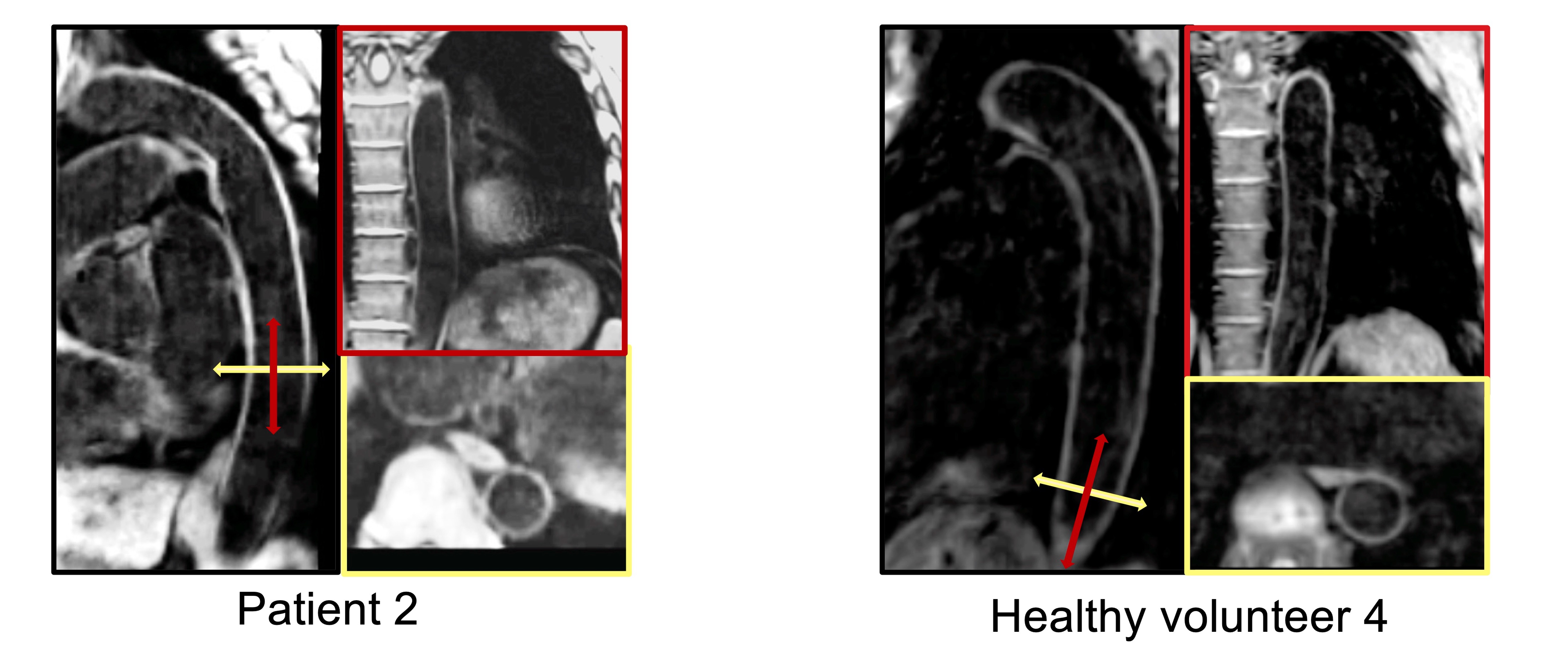
In-vivo 3D acquisitions were successful in healthy subjects and in patients with aortopathy with a mean±SD scan time of 6.6±0.9 min. The proposed DANTE moving spin suppression module demonstrated good quality depiction of the reformatted sagittal oblique and axial views of the thoracic aorta respectively, versus the conventional 2D TSE dark-blood image (Fig. 2 and 3). High-resolution imaging of thoracic aortic wall is demonstrated in the 3D DANTE images with homogenous blood and fat suppression throughout the field of view. 3D acquisition allows reformatting of the aorta in all imaging planes, enabling increased volumetric coverage compared to the 2D TSE imaging (Fig. 4). Bland-Altman analysis demonstrated very good agreement in the co-axial aortic measurements of the mid ascending and mid descending aorta between the DANTE and the corresponding clinical TSE dataset, with a negligible bias across these landmarks (Fig. 5).Discussion
This study validated a 3D dark-blood DANTE-prepared water/fat sequence for thoracic and abdominal aorta imaging that has high isotropic resolution, good blood suppression, and a clinically acceptable scan time. The proposed sequence has the potential for aortic wall imaging of the thoracic and abdominal aorta in healthy volunteers and patients with aortopathy and allows for reliable measurement of the aortic dimensions in a free-breathing manner.Conclusion
Future work including validation of the sequence in larger patient cohorts with aortic pathologies is warranted.Acknowledgements
This work was supported by the following grants: (1) EPSRC P/V044087/1; (2) BHF programme grant RG/20/1/34802, (3) Wellcome/EPSRC Centre for Medical Engineering (WT 203148/Z/16/Z), (4) Millennium Institute for Intelligent Healthcare Engineering ICN2021_004, (5) FONDECYT 1210637 and 1210638, (6) IMPACT, Center of Interventional Medicine for Precision and Advanced Cellular Therapy, ANID FB210024.References
1. Mihai G, Chung YC, Merchant A, Simonetti OP, Rajagopalan S. T1-weighted-space dark blood whole body magnetic resonance angiography (DB-WBMRA): Initial experience. J Magn Reson Imaging 2010;31:502–9.
2. Koktzoglou I, Li D. Diffusion-prepared segmented steady-state free precession: Application to 3D black-blood cardiovascular magnetic resonance of the thoracic aorta and carotid artery walls. J Cardiovasc Magn Reson 2007;9:33–42.
3. Li L, Miller KL, Jezzard P. DANTE-prepared pulse trains: A novel approach to motion-sensitized and motion-suppressed quantitative magnetic resonance imaging. Magn Reson Med 2012;68:1423–38.
4. Zhu C, Haraldsson H, Faraji F, Owens C, Gasper W, Ahn S, et al. Isotropic 3D black blood MRI of abdominal aortic aneurysm wall and intraluminal thrombus. Magn Reson Imaging 2016;34:18–25.
5. Fotaki, A., Munoz, C., Emanuel, Y. et al. Efficient non-contrast enhanced 3D Cartesian cardiovascular magnetic resonance angiography of the thoracic aorta in 3 min. J Cardiovasc Magn Reson 24, 5 (2022).
6. Mihai G, Chung YC, Merchant A, Simonetti OP, Rajagopalan S. T1-weighted-SPACE dark blood whole body magnetic resonance angiography (DB-WBMRA): initial experience. J Magn Reson Imaging. 2010;31:502–509.
7. Prieto C, Doneva M, Usman M, Henningsson M, Greil G, Schaeffter T, et al. Highly efficient respiratory motion compensated free-breathing coronary MRA using golden-step Cartesian acquisition. J Magn Reson Imaging 2015;41:738–46.
8. Munoz C, Bustin A, Neji R, Kunze KP, Forman C, Schmidt M, et al. Motion-corrected 3D whole-heart water-fat high-resolution late gadolinium enhancement cardiovascular magnetic resonance imaging. J Cardiovasc Magn Reson 2020;22:53.
Figures