0329
Myocardial Infarction: An Investigation of Mesostructure using Diffusion Tensor Imaging and Voxel-Based Analysis1Radiological Sciences Laboratory, Stanford University, Stanford, CA, United States, 2Division of Radiology, Veterans Administration Health Care System, Palo Alto, CA, United States, 3Cardiovascular Institute, Stanford University, Stanford, CA, United States, 4Department of Mechanical and Aerospace Engineering, University of Central Florida, Orlando, FL, United States
Synopsis
Keywords: Myocardium, Diffusion Tensor Imaging
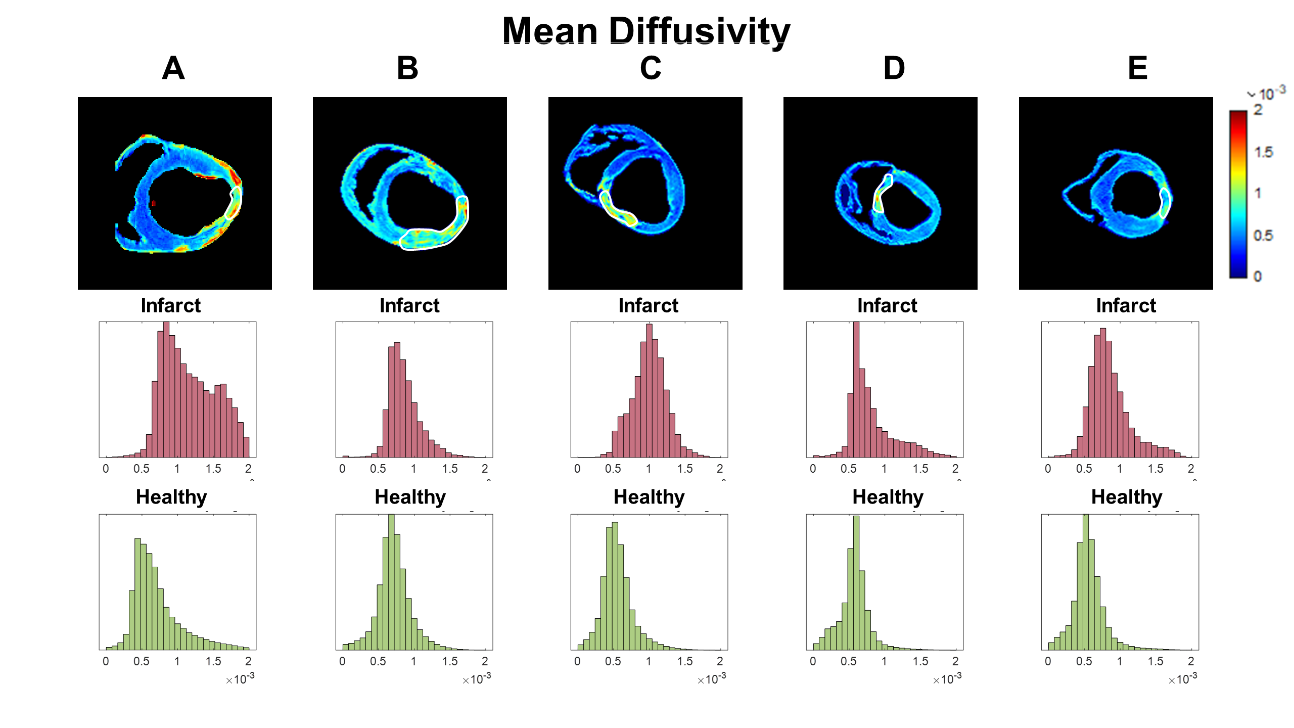
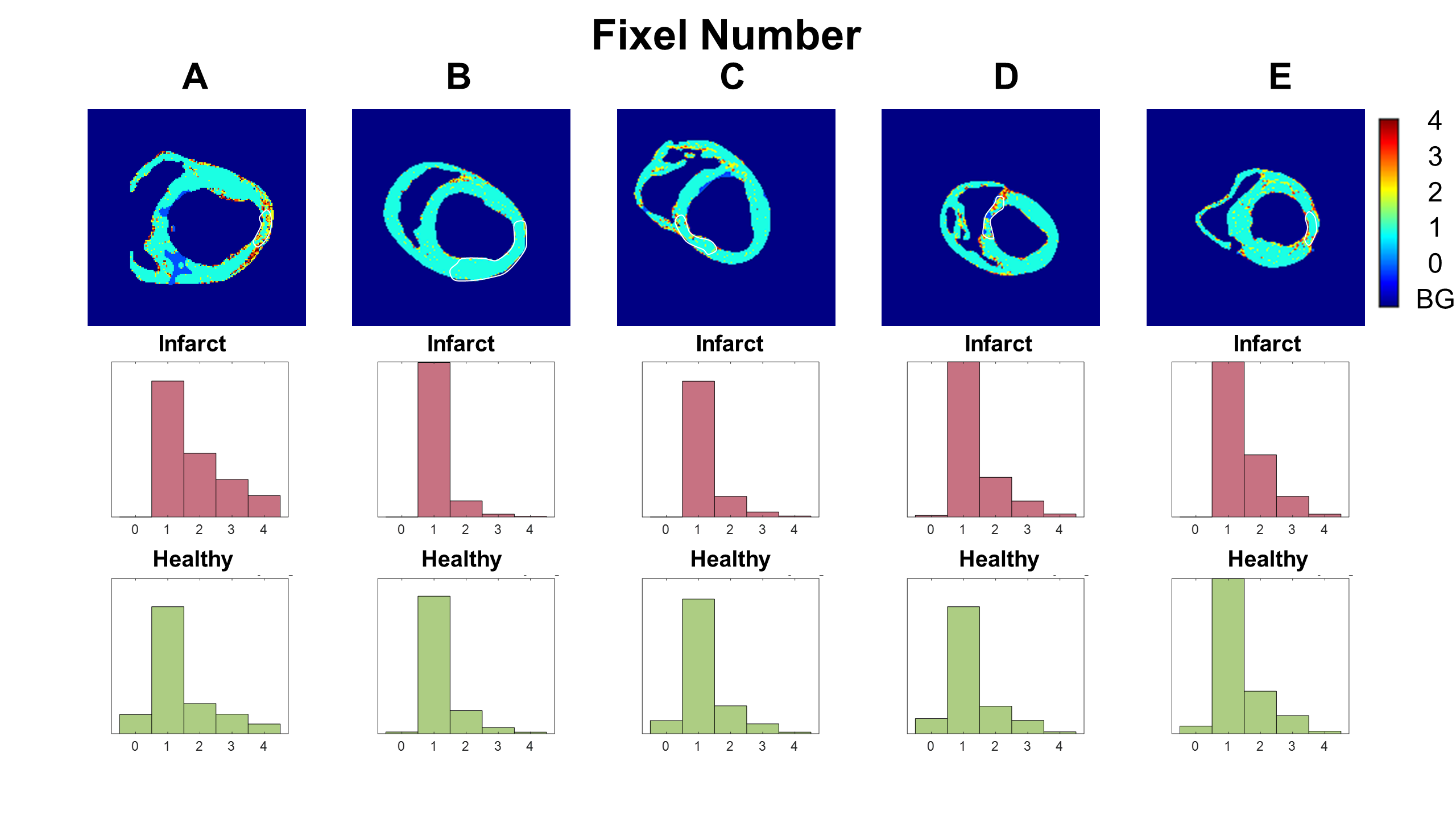
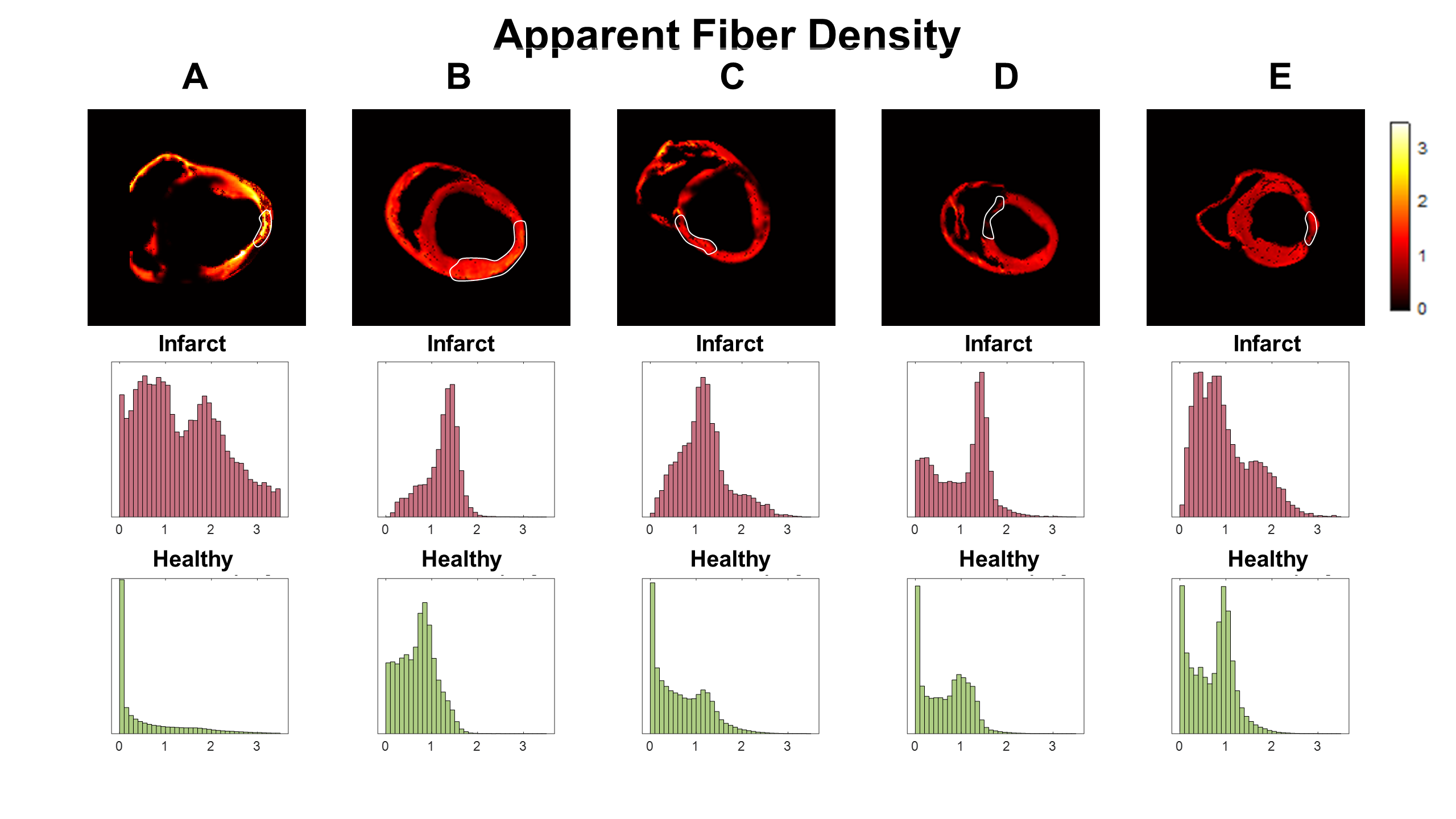
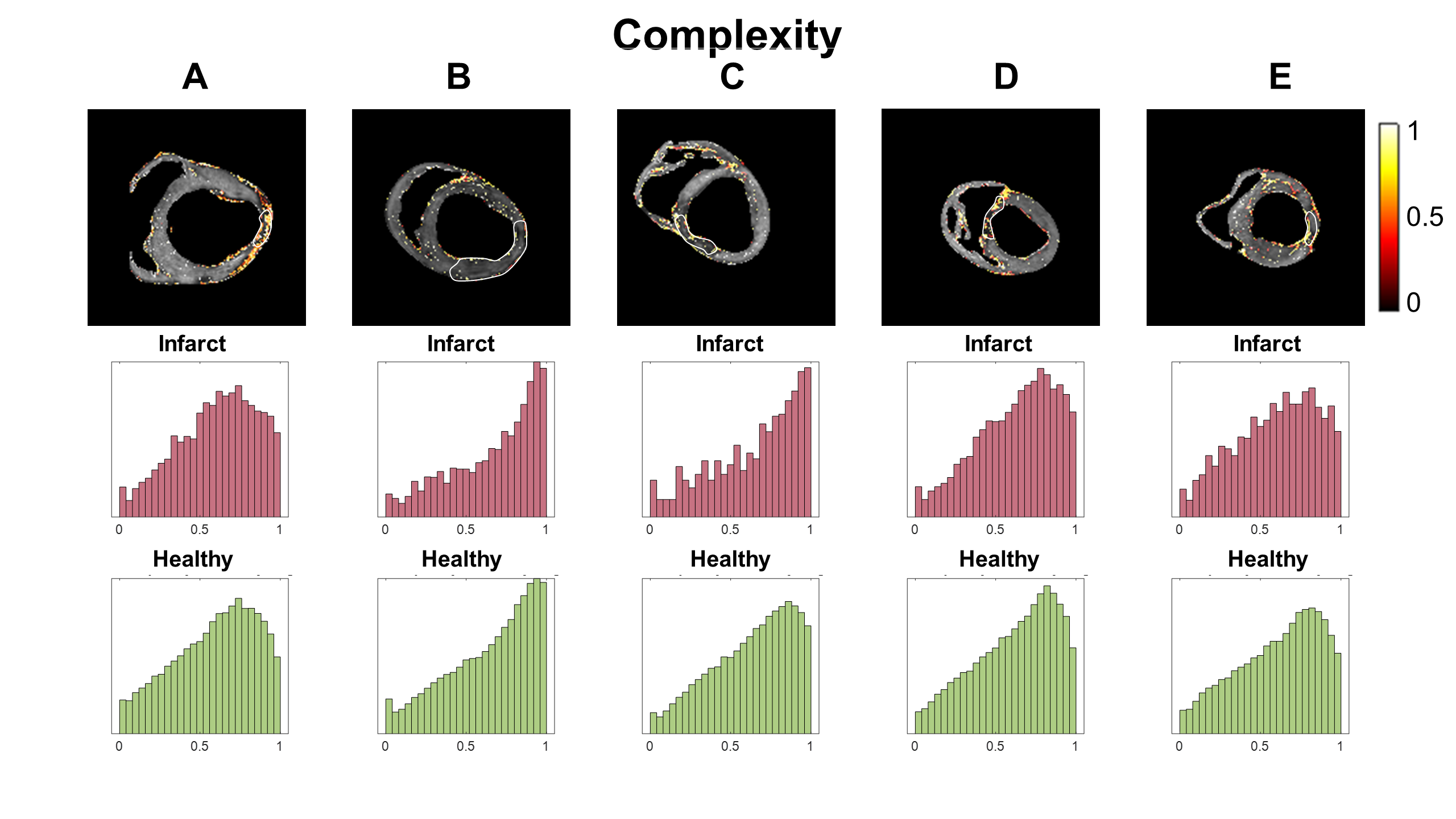
Ex vivo DTI was performed in hearts suffering from myocardial infarction. Data was reconstructed using both tensor-based and voxel-based analysis. The fixel number (FN) consistently showed that there was a single fiber population (FN=1) in both infarcted and healthy myocardium. The complexity (CX) indicates the spread and magnitude of diffusion along different directions, but was not significantly different between infarct and healthy regions. The apparent fiber density (AFD) reveals the density of each fiber population within a voxel, and curiously increased in the infarct region, which may indicate the compaction of fiber populations running through the infarct.
Background
Myocardial infarction results in cellular death and replacement fibrosis. In the infarct region, the myocardium undergoes significant mesostructural remodeling. Diffusion tensor imaging (DTI) allows for the measurement of tissue mesostructure. Reconstructing the diffusion data using the tensor model allows for the extraction of tensor invariants mean diffusivity (MD) and fractional anisotropy (FA). In the infarct region, previous studies have reported an increase in MD, thought to be a result of the expansion of the extracellular matrix, and a decrease in FA attributed to the “disarray” of myocyte organization1,2. However, there is evidence that replacement collagen structures align with the original myocyte direction3 and a degree of structural order is retained.Voxel-based analysis of diffusion data produces a number of “advanced” diffusion metrics that are particularly suited to voxels containing crossing fiber populations4. For example, the fixel number (FN) indicates the number of fiber populations within a voxel. For myocardium we expect voxels to predominantly have a single cardiomyocyte population (FN=1). The voxel complexity (CX) changes when diffusion is spread over a range of directions, with the magnitude of diffusion also varying across directions. The apparent fiber density (AFD) reveals the density of each fiber population within a voxel.
Herein, we use induce myocardial infarction in porcine hearts (n=5) and image them with high-resolution ex vivo DTI. The diffusion data is then reconstructed using both tensor and voxel-based analysis approaches. The objective of this work is to test whether the tissue mesostructure within an infarct is significantly different from healthy myocardium using voxel-based analysis.
Methods
Myocardial infarction was induced in healthy porcine hearts (n=5) by injecting a volume (2.5–3.0mL) of microspheres (90 µm Polystyrene) into the left circumflex (LCx) or left anterior descending (LAD) artery5. Six to ten weeks after myocardial infarction, pigs were anaesthetized, injected with gadolinium-based contrast (0.6mL/kg gadopentetate dimeglumine with 10 mL saline), and euthanized 10 minutes after contrast injection. Hearts were excised, submerged in fomblin to minimize background signal and imaged using a Siemens 3T Prisma scanner within a 15-channel knee coil. For each heart, one post-contrast high-resolution T1-weighted anatomical image (T1) was acquired using a three-dimensional fast low angle shot (3D–FLASH) sequence. T1 imaging parameters were: TR=12ms, TE=3.15ms, flip angle=25°, voxel size=1×1×1mm3. Diffusion images were acquired using a multi-shot readout-segmented spin-echo sequence. Diffusion imaging parameters were: Navg=5, TR=15560ms, TE=62 ms, flip angle=180°, voxel size=1×1×1 mm3, 128 axial slices, b=1000s/mm2, Ndir=30. Regions of infarct were manually segmented from the contrast-enhanced regions of the T1 images. Using FSL6, the diffusion data was co-registered and resampled to anatomical image space, shown to improve the reproducibility of the DTI analysis7. The voxel-based analysis was performed using the Mrtrix3 software package8. For each measurement median values from the infarct and healthy region are taken for each heart. The median values are then compared using the Wilcoxon rank sum test. A linear regression was performed between all pairs of the FA, MD, AFD, CX and FN to check for data redundancy.Results
On the T1 images, infarct regions show post-contrast enhancement compared with healthy myocardium. Median values of FA were not significantly different between infarct and healthy regions (Figure 1). MD was significantly greater in the infarct regions compared with healthy regions (P<0.01, Figure 2). Median FN was identical for infarct and healthy regions, with FN=1 for both regions of the myocardium for all hearts (Figure 3). The median values of AFD were significantly larger in infarct compared with healthy regions (P < 0.01, Figure 4). CX showed a similar distribution between infarct and healthy regions in all hearts (Figure 5), with no significant difference between median values. From the regression analysis, FN and CX showed a moderate correlation (r=0.77), AFD and CX showed a mild inverse correlation (r=-0.48) and MD and FA had a mild inverse correlation (r=-0.46).Discussion
Voxel-based analysis allowed for the comparison of infarct and healthy myocardial regions using FN, AFD and CX, which to our knowledge are novel measurements. Unlike previous literature, FA values were not significantly different in the infarcted myocardium, perhaps due to the inclusion of the infarct border zone within the healthy region, as opposed to comparing the infarct region with a remote region. Using these voxel-based analysis metrics, the AFD in the infarct region significantly increased. This may be indicative of fiber populations from healthy regions being funneled into the smaller infarct region, leading to an increase in density. Alternatively, the AFD may be measuring properties of the aligned3 replacement collagen fibers in the infarct.Conclusion
Voxel-based analysis of ex vivo DTI performed in porcine hearts (n=5) is consistent with a retention of structure within infarcted myocardium.Acknowledgements
This work was supported, in part, by American Heart Association Grant 19IPLOI34760294 (to D.B.E.) and National Heart, Lung, and Blood Institute Grants R01-HL131823 (to D.B.E.) and R01-HL152256 (to D.B.E.)References
[1] Pop, M., Ghugre, N.R., Ramanan, V., Morikawa, L., Stanisz, G., Dick, A.J. and Wright, G.A., 2013. Quantification of fibrosis in infarcted swine hearts by ex vivo late gadolinium-enhancement and diffusion-weighted MRI methods. Physics in Medicine & Biology, 58(15), p.5009.
[2] Kung, G.L., Ajijola, O., Ramirez, R.J., Gahm, J.K., Zhou, W., Wisniewski, N., Mahajan, A., Garfinkel, A., Shivkumar, K. and Ennis, D., 2012. Microstructural remodeling in the post-infarct porcine heart measured by diffusion tensor MRI and T1-weighted late gadolinium enhancement MRI. Journal of Cardiovascular Magnetic Resonance, 14(1), pp.1-3.
[3] Pashakhanloo, F., Herzka, D.A., Mori, S., Zviman, M., Halperin, H., Gai, N., Bluemke, D.A., Trayanova, N.A. and McVeigh, E.R., 2017. Submillimeter diffusion tensor imaging and late gadolinium enhancement cardiovascular magnetic resonance of chronic myocardial infarction. Journal of cardiovascular magnetic resonance, 19(1), pp.1-14.
[4] Raffelt, D.A., Smith, R.E., Ridgway, G.R., Tournier, J.D., Vaughan, D.N., Rose, S., Henderson, R. and Connelly, A., 2015. Connectivity-based fixel enhancement: Whole-brain statistical analysis of diffusion MRI measures in the presence of crossing fibres. Neuroimage, 117, pp.40-55.
[5] Rahman, T., Moulin, K. and Perotti, L.E., 2022. Cardiac Diffusion Tensor Biomarkers of Chronic Infarction Based on In Vivo Data. Applied Sciences, 12(7), p.3512.
[6] Jenkinson, M., Beckmann, C.F., Behrens, T.E., Woolrich, M.W. and Smith, S.M., 2012. Fsl. Neuroimage, 62(2), pp.782-790.
[7] Kang, X., Yund, E.W., Herron, T.J. and Woods, D.L., 2007. Improving the resolution of functional brain imaging: analyzing functional data in anatomical space. Magnetic resonance imaging, 25(7), pp.1070-1078.
[8] Tournier, J.D., Smith, R., Raffelt, D., Tabbara, R., Dhollander, T., Pietsch, M., Christiaens, D., Jeurissen, B., Yeh, C.H. and Connelly, A., 2019. MRtrix3: A fast, flexible and open software framework for medical image processing and visualisation. Neuroimage, 202, p.116137.
Figures

Figure 1: Fractional Anisotropy (FA). Representative short-axis slices from the five porcine hearts (columns A through E, top row). Frequency histogram of FA from the infarct region (middle row, red), and the healthy region (bottom row, green). The infarct region shows lower FA compared with healthy regions for hearts A,C and E. Hearts B and D showing similar FA distributions between the infarct and healthy regions.