0321
Multi-echo Diffusion-weighted Imaging for Quantifying Luminal Water Fraction in the Prostate1Center for MR Research, University of Illinois at Chicago, Chicago, IL, United States, 2Department of Biomedical Engineering, University of Illinois at Chicago, Chicago, IL, United States, 3Departments of Radiology and Neurosurgery, University of Illinois at Chicago, Chicago, IL, United States
Synopsis
Keywords: Prostate, Quantitative Imaging
Prostate cancer remains one of the leading causes of cancer-related deaths in men. Reduced luminal water fraction has been observed in prostate cancer using diffusion MRI, but the image acquisition takes a long time. We herein introduce a multi-echo DWI sequence that is capable of quantifying luminal water fraction with a substantially improved time efficiency. The sequence incorporated multiple readout echo-trains into diffusion-weighted EPI, together with a 2D RF excitation pulse to reduce the FOV, thereby enabling multiple TEs in one shot. With this sequence, we have evaluated the volume fractions of lumen and non-lumen tissues in the human prostate.Introduction
Prostate cancer remains a major cause of morbidity and mortality among men worldwide 1. It has been reported that the luminal water volume fraction reduces in comparison with normal or benign tissue of the prostate, which offers an alternative way for prostate cancer detection 2,3. Over the past few years, several quantitative imaging techniques have been proposed to estimate the luminal water fraction 2-4. In a method known as hybrid multidimensional imaging 4, diffusion-weighted images with multiple b-values and multiple TEs are successively acquired to estimate the luminal water fraction by exploiting both ADC and T2 values associated with different tissue components. This approach, however, requires multiple acquisitions at different TEs, which inevitably lengthens the scan times and leads to increased vulnerability to subject motion. We herein report a time-efficient pulse sequence – multi-echo diffusion-weighted imaging (DWI) – to acquire diffusion-weighted signals at multiple TEs after a single excitation. This sequence, in conjunction with a two-compartment model, has been demonstrated on prostate to differentiate between lumen and non-lumen tissues.Methods
Multi-echo DWI sequence design: Unlike the conventional diffusion-weighted spin-echo EPI sequence, the multi-echo DWI sequence incorporated multiple (e.g., 4) EPI readout echo-trains after a Stejskal-Tanner diffusion preparation module (Figure 1). Each EPI readout echo-train corresponded to a distinct effective TE. The first echo-train coincided with the nominal TE (TE0). The subsequent echo-trains were acquired following each 180° RF refocusing pulse. To reduce the length of each individual echo-train without compromising the spatial resolution, the FOV was restricted to the prostate by using a 2D RF pulse, enabling fewer phase-encoding steps (i.e., shorter echo-train length). The 2D RF pulse was designed by employing a fly-back EPI-like excitation k-space trajectory. Eleven sub-pulses with a time-bandwidth product (TBP) of 3.01 were modulated by an envelope pulse whose TBP was 3.53 and pulse width was 14.7 ms. A tilted excitation k-space strategy was employed to enable multi-slice imaging 5,6.Two-compartment model: It has been reported that lumen has longer T2 and higher ADC than the other compartments in the prostate 4. The corresponding MRI signal can be described by the following equation:$$S\left ( b,TE \right )/S_{0}=fexp\left ( -TE/T2_{l} \right )exp\left ( -b\times ADC_{l} \right )+\left ( 1-f \right )exp\left ( -TE/T2_{nl} \right )exp\left ( -b\times ADC_{nl} \right )$$where S0 is the signal intensity with b-value = 0 s/mm2 and TE = 0 ms, f is the volume fraction of lumen, and the subscripts l and nl indicate the luminal and non-luminal compartments, respectively, for T2 or ADC.
Experiments: The multi-echo DWI sequence was implemented on a GE MR750 3T scanner. To demonstrate the technique, axial prostate MRI scans were performed using a 32‐channel cardiac coil on healthy volunteers with the following parameters: FOV = 180 × 90 mm2, imaging matrix = 64 × 32, reconstruction matrix = 128 × 64, number of slices = 12, slice thickness = 4 mm, slice spacing = 0.5 mm, TR = 4000 ms, number of echo-trains = 4, TEs = 60.8/91.8/122.8/153.8 ms, b-valuesNEX = 04/2506/50010/75015/100020 s/mm2, and the scan time = 11 min and 4 s.
Data analysis: Diffusion-weighted images acquired at different TEs were individually reconstructed. The two-compartment model fitting was performed on the set of 20 images (5 b-values × 4 TEs) using a non-linear least-squares fitting algorithm in MATLAB 2021a (MathWorks, Inc.). To avoid local minima, the fitting was repeated 100 times with randomized initial parameters. Regions of interest (ROIs) were drawn in the peripheral zone (PZ) by avoiding the partial-volume effects from urethra (Figure 2).
Results
Figures 2 displays a set of two-dimensional diffusion-weighted and T2-weighted prostate images from a 35-year-old representative healthy male subject, with five b-values in one dimension (vertical) and four TEs in the other dimension (horizontal). Parameter maps estimated using the two-compartment diffusion model were shown in Figure 3. ADC, T2, and volume fraction were demonstrated for lumen and non-lumen tissues, respectively. Quantitative measurement of luminal water fraction revealed a value of approximately 24% in the PZ. Figure 4 demonstrates that the two-compartment diffusion model fits the measured image intensities very well, with an RSE (relative squared error) < 0.01.Discussion and Conclusion
We have demonstrated a novel multi-echo DWI sequence which is capable of producing diffusion-weighted images at different TEs following an excitation without lengthening the scan times. This approach resulted in a set of spatially co-reregistered images across multiple b-values and TEs, enabling two-compartmental analysis that accounted for both diffusion and T2 relaxation effects in the prostate. This technique produced volume fraction of lumen and non-lumen tissues that were in general agreement with the literature results 4 obtained with techniques requiring a much longer scan time. Compared with the results from a previous study 3, the lower T2 values (T2l: ~90 ms and T2nl: ~20 ms) in our study were likely caused by the imperfect 180° refocusing pulses. Nonetheless, the multi-echo DWI sequence and its associated compartmentalized analysis are expected to assist with characterization for the prostate cancer and other cancerous tissues.Acknowledgements
References
1. Wang L, Lu B, He M, Wang Y, Wang Z, Du L. Prostate Cancer Incidence and Mortality: Global Status and Temporal Trends in 89 Countries From 2000 to 2019. Front Public Heal. 2022;10:811044.
2. Sabouri S, Fazli L, Chang SD, et al. MR measurement of luminal water in prostate gland: Quantitative correlation between MRI and histology. J Magn Reson Imaging. 2017;46(3):861-869.
3. Sabouri S, Chang SD, Savdie R, et al. Luminal water imaging: A new MR imaging T2 mapping technique for prostate cancer diagnosis. Radiology. 2017;284(2):451-459.
4. Chatterjee A, Bourne RM, Wang S, et al. Diagnosis of prostate cancer with noninvasive estimation of prostate tissue composition by using hybrid multidimensional MR imaging: A feasibility study. Radiology. 2018;287(3):864–873.
5. Finsterbusch J. Fast-spin-echo imaging of inner fields-of-view with 2D-selective RF excitations. J Magn Reson Imaging. 2010;31(6):1530-1537.
6. Zhong Z, Merkitch D, Karaman MM, et al. High-spatial-resolution diffusion MRI in Parkinson disease: lateral asymmetry of the substantia nigra. Radiology. 2019;291(1):149-157.
Figures
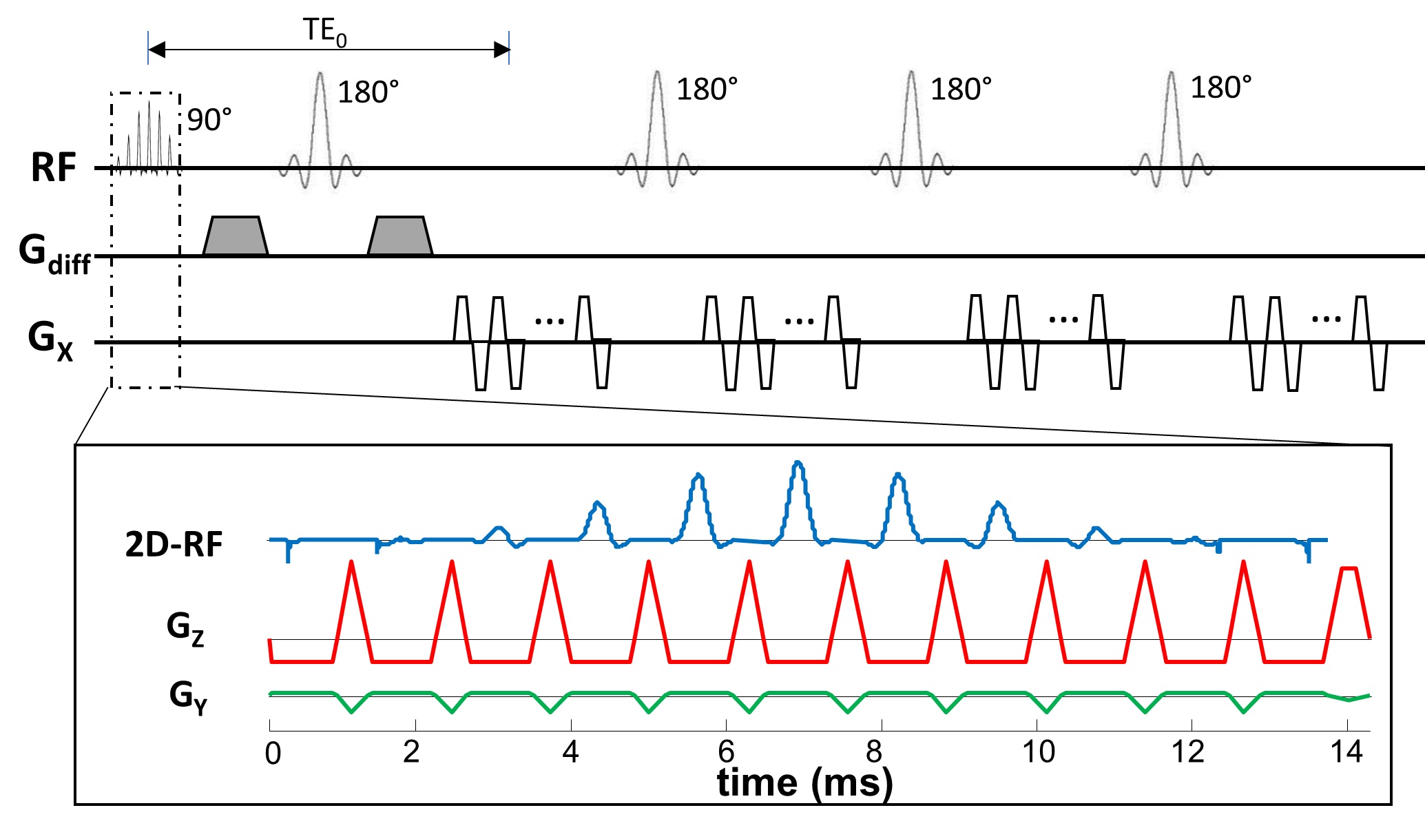
Figure 1: A diagram of the multi-echo DWI pulse sequence. Multiple EPI echo-trains (or readout trains) are incorporated into the sequence, each following a 180° refocusing pulse and with a specific effective TE. A 2D RF excitation pulse is employed to restrict the FOV and shorten the echo train length, enabling multiple echo-trains to be acquired.
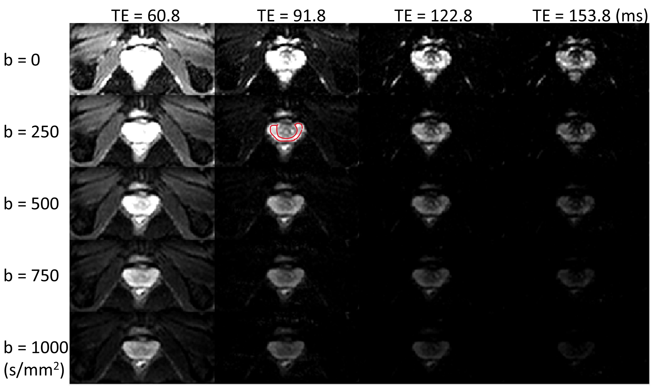
Figure 2: A set of multi-echo diffusion-weighted images of the prostate with five b-values and four TEs from a 35-year-old healthy male subject, resulting in twenty images as shown. A region of interest (ROI; red contour) was drawn on the diffusion-weighted image with b = 250 s/mm2 and TE = 91.8 ms.
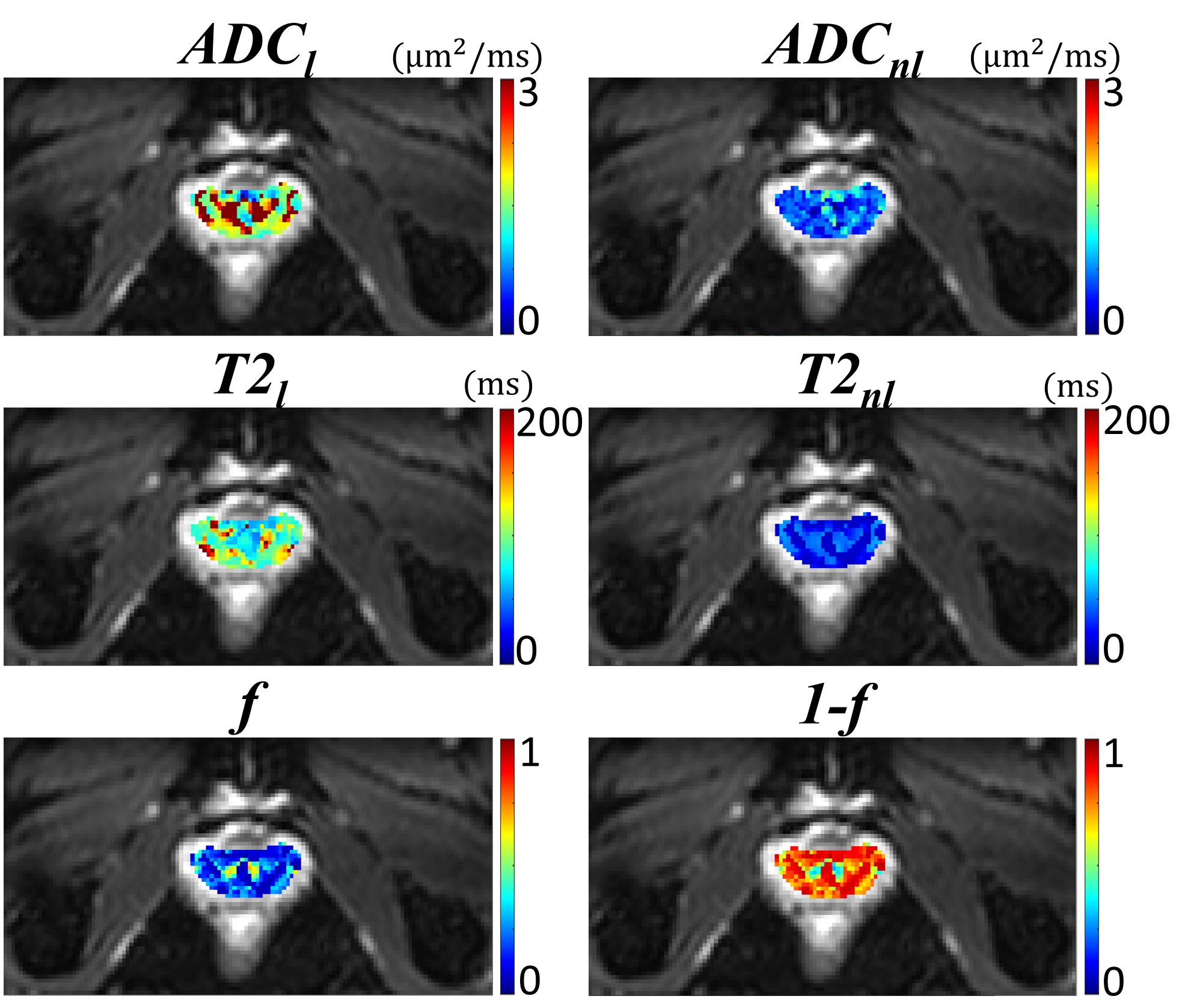
Figure 3: A set of representative parameter maps of ADCl, ADCnl, T2l, T2nl, f, and (1-f) calculated from the multi-compartment model using the 20 diffusion-weighted images acquired with 5 b-values and 4 TEs.
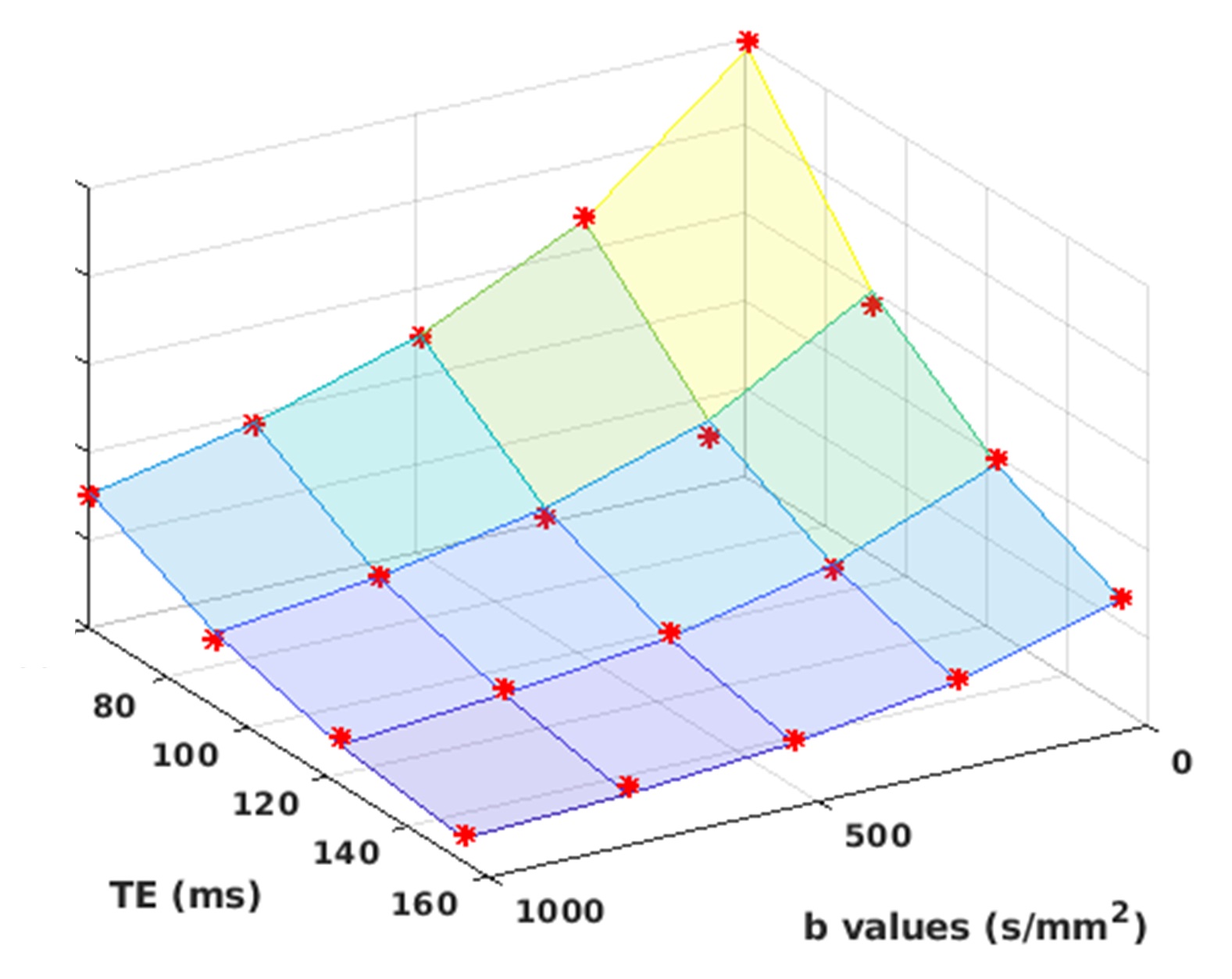
Figure 4: Experimental data (red asterisks) and the fitting results (colored lines) for a randomly selected voxel in the prostate images. The relative squared error (RSE) was smaller than 0.01, indicating excellent fitting. The multi-echo DWI sequence allows investigation of coupling between diffusion and T2-relaxation effects using a two-compartment model.