0302
Full-scale imaging system for brain MRI and magnetoencephalography
Koos Zevenhoven1, Marko Havu1, Iiro Lehto1, Antti Mäkinen1, Juho Luomahaara2, Petteri Laine3, Mikko Kiviranta2, and Risto J Ilmoniemi1
1Aalto University, Espoo, Finland, 2VTT, Espoo, Finland, 3MEGIN Oy, Espoo, Finland
1Aalto University, Espoo, Finland, 2VTT, Espoo, Finland, 3MEGIN Oy, Espoo, Finland
Synopsis
Keywords: Low-Field MRI, New Devices, SQUID, MEG
We have built the first full-scale brain scanner performing both MRI and neuromagnetic measurements. Many improvements have been made compared to previous-generation systems. Parts of the system include a helmet-shaped array of 120 superconducting sensors and a pulsed superconducting polarizing coil. An open-geometry coil system provides unobstructed access, and MRI signal acquisition is performed at ultra-low field. The first MEG and MRI data have been successfully obtained with the device. The new prototype is an important milestone on the way towards a commercial implementation.Background
In magnetoencephalography (MEG), the electrical activity of the brain is studied via the weak magnetic fields it generates. The magnetic field signal is detected using an array of extremely sensitive magnetic sensors inside a magnetically shielded room. From the multichannel signal, brain dynamics can be studied at millisecond time resolution and it is possible to localize brain activity using a number of source reconstruction techniques. For the spatial analysis, structural information – typically from MRI – is used. When MRI data is not available, analyses are often conducted without a proper spatial analysis, limiting the full potential of the technology. Then again, if MRI is available, the process of combining the two modalities is subject to error.For these reasons, an attractive goal is to build a hybrid brain scanner capable of both MEG and MRI. Additional spatial accuracy benefits are obtained when the two signals are detected with the same sensor array. Indeed, it has been shown that magnetic resonance can be detected with sensors based on superconducting quantum-interference devices (SQUIDs), while also able to detect MEG1. However, significant effort has been necessary to move from early demonstrations towards a next generation of scanners closer to being commercializable.
Methods
In this work, we have designed and built a full-scale hybrid MEG–MRI prototype. Our MRI implementation uses a combination of pulsed prepolarization and subsequent SQUID detection in a microtesla-range readout field2, namely 50 µT, which is on the order of Earth's field. The structure of the system is depicted in Fig. 1; all parts differ from conventional MRI.A liquid-helium bath at 4.2 K is shared by a superconducting polarizing coil and a helmet-shaped array of 120 SQUID magnetometers around the head (Fig. 2). The SQUID magnetometers were fabricated with all superconducting structures narrower than 2 µm in order to avoid flux trapping in superconducting vortices. In addition, a number of tricks are applied to recover the SQUIDs within MRI pulse sequences. Instead of gradiometric sensors which are easier to operate, our sensors are magnetometers, which have a better sensitivity to the signal but are directly subject to the applied magnetic fields. This poses additional challenges to our electronics, which needs to provide extremely low-noise currents.
The pulsed polarizing coil was wet-wound of multi-filament superconducting wire with an automatic winding machine onto a bobbin made of cotton-reinforced phenolic sheets. The coil was impregnated with Emerson-Cuming Stycast2850FT cryogenic epoxy, designed for low thermal expansion and good thermal conductivity. Additional passive turns that carry no current create a field symmetry within the coil that significantly reduces the stray field caused by trapped flux. The coil is capable of generating over 100 mT of field over the human neocortex.
The coil system further has an unobstructed geometry: the main readout field and gradient fields are produced by a set of biplanar coils around the helmet configuration, and the excitation field is produced by a single planar square coil with a circular hole to accommodate the helmet.
Results
The new prototype is functional and now produces both MEG and MRI data. As a demonstration of MEG, a test subject was shown as visual stimulus a checkerboard pattern with colors reversing every 500 ms. The averaged responses after every other reversal are visualized in Fig. 3. Regarding MRI – after many issues concerning the acquisition of reliable signals after MRI field ramps – the system has now finally produced its first MR images (Fig. 4), paving the way for further MRI studies.Discussion
To be useful, the MRI obtained from the device need not have a resolution or signal-to-noise ratio comparable to a high-field MRI system. The most important aspect is that the images are geometrically accurate, which is indeed one of the promises of ultra-low-field MRI. Our spatial calibration method achieves sub-millimeter accuracy3, essentially bringing MEG and MRI into the same coordinate system.A number of expected and unexpected challenges were faced when building and integrating the prototype. While SQUID sensors are the most sensitive existing technology for measuring magnetic field signals, their use within an MRI pulse sequence poses a number of challenges. Our new generation of SQUIDs provides solutions to some of the shortcomings of earlier generations of sensors, but also introduces problems not previously present, mainly related to recovery after MRI field ramps and pickup of external electromagnetic interference. However, these problems have shown to be at least partially solvable with the present hardware and, after rejection of interference by multichannel signal processing, clean MEG evoked responses were obtained. MRI has been more problematic, but finally, the first successful MRI has also been obtained, with more expected to follow in the near future.
Conclusion
A full-scale brain scanner was built, detecting both MEG and MRI with the same array of SQUID sensors with 'whole-head' coverage. The first MEG and MRI data with the device has been obtained. Next steps include further imaging studies and tweaks to improve signal and image quality. The new prototype is an important milestone on the way towards a commercial hybrid MEG–MRI system.Acknowledgements
The research leading to these results has received funding from the European Union’s Horizon 2020 research and innovation programme under grant agreements No 686865 and No 852111. Funding for this work was also received from the Finnish Cultural Foundation, from the International Doctoral Programme on Biomedical Engineering and Medical Physics (iBioMEP), and from Business Finland.References
1. Volegov P, Matlachov AN, Espy MA, George JS, Kraus RH Jr. Simultaneous magnetoencephalography and SQUID detected nuclear MR in microtesla magnetic fields. Magn Reson Med. 2004;52(3):467-470.
2. Clarke J, Hatridge M, Mössle M. SQUID-detected magnetic resonance imaging in microtesla fields. Annu Rev Biomed Eng. 2007;9:389-413.
3. Makinen AJ, Zevenhoven KCJ, Ilmoniemi RJ. Automatic Spatial Calibration
of Ultra-Low-Field MRI for High-Accuracy Hybrid MEG-MRI. IEEE Trans Med Imaging. 2019;38(6):1317-1327.
Figures
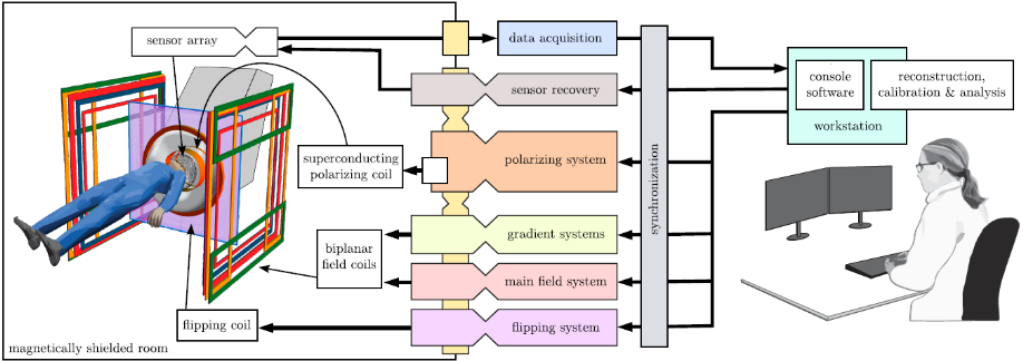
Figure 1: Structure of the hybrid MEG–MRI system.
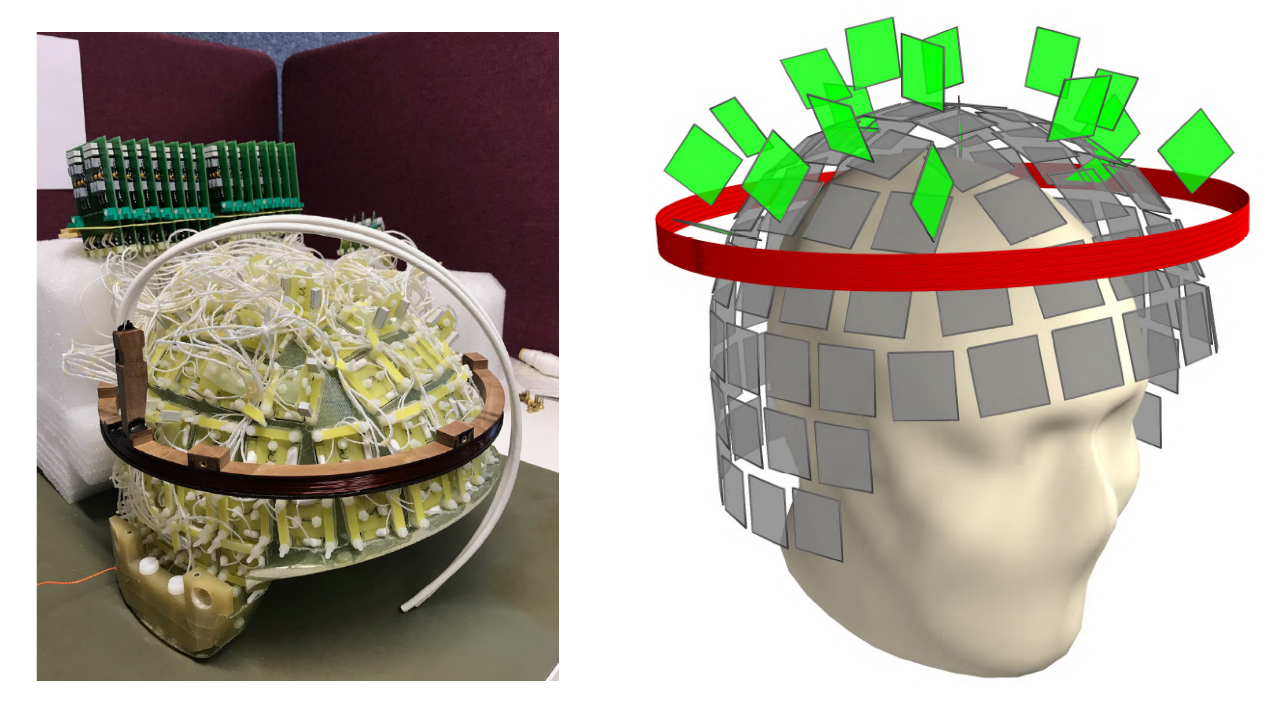
Figure 2: The sensor array and the polarizing coil (red). The sensor array consists of 120 sensors in a helmet shape around the head. 102 sensors (grey) are primarily for measuring the MEG and MRI signals, and 18 tangential sensors (green) are primarily to help interference suppression in multichannel signal analysis.
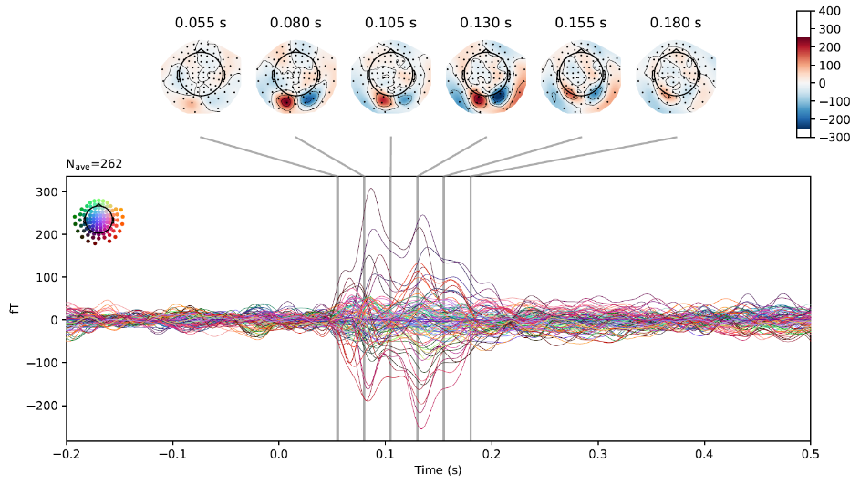
Figure 3: MEG results: responses to a visual stimulus (reversing checkerboard) from sensor channels around the head. Topographies of the signal at different time points after the reversal are shown, projected onto a plane above the head.
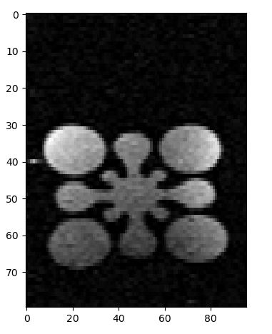
Figure 4: Initial spin-echo MRI image of a water phantom with a 8.3-mm side length. The image has an in-plane resolution of approximately 1x2 mm2. Further imaging results are expected.
DOI: https://doi.org/10.58530/2023/0302