0287
Depression Related Structural Changes Based on 3D Brain MRI and Neural Fingerprint Assisted Segmentation1Radiology, Affiliated Longhua People's Hospital, the Third School of Clinical Medicine, Southern Medical University, Shenzhen, China, 2Clinical Psychology, Affiliated Longhua People's Hospital, the Third School of Clinical Medicine, Southern Medical University, Shenzhen, China, 3Central lab, Affiliated Longhua People's Hospital, the Third School of Clinical Medicine, Southern Medical University, Shenzhen, China
Synopsis
This study focused on the slight quantitative changes of brain structures based on the high-resolution brain MRI and automatic segmentation with neural fingerprint technology for whole-brain of depressive patients. We found the volume changes of specific regional sub-structures, which were related to emotion and spirit, such as the cingulate gyrus, cuneiform lobe, hippocampus, et al before clinical diagnosis. Depression revealed the gender difference. The male depressions were more sensitive to hippocampus, while female’s were more sensitive to entorhinal cortex. It might indicate ovarian hormone fluctuations modulate women’s susceptibility to stress, brain structure/function, and inflammatory activity and reactivity.
Purpose
Depressive patients often suffer from the neuroinflammation that may affect their brain functions and even brain structure. To explore the feasibility of using high-resolution brain MRI and artificial intelligence based neural fingerprint technology to segment and quantify the whole-brain of the slight changes of brain structures in depressive patients.Methods
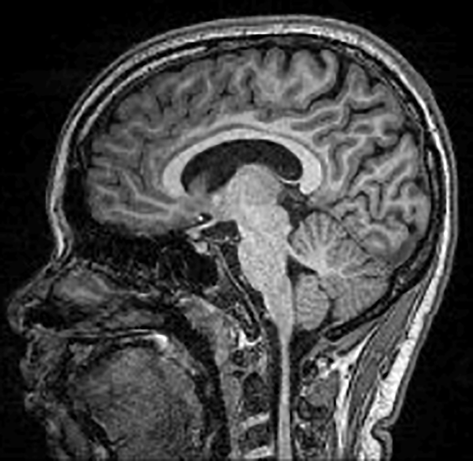
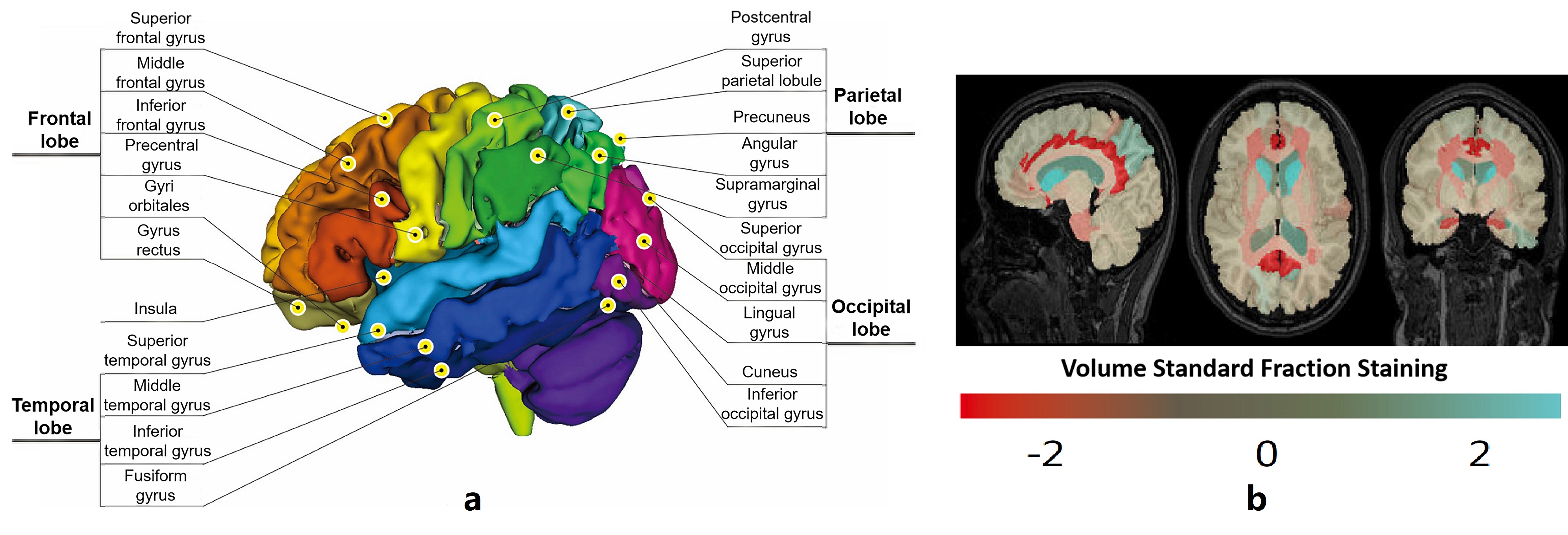
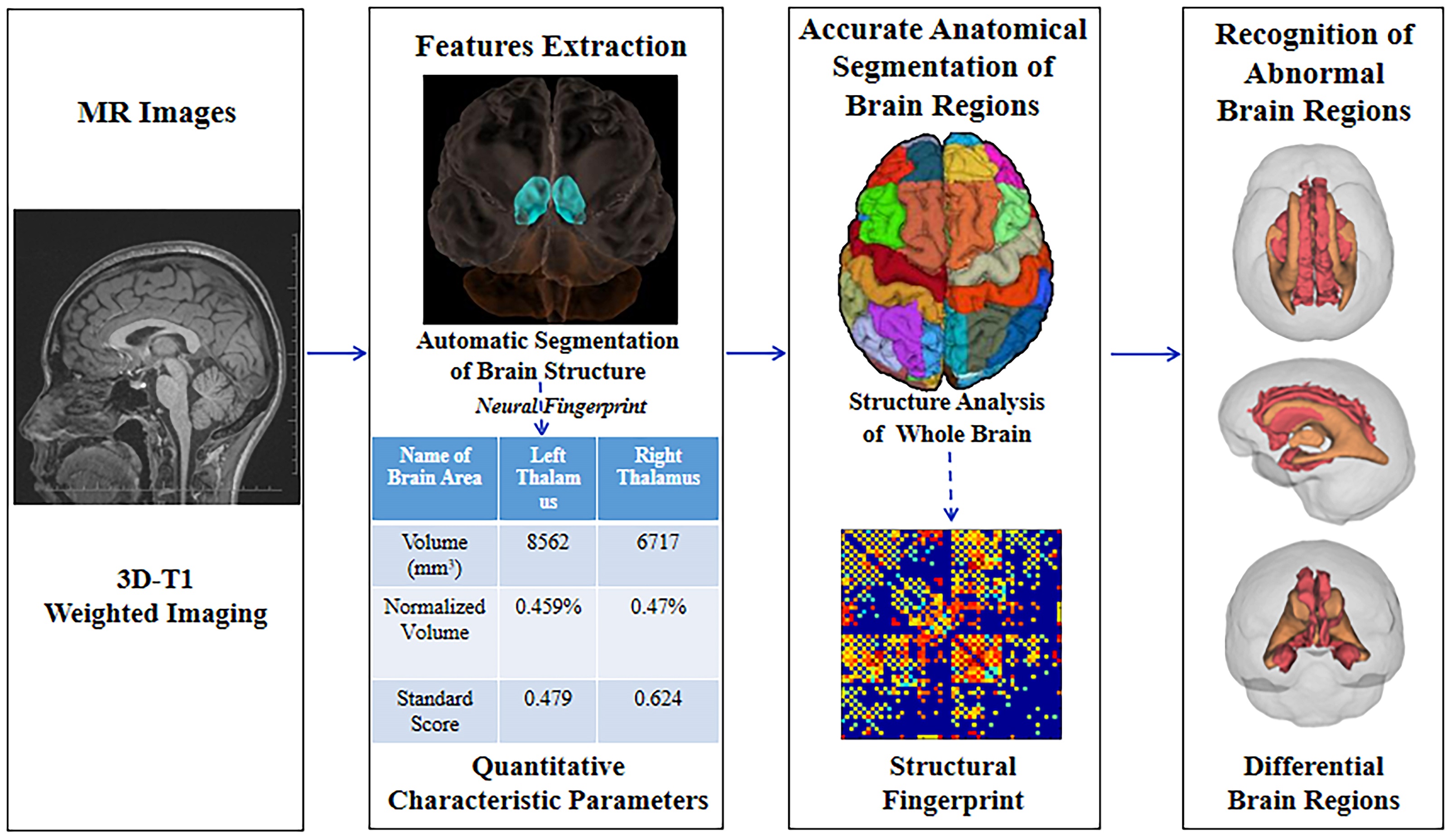
This study was approved by the Institutional Review Board and obtained the written informed consent from all subjects or their legal custodians. Fifty-one patients, including 21 males and 30 females with mild or moderate depression and 48 age and gender matched healthy controls, including 20 males and 28 females were recruited in this study. All the participants completed the Hamilton Depression and Anxiety Scale (HAMD and HAMA), the Self-Rating Depression Scale (SDS) and the Self-Rating Anxiety Scale (SAS) without any abnormal blood tests or other brain diseases. Subsequently, they received the brain MRI and high-resolution 3D-T1-weighted imaging with 1 mm slice thickness in 3T MR scanner using a clinic routine MRI protocol [Figure 1]. Using BrainLabel brain image analysis platform which also includes neural fingerprint technology, we segmented the brain regions and whole brain, followed by quantifying all volumes of whole brain and sub-structure regions in all subjects [Figure 2]. The work flow of process was showed in Figure 3. Statistical software, i.e., SPSS 20.0 was used for comparison analyses of gender, age, education level, and quantitative image data, including the brain volumes of the whole brain and different sub-structure brain regions in both patients and controls. Further, these brain regions with statistic differences were correlated with all scores of HAMD, HAMA, SDS, and SAS. The data about specific relevance on individual variables with abnormal distribution were analyzed by Spearman from SPSS 20.0. P < 0.05 was considered statistically significant.Results
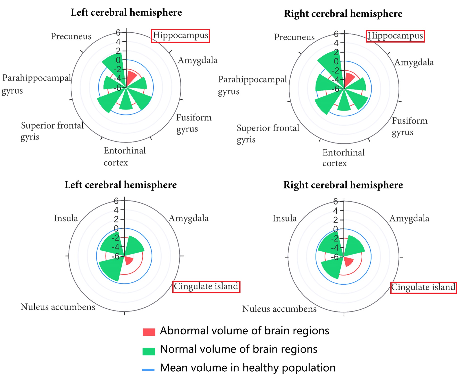
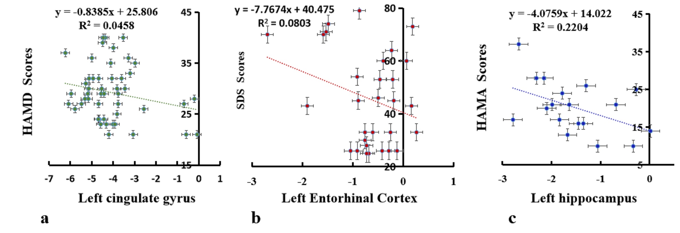
Even the blood test did not indicate any inflammatory reactions, we observed significant brain structural differences (P<0.05) in bilateral cingulate gyrus and cuneiform lobe, right inferior temporal, middle frontal, straight gyrus, superior parietal lobule, left hippocampus, superior and middle occipital, and lingual gyrus between the depression group and the normal controls. One example was showed in Figure 4. The volume of left cingulate gyrus was negatively correlated with HAMD and HAMA scores (P<0.05) without SDS and SAS scores by correlation analysis in Figure 5a. Interesting, it was found there was a statistical difference in the total volume of the ventricular and cerebrospinal fluid (P<0.05), but significant difference was not found in the whole volume of brain tissue between the two groups. Moreover, comparing with the male depressions and the matched normal controls, the female subjects in the depression group showed changes in brain volumes (P<0.05) in more brain regions, included bilateral cingulate, superior and inferior temporal, fusiform, straight, supra and middle occipital, lingual gyrus, cuneiform lobe, right entorhinal cortex, middle frontal and temporal, superior parietal gyrus, and the left orbital gyrus. In addition, the female depression group also had different brain volume changes (P<0.05) in the bilateral cerebral cortex, lower white matter, and right posterior white matter, left thalamic sub-structure gyrus of brain regions, while the male depressive group had brain volume changes only in bilateral cingulate gyrus, hippocampus and left anterior central gyrus (P<0.05) comparing with the matched normal controls. When we examine whether the changes in the brain regional volumes correlated with evaluation questionnaire scales. We also found that negative correlations between the volume of left cingulate gyrus and entorhinal cortex with SDS score (P<0.05) in the female patients [Figure 5b]. However, there only was negative correlation between volumes of the cingulate gyrus and left hippocampus with HAMA scores (P<0.05) in the male depression group [Figure 5c].Conclusions
1. Our study indicated that there was no significant changes of the whole brain volume in depressive patients, but the volume changes of specific regional sub-structures were found significantly, especially in the brain regions related to emotion and spirit, such as the cingulate gyrus, cuneiform lobe, hippocampus. Although no clinically measured inflammatory was observed in this cohort of the depressed patients, we identified early changes of brain volume in those regions might service as the objective markers for diagnosis of depression. 2. The female depression is different from the male. The male depressions were more sensitive to hippocampus, while female depressions were more sensitive to entorhinal cortex and more brain regions of volume changes. 3. It might indicate that female was more sensitive to emotion than male in depression and anxiety from the results of correlation analysis.Acknowledgements
This work was supported by Shenzhen Fundamental Research Program (JCYJ20190808095413252), Department of Innovation of Science and Technology of Longhua, Shenzhen, and Hangzhou Pujian Medical Technology Co., LTD, China.References
[1] WHO. International Classification of Diseases 11th Revision [J]. https://icdwhoint/en (accessed Jan 31, 2023), 2019.
[2] BUKSTEIN O G. Screening for Adolescent Depression and Suicide Risk [J]. Jama, 2022, 328(15): 1504-5.
[3] WEAVERS B, HERON J, THAPAR A K, et al. The antecedents and outcomes of persistent and remitting adolescent depressive symptom trajectories: a longitudinal, population-based English study [J]. The lancet Psychiatry, 2021, 8(12): 1053-61.
[4] YU M, CULLEN N, LINN K A, et al. Structural brain measures linked to clinical phenotypes in major depression replicate across clinical centres [J]. Mol Psychiatry, 2021, 26(7): 2764-75.
Figures