0280
Quantitative Comparison of Bone-Selective MRI Techniques for Craniofacial Imaging1Department of Radiology, Perelman School of Medicine, University of Pennsylvania, Philadelphia, PA, United States, 2Department of Bioengineering, University of Pennsylvania, Philadelphia, PA, United States, 3School of Electronics Engineering, Kyungpook National University, Daegu, Korea, Republic of, 4Division of Plastic, Reconstructive, and Oral Surgery, Children's Hospital of Philadelphia, University of Pennsylvania, Philadelphia, PA, United States, 5Department of Orthopaedic Surgery, University of Pennsylvania, Philadelphia, PA, United States
Synopsis
Keywords: Quantitative Imaging, Bone, Skull imaging
Risk of ionizing radiation from CT remains a concern for pediatric patients, such as craniosynostosis patients who may require multiple scans at a young age. We quantitatively compared three MRI methods for imaging the skull by scanning healthy adults using our solid-state bone-selective ultrashort echo subtraction sequence, along with zero echo time and gradient-echo sequences. We demonstrate the similarities among the 3D rendered bone segmentations in terms of Dice similarity coefficient and craniometric measurements across all imaging methods. Our UTE echo subtraction technique shows clear visualization of craniofacial structures near the bone-air boundaries and superior signal suppression of soft-tissues.Summary of Main Findings
Craniofacial imaging with ultrashort echo time and zero echo time sequences demonstrate enhanced bone-contrast and superior suppression of soft-tissues and air compared to gradient echo sequences.Introduction
Craniosynostosis is the premature fusion of the cranial sutures causing skull shape deformities in 1 in 2000 infants.1 Computed Tomography (CT) is the clinical standard imaging modality for evaluating craniofacial skeletal pathologies. However, ionizing radiation exposure remains a problem, especially for children and infants who may require multiple scans at a very young age.2,3 There have been few approaches utilizing magnetic resonance imaging (MRI) for high-resolution skull imaging as a radiation-free alternative to CT,4-7 including some that where evaluated in pediatric patients.8-12 “Black-bone” MRI is the most common technique done using a conventional 3D gradient-echo (GRE) sequence with very low flip angles6, but it cannot distinguish between air and bone (e.g. sinuses) complicating skull tissue segmentation. Zero-echo time (ZTE) sequences have been demonstrated to resolve the bone-air issue interface5,8,13; however, the technique is limited by the need for post-processing algorithms to suppress soft-tissues and differentiate bone from air. Alternatively, our lab developed an ultrashort echo-time (UTE) solid-state technique that generate bone-selective images by means of echo subtraction.4 The dual-RF, dual-echo, three-dimensional ultrashort echo (DURANDE) sequence exploits the sensitivity of bone proton magnetization to both T2 and RF pulse duration.4 It has previously demonstrated enhanced bone-signal and suppression of soft-tissues and air, and has been evaluated against CT both ex vivo and in vivo.14,15 Our goal is to quantitatively assess our proposed sequence (DURANDE) to other existing sequences (ZTE and GRE) to demonstrate the advantages of using MRI as an alternative to CT for craniofacial imaging.Methods
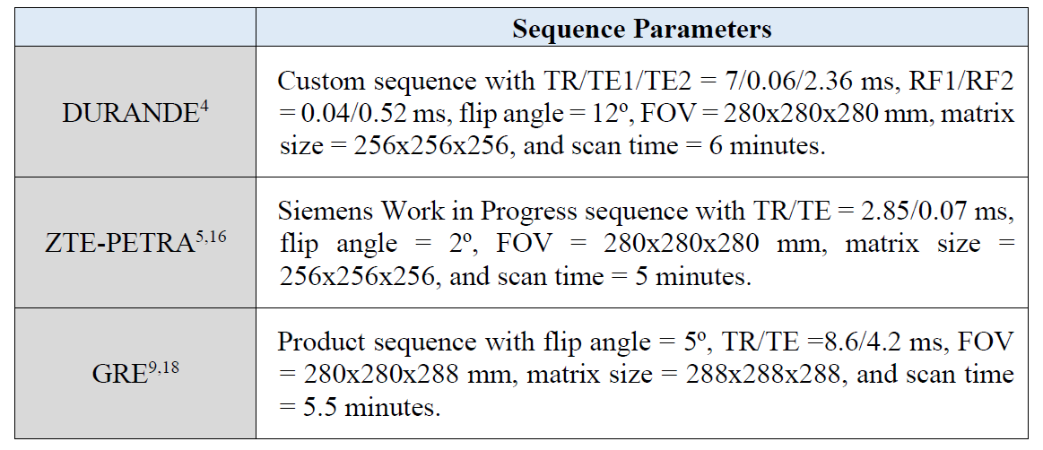
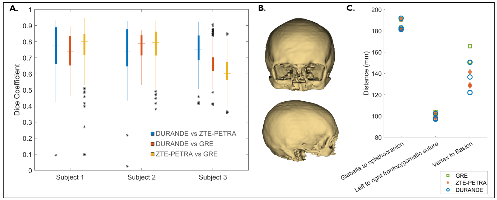
Three healthy participants (ages 23-27 years old) were imaged at 3.0 T (Prisma, Siemens, Erlangen, Germany) with a 20-channel head/neck coil using three sequences. Imaging parameters for DURANDE4, ZTE-PETRA16, and GRE6,9 are in Table 1. DURANDE bone-specific images are generated by weighted echo subtraction of the short- and long T2 images [Imagebone = (Imageecho1 – Imageecho2) / (Imageecho1 + Imageecho2)]. For ZTE-PETRA and GRE, we applied bias-field correction using the nonparametric N4ITK method.17 Soft-tissue suppression of ZTE-PETRA was done by logarithmic inversion to yield bright-bone images.5,8,13 All images were manually segmented and 3D rendered. Six craniometric landmarks were identified in each scan to calculate three distances: glabella to opisthocranion, left frontozygomatic to right frontozygomatic sutures, and vertex to basion. After registering the ZTE-PETRA and GRE masks to the DURANDE mask, the segmented skulls were then manually cropped to include only the cranial vault, orbit, and part of the maxilla. Dice similarity coefficient (DSC) was calculated to quantitatively compare the similarity of the segmented skulls among the three sequences.Results
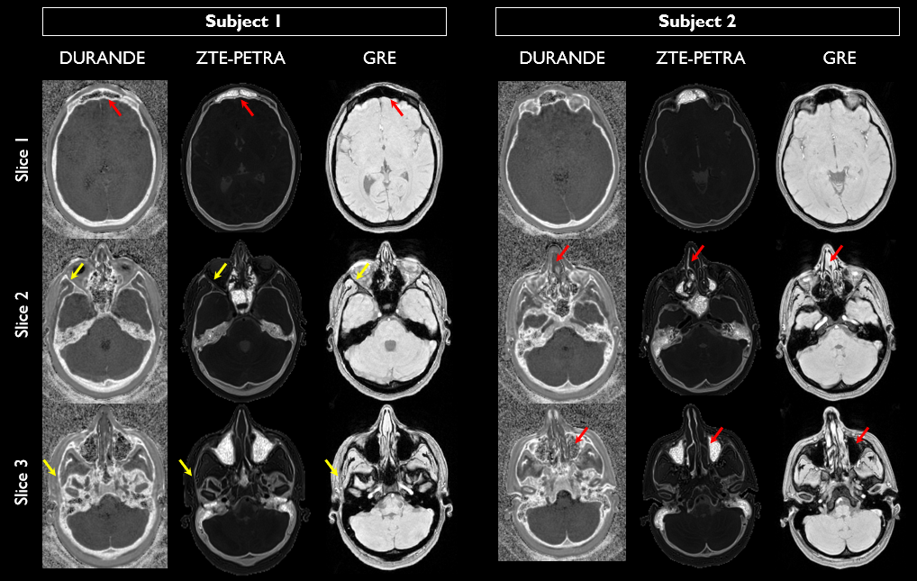
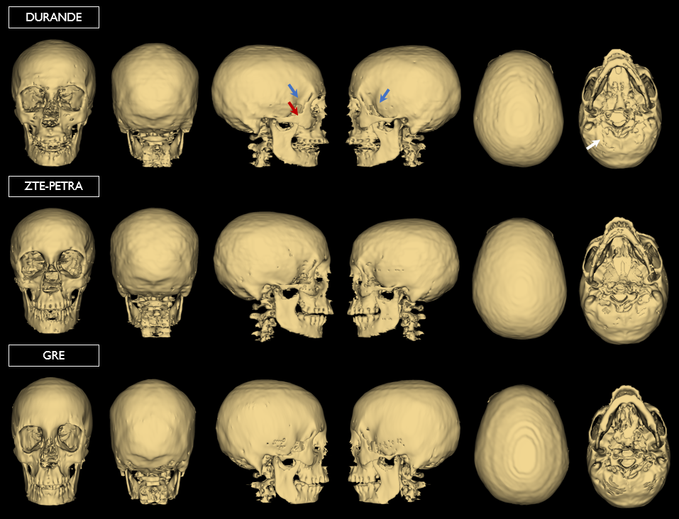
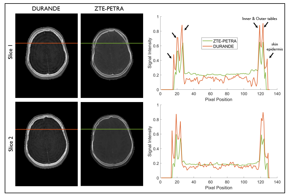
Sample bone slices from DURANDE, ZTE-PETRA and low-flip-angle short-TE GRE (referred to as “black bone”6) are shown in Figure 1, along with whole- skull 3D renderings in Figure 2. Comparison of the bone signal intensity between DURANDE and ZTE-PETRA reveal clear depiction of the outer and inner tables of the skull (Figure 3), with DURANDE showing greater contrast between bone and soft tissues in comparison to ZTE-PETRA. GRE has minimal (or no) differentiation of the skull bone tables. The DSC among the three scans varied considerably depending on the slice location (Figure 4A-C). In Figure 4D, the craniometric distances are similar across the three methods for two of the measured distances (glabella to opisthocranion, and left to right frontozygomatic sutures). However, there is a larger variability in the distance between the vertex and basion.Discussion
We evaluated three MRI sequences for craniofacial imaging by assessing the similarities among their skull segmentations and craniometrics measurements in healthy adults. We observe overall good agreement in skull masks among all three methods, however, this agreement is dependent on slice location. DSC is higher for superior slices in the cranial vault, and lower in inferior slices containing sinuses, and occipital bones. Unlike DURANDE and ZTE-PETRA, GRE does not differentiate between bone and air. This results in differences in the segmented skulls in regions with bone-air boundaries. Moreover, there is a larger variability in the craniometric distance between the vertex and basion when calculated from the three imaging methods (Figure 4D). This can be due to observed differences in segmented masks at the occipital bone region of the skull (Figure 2), making it more challenging to accurately identify the basion landmark. Both DURANDE and ZTE-PETRA are superior to GRE because they are solid-state MRI techniques that capture short T2 signal from bone (Figure 1). However, the main limitation of ZTE-type sequences is the need for bias-field correction and further post-processing algorithms to suppress soft-tissues and separate bone from air,5,8,13 the latter is further limited by the overlap of the histogram of bone and air. In comparison, UTE-type subtraction techniques such as DURANDE are self-normalized and are designed to generate bone-selective images with soft-tissue and air suppression. Future work will examine the three techniques against gold standard CT since our current work is limited to assessing the MRI sequences against each other only.Conclusion
All three MRI techniques yielded comparable craniometric measurements, with DURANDE and ZTE-PETRA having superior air-bone contrast and soft-tissue suppression compared to black-bone GRE.Acknowledgements
NIH T32 EB020087; NIH R21 DE028417References
1. Johnson D, Wilkie AO. Craniosynostosis. Eur J Hum Genet 2011;19(4):369-76. (In eng). DOI: 10.1038/ejhg.2010.235.
2. Pearce MS, Salotti JA, Little MP, et al. Radiation exposure from CT scans in childhood and subsequent risk of leukaemia and brain tumours: a retrospective cohort study. Lancet 2012;380(9840):499-505. (In eng). DOI: 10.1016/s0140-6736(12)60815-0.
3. Mathews JD, Forsythe AV, Brady Z, et al. Cancer risk in 680,000 people exposed to computed tomography scans in childhood or adolescence: data linkage study of 11 million Australians. BMJ (Clinical research ed) 2013;346:f2360. (In eng). DOI: 10.1136/bmj.f2360.
4. Lee H, Zhao X, Song HK, Zhang R, Bartlett SP, Wehrli FW. Rapid dual-RF, dual-echo, 3D ultrashort echo time craniofacial imaging: A feasibility study. Magnetic resonance in medicine 2019;81(5):3007-3016. (In eng). DOI: 10.1002/mrm.27625.
5. Wiesinger F, Sacolick LI, Menini A, et al. Zero TE MR bone imaging in the head. Magnetic resonance in medicine 2016;75(1):107-14. (In eng). DOI: 10.1002/mrm.25545.
6. Eley KA, McIntyre AG, Watt-Smith SR, Golding SJ. "Black bone" MRI: a partial flip angle technique for radiation reduction in craniofacial imaging. The British journal of radiology 2012;85(1011):272-278. (In eng). DOI: 10.1259/bjr/95110289.
7. Patel KB, Eldeniz C, Skolnick GB, et al. 3D pediatric cranial bone imaging using high-resolution MRI for visualizing cranial sutures: a pilot study. J Neurosurg Pediatr 2020;26(3):311-317. (In eng). DOI: 10.3171/2020.4.Peds20131.
8. Lu A, Gorny KR, Ho ML. Zero TE MRI for Craniofacial Bone Imaging. AJNR Am J Neuroradiol 2019;40(9):1562-1566. (In eng). DOI: 10.3174/ajnr.A6175.
9. Eley KA, Watt-Smith SR, Sheerin F, Golding SJ. "Black Bone" MRI: a potential alternative to CT with three-dimensional reconstruction of the craniofacial skeleton in the diagnosis of craniosynostosis. Eur Radiol 2014;24(10):2417-26. (In eng). DOI: 10.1007/s00330-014-3286-7.
10. Kuusela L, Hukki A, Brandstack N, Autti T, Leikola J, Saarikko A. Use of black-bone MRI in the diagnosis of the patients with posterior plagiocephaly. Childs Nerv Syst 2018;34(7):1383-1389. (In eng). DOI: 10.1007/s00381-018-3783-0.
11. Kralik SF, Supakul N, Wu IC, et al. Black bone MRI with 3D reconstruction for the detection of skull fractures in children with suspected abusive head trauma. Neuroradiology 2019;61(1):81-87. (In eng). DOI: 10.1007/s00234-018-2127-9.
12. Saarikko A, Mellanen E, Kuusela L, et al. Comparison of Black Bone MRI and 3D-CT in the preoperative evaluation of patients with craniosynostosis. J Plast Reconstr Aesthet Surg 2020;73(4):723-731. (In eng). DOI: 10.1016/j.bjps.2019.11.006.
13. Delso G, Wiesinger F, Sacolick LI, et al. Clinical evaluation of zero-echo-time MR imaging for the segmentation of the skull. J Nucl Med 2015;56(3):417-22. (In eng). DOI: 10.2967/jnumed.114.149997.
14. Zhang R, Lee H, Zhao X, et al. Bone-Selective MRI as a Nonradiative Alternative to CT for Craniofacial Imaging. Academic Radiology 2020;27(11):1515-1522. DOI: https://doi.org/10.1016/j.acra.2020.03.001.
15. Zimmerman CE, Khandelwal P, Xie L, et al. Automatic Segmentation of Bone Selective MR Images for Visualization and Craniometry of the Cranial Vault. Acad Radiol 2021 (In eng). DOI: 10.1016/j.acra.2021.03.010.
16. Li C, Magland JF, Seifert AC, Wehrli FW. Correction of excitation profile in Zero Echo Time (ZTE) imaging using quadratic phase-modulated RF pulse excitation and iterative reconstruction. IEEE transactions on medical imaging 2014;33(4):961-9. (In eng). DOI: 10.1109/tmi.2014.2300500.
17. Tustison NJ, Avants BB, Cook PA, et al. N4ITK: Improved N3 bias correction. IEEE transactions on medical imaging 2010;29(6):1310-1320. (Article). DOI: 10.1109/TMI.2010.2046908.
18. Eley KA, Watt-Smith SR, Golding SJ. "Black bone" MRI: a potential alternative to CT when imaging the head and neck: report of eight clinical cases and review of the Oxford experience. The British journal of radiology 2012;85(1019):1457-64. (In eng). DOI: 10.1259/bjr/16830245.
Figures