0267
Identification of IDH and tumor subtype of adult-type diffuse gliomas based on WHO CNS5 using standard, high and ultra-high b value DWI1School of Medical Imaging, Fujian Medical University, Fujian, China, 2School of Basic Medical Sciences, Fujian Medical University, Fuzhou, Fujian, China, 3School of Medical Imaging, Fujian Medical University, Fuzhou, Fujian, China, 4SIEMENS Healthcare, Diagnostic Imaging, Shanghai, China, 5Radiology, Fujian Medical University Union Hospital, Fuzhou, Fujian, China
Synopsis
Keywords: Tumors, Nervous system
To date, few studies have investigated the association between ADC values and tumor subtypes of adult-type diffuse gliomas based on WHO CNS5. By comparing standard, high and ultra-high b value DWI, our research found that all ADC values can differentiate IDH genotypes with ADC8,000 having the highest AUC of 0.827, and mostly ADC values can differentiate the tumor subtypes of gliomas, with ADC8,000 and ADC4,000 having the highest AUC of 0.780 and 0.902, respectively. High and ultra-high b value DWI, is a potent approach in identifying IDH genotype and tumor subtype of adult-type diffuse gliomas based on WHO CNS5.
Introduction
Gliomas are primary brain tumors characterized by high morbidity and mortality1. Isocitrate dehydrogenase (IDH) is related to the internal heterogeneity and biological behavior of glioma. Compared with IDH-wildtype gliomas, IDH-mutant gliomas have better survival, regardless of the histological grade2; 3. In 2021, the fifth edition of the WHO Classification of Tumors of the Central Nervous System (WHO CNS5) which places greater emphasis on genotyping was published. The adult-type diffuse gliomas are currently divided into three subtypes: (1) glioblastoma, IDH-wildtype, (2) astrocytoma, IDH-mutant, and (3) oligodendroglioma, IDH-mutant and 1p/19q-codeleted4. However, biopsy or localized resection may not be representative of the tumor as a whole. Recently, diffusion-weighted imaging (DWI) has been widely used for calculating a major parameter for quantifying water diffusion called the apparent diffusion coefficient (ADC) to noninvasively reflect the diffusion of water molecules in biological tissues. Moreover, high and even ultra-high b values DWI have been applied to clinical practice more frequently which gives more access to the intracellular space and membrane interactions5-8. However, the relationship between ADC values and tumor subtypes of adult-type diffuse gliomas based on WHO CNS5 has not been discussed up to now. Therefore, the purpose of this study was to investigate the effect of ultra-high b value DWI in preoperatively identifying IDH genotype and classifying the tumor subtype of adult-type diffuse gliomas based on WHO CNS5 and to compare its diagnostic efficiency with standard and high b value DWI.Methods
Fifty patients (26 males and 24 females) with adult-type diffuse gliomas, who underwent preoperative multi-b value DWI at 3T MR scanner (MAGNETOM Prisma, SIEMENS Healthcare, Erlangen, Germany) with a 64-channel receive-only head coil, were enrolled. A single-shot echo planer imaging sequence was used for the multiple b value DWI imaging with the following parameters: TR/TE = 3,800ms/74ms; slice thickness = 5mm, gap = 1mm; field of view (FOV) = 23cm×23cm; acquisition matrix = 128×128, reconstruction matrix = 256×256; accelerating factor with GRAPPA = 2; slice acceleration factor, 2; and pixel bandwidth = 2055Hz/pixel. The apparent diffusion coefficient (ADC) values including ADC1,000, ADC2,000, ADC3,000, ADC4,000, ADC6,000, ADC8,000, and ADC10,000 in tumor parenchyma (TP) and contralateral normal-appearing white matter (NAWM) were calculated. The Mann-Whitney U test was performed to compare the differences in ADC values between tumors with different IDH genotypes as well as the ADC values across patients with glioblastoma, IDH-wildtype, astrocytoma, IDH-mutant and oligodendroglioma, IDH-mutant, and 1p/19q-codeleted. And the diagnostic performances were assessed using receiver operating characteristic (ROC) curves and the area under curve (AUC).Results
ADC1,000, ADC2,000, ADC3,000, ADC4,000, ADC6,000, ADC8,000, and ADC10,000 were significantly higher in the IDH-mutant gliomas than those in the wildtype gliomas (p < 0.05 for all), and ADC8,000 having the highest diagnostic performance (AUC[95%CI]=0.827 [0.694; 0.919], specificity=84.62% and sensitivity=70.83%). ADC2,000, ADC3,000, ADC4,000, ADC6,000, ADC8,000, and ADC10,000 were significantly lower in the glioblastoma, IDH-wildtype, than those in the astrocytoma, IDH-mutant, and those in the oligodendroglioma, IDH-mutant and 1p/19q-codeleted (p < 0.05 for all), with ADC8,000 and ADC4,000 showing the highest diagnostic performance (AUC[95%CI]=0.780 [0.621; 0.895], specificity=81.82% and sensitivity=66.67%), (AUC[95%CI]=0.902 [0.729; 0.981], specificity=95.45% and sensitivity=83.33%). No significant difference was found for all ADC values between astrocytoma, IDH-mutant and oligodendroglioma, IDH-mutant, and 1p/19q-codeleted (p > 0.05 for all). At the same time, all ADC values were significantly lower in glioblastoma, IDH-wildtype compared with gliomas of IDH-mutant (p < 0.05 for all).Discussion
Our research found that all ADC values were able to well differentiate IDH-mutant from IDH-wildtype gliomas. Compared with IDH-wildtype gliomas, all ADC values were significantly higher in IDH-mutant gliomas. This may be explained by the smaller tumor cells and lower tumor cell density within IDH-mutant than IDH-wildtype. We found that all ADC values, except ADC1,000, were able to differentiate the glioblastoma, IDH-wildtype from astrocytoma, IDH-mutant and oligodendroglioma, IDH-mutant, and 1p/19q-codeleted by delineating the diffusion difference of water molecules, and those ADC values were significantly lower in glioblastoma, IDH-wildtype. The possible explanation is that glioblastoma, IDH-wildtype is a mitotically active tumor, which can restrict movement of the water molecule. In contrast, astrocytoma, and IDH-mutant are characterized by mild hypercellularity and nuclear atypia and oligodendroglioma, IDH-mutant, and 1p/19q-codelete consist of more homogeneous and looser cell composition, indicating that diffusion of the water molecule is not restricted in the tissue. By comparing the diagnostic performance of different ADC values, we found that as the b values increased, both diagnostic performance of the corresponding ADC values in identifying IDH genotype and differentiating glioblastoma, IDH-wildtype from astrocytoma, IDH-mutant seemed to increase in parallel, with the ADC values of ultra-high b values DWI having the highest diagnostic AUCs. Similarly, in differentiating glioblastoma, IDH-wildtype from oligodendroglioma, IDH-mutant, and 1p/19q-codeleted, the ADC values of high and ultra-high b values DWI showed higher diagnostic AUC. In contrast, ADC1,000 of standard b value DWI showed poor diagnostic performance in this study. Theoretically, as the b value rises, a higher b value DWI provides better contrast with its reflection of more tissue diffusivity and less T2 shine-through effect9.Conclusion
DWI is a potent approach in identifying tumor subtypes of adult-type diffuse gliomas based on WHO CNS5, with higher b value providing better diagnostic performance.Acknowledgements
No acknowledgement found.References
1 Weller M, Wick W, Aldape K et al. Glioma. Nat Rev Dis Primers. 2015;1:15017.
2 Suh CH, Kim HS, Jung SC, Choi CG, Kim SJ. Perfusion MRI as a diagnostic biomarker for differentiating glioma from brain metastasis: a systematic review and meta-analysis. Eur Radiol. 2018;28(9):3819-3831.
3 Sun H, Yin L, Li S et al. Prognostic significance of IDH mutation in adult low-grade gliomas: a meta-analysis. J Neurooncol. 2013;113(2):277-284.
4 Louis DN, Perry A, Wesseling P et al. The 2021 WHO Classification of Tumors of the Central Nervous System: a summary. Neuro Oncol. 2021;23(8):1231-1251.
5 Le Bihan D. Apparent diffusion coefficient and beyond: what diffusion MR imaging can tell us about tissue structure. Radiology. 2013;268(2):318-322.
6 Lettau M, Laible M. 3-T high-b-value diffusion-weighted MR imaging in hyperacute ischemic stroke. J Neuroradiol. 2013;40(3):149-157.
7 Huang X, Xu X, Sun Y et al. Ultra-high b value DWI in distinguishing fresh gray matter ischemic lesions from white matter ones: a comparative study with routine and high b value DWI. Quant Imaging Med Surg. 2021;11(11):4583-4593.
8 Xueying L, Zhongping Z, Zhoushe Z et al. Investigation of Apparent Diffusion Coefficient from Ultra-high b-Values in Parkinson's Disease. Eur Radiol. 2015;25(9):2593-2600.
9 Burdette JH, Durden DD, Elster AD, Yen YF. High b-value diffusion-weighted MRI of normal brain. J Comput Assist Tomogr. 2001;25(4):515-519.
Figures
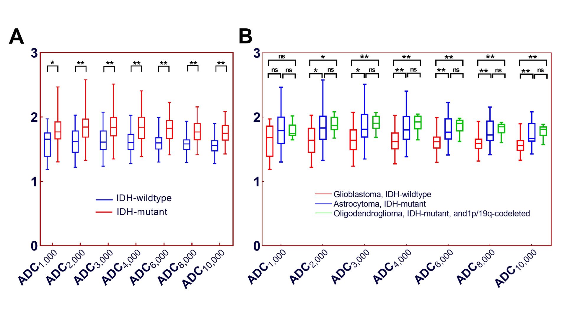
Figure 1. Box and whisker plot of ADC values in TP/NAWM in the evaluation of IDH genotype and tumor subtype. Box and whisker plot shows the distribution of ADC values in TP/NAWM that could be used to identify (A) the IDH genotype and (B) the tumor subtype of adult-type diffuse gliomas based on WHO CNS5. * p < 0.05, and ** p < 0.01. IDH = isocitrate dehydrogenase, ns = no significance.
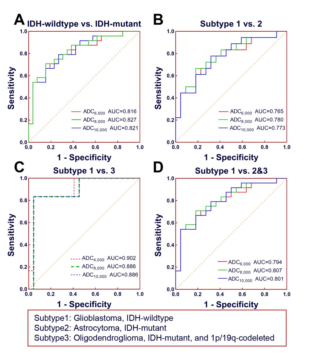
Figure 2. ROC curves with top 3 AUCs of different ADC values in TP/NAWM in identifying IDH genotypes and the tumor subtypes of adult-type diffuse gliomas based on WHO CNS5. ROC curves for the ADC values in TP/NAWM with top 3 AUCs in differentiating (A) IDH-wildtype from IDH-mutant gliomas, (B) glioblastoma, IDH-wildtype from astrocytoma, IDH-mutant, (C) glioblastoma, IDH-wildtype from oligodendroglioma, IDH-mutant, and 1p/19q-codeleted, (D) and glioblastoma, IDH-wildtype from gliomas of IDH-mutant. AUC = area under curve, ADC = apparent diffusion coefficient.
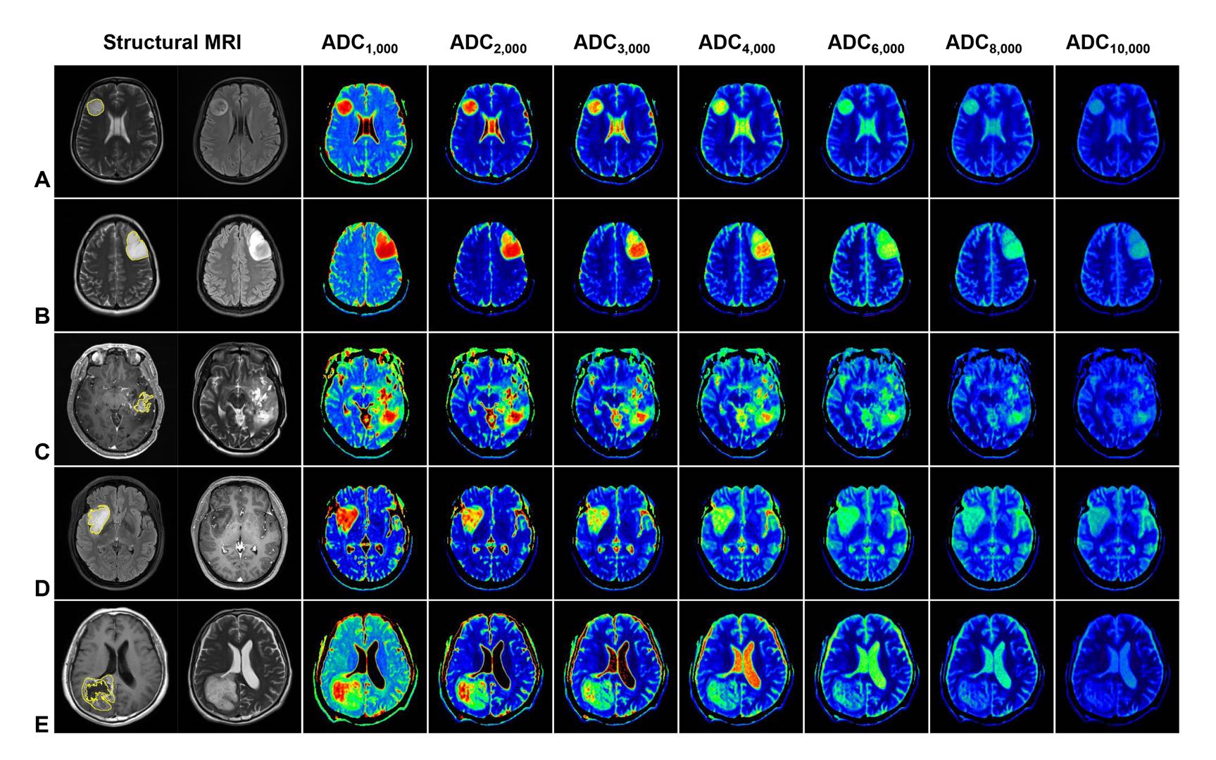
Figure 3. ROI delineation of representative cases and the role of ADC values in evaluation of the IDH genotype and tumor subtype. They are a 52-year-old female patient with astrocytoma, IDH-mutant (grade 2) (A), a 31-year-old female patient with astrocytoma, IDH-mutant (grade 3) (B), a 31-year-old male patient with astrocytoma, IDH-mutant (grade 4) (C), a 47-year-old female patient with oligodendroglioma , IDH-mutant, and 1p/19q-codeleted (grade 2) (D), and a 62-year-old male patient with glioblastoma, IDH-wildtype (E), respectively.