0256
The Comparison of Diffusion Weighted Imaging and Advanced Diffusion Techniques in Predicting Postoperative Recurrence of Meningioma1Fujian Medical University Union Hospital, Fuzhou, China, 2MR Scientific Marketing, Siemens, Healthineers Ltd., Shanghai, China
Synopsis
Keywords: Tumors, Diffusion/other diffusion imaging techniques
Prior investigators have found that diffusion weighted imaging (DWI) contributes to improving the prediction of postoperative recurrence of meningioma. This study evaluated the feasibility of intravoxel incoherent motion (IVIM) and diffusional kurtosis imaging (DKI) in predicting postoperative recurrence of meningioma and compared them with DWI. Histogram metrics derived from DWI, IVIM, and DKI parameters were compared between the recurrent group and non-recurrent group. It was found that the lower ADC, D, f value and the higher D*, K value, the higher risk of postoperative recurrence of meningioma. However, IVIM and DKI derived parameters had similar diagnostic performance compared with DWI.Background
Prior studies have found that diffusion weighted imaging (DWI) contributes to the differentiation of the inter- and intra-layer variability of the pathological grade of meningioma, thereby improving the prediction of postoperative recurrence of meningioma (1-3). But tumor heterogeneity cannot be captured when calculating average diffusion parameters from single-slice regions of interest (ROI) because of the spatial variation of tumor cellularity and vascularity at histopathology (4, 5). Histogram analysis of the whole tumor shows its effectiveness for better and more systematic evaluation of inter- and intra-tumor heterogeneity (6). Several studies have demonstrated that intravoxel incoherent motion (IVIM) and diffusion kurtosis imaging (DKI) have high accuracy in reflecting tumor biological behavior and predicting tumor grade (7). However, the use of IVIM and DKI to assess the prognosis of meningioma is still lacking. Here, we hypothesize that multi-model diffusion techniques (DWI, IVIM, and DKI) could help predict postoperative recurrence of meningioma, and histogram analysis of the entire tumor volume could solve the issues of tumor heterogeneity. Thus, this study aims to explore and compare the feasibility and accuracy of DWI, IVIM, and DKI derived histogram metrics in predicting postoperative meningioma recurrence.Methods
Forty patients with histologically confirmed meningioma who underwent DWI, IVIM, and DKI were enrolled in this retrospective study. MRI was performed using a 3T MRI system equipped with a gradient system with a maximum amplitude of 50 mT/m and an eight-channel receiver head coil. DWI used a spin echo (SE)-echo planar imaging (EPI) diffusion sequence in the axial plane (TR=5,000ms, TE=84.6ms, section thickness=5mm, intersection gap=0mm, FOV=24cm, matrix=192×192, number of sections=30). Twelve b-values from 0 to 3,000 s/mm2 (0, 50, 100, 150, 200, 300, 500, 800, 1,000, 1,500, 2,000, and 3,000 s/mm2; with number of excitations [NEX]=1 for b=0-500 s/mm2, two NEX for b=800-1,000 s/mm2, three NEX for b=1,500 s/ mm2, four NEX for b=2000 s/mm2, and six NEX for b=3000 s/mm2) were used in three orthogonal directions. DKI used a spin-echo DW echo-planar imaging sequence (TR=6000ms, TE=94ms, sections thickness=5mm, intersection gap=0mm, FOV=24cm, matrix=128×128, NEX=30, three images of b0, b-values=1000 and 2000 s/mm, gradient directions=30 for each NEX). The DWI data were obtained and transferred to a workstation (Advantage Workstation 4.6) for processing. Parameter maps were generated by the MADC program in the Functool software for each model. Tumor segmentation was manually delineated by using the ITK-SNAP program. On contrast-enhanced T1-weighted imaging, ROIs were drawn on the solid region of every tumor section. The DWI (ADC), IVIM (perfusion-related diffusion fraction, f; slow diffusion coefficient, D; fast diffusion coefficient, D*) and DKI (kurtosis, K; diffusivity, Dk) histogram metrics were obtained by using whole-tumor volume. Histogram metrics derived from DWI, IVIM, and DKI parameters were compared between the recurrent group and non-recurrent group by using the Mann-Whitney U test. The diagnostic performance of statistically different histogram metrics was further assessed using receiver operating characteristic (ROC) curves. Delong test was used to compare the areas under curve. P values <0.05 were considered statistically significant.Results
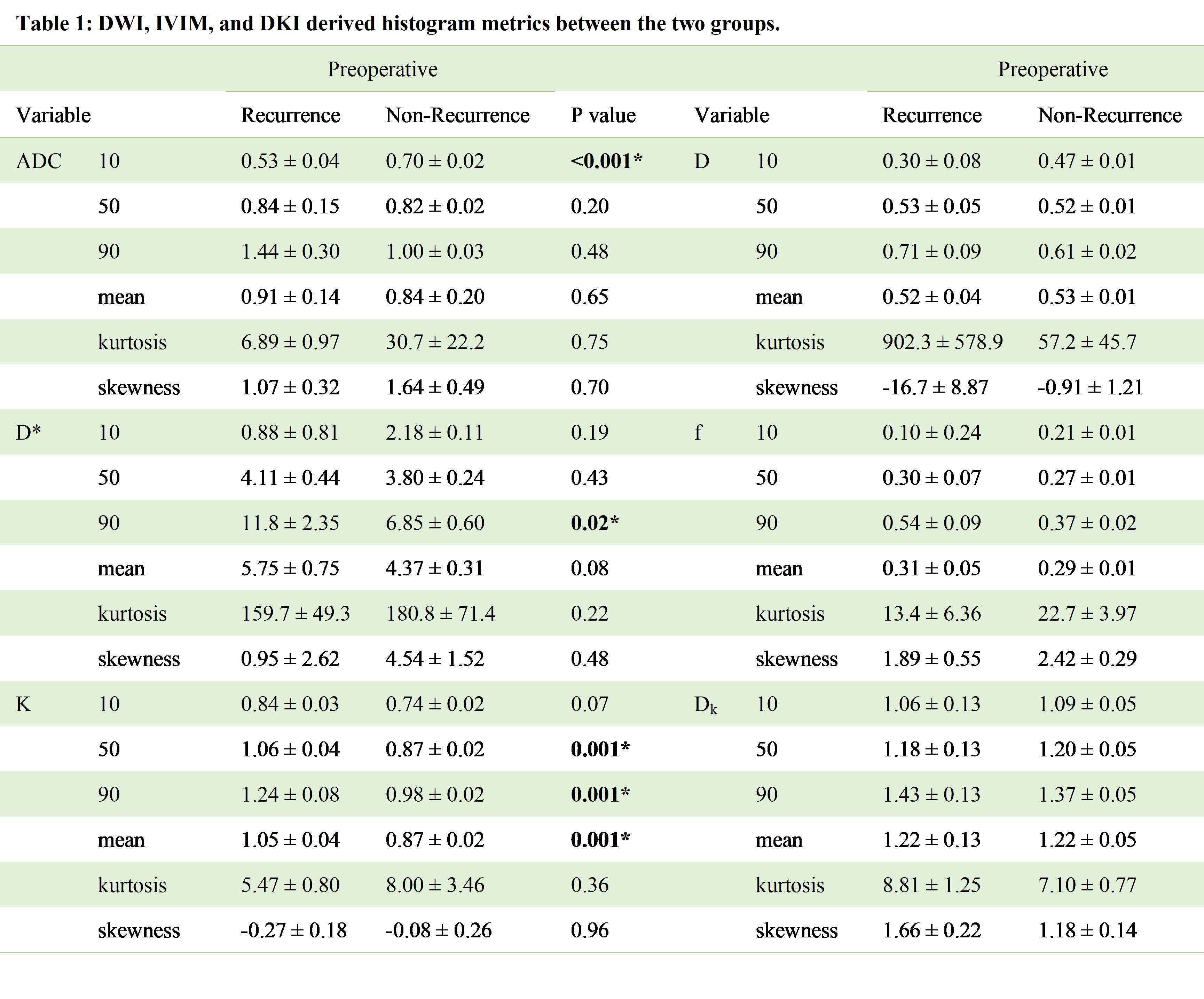
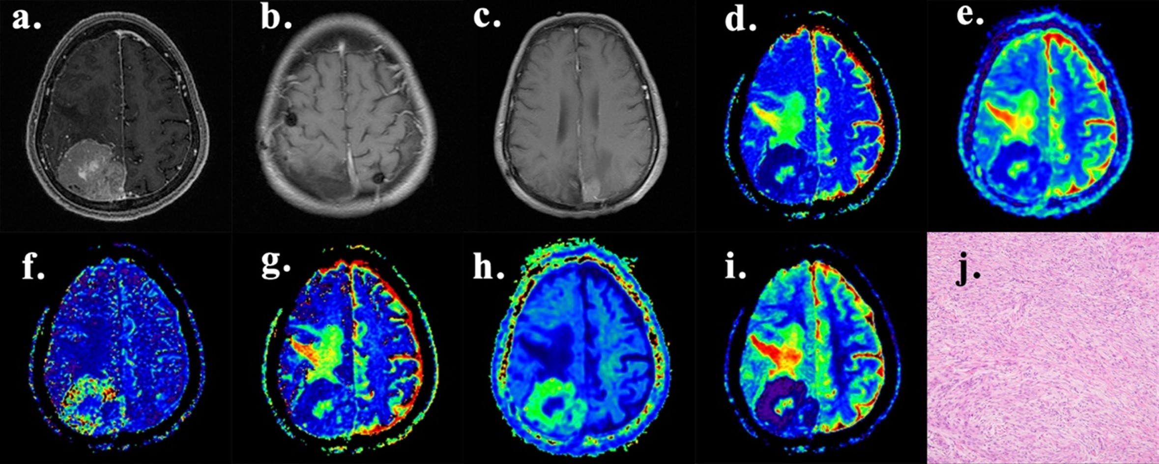
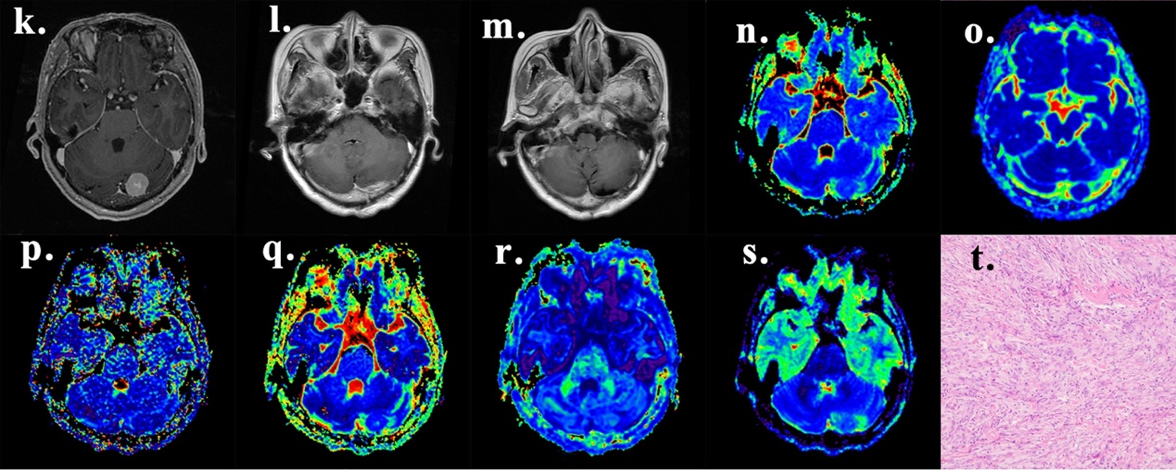
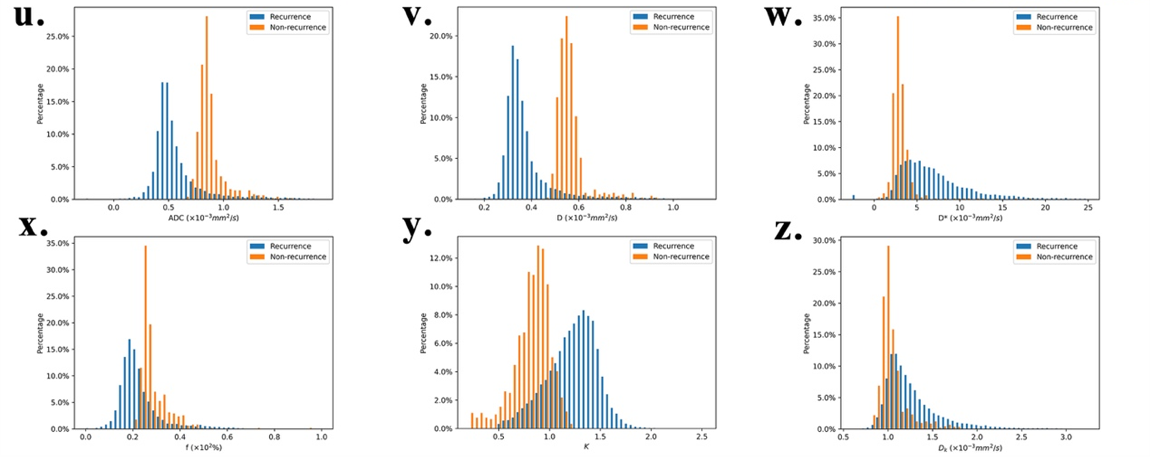
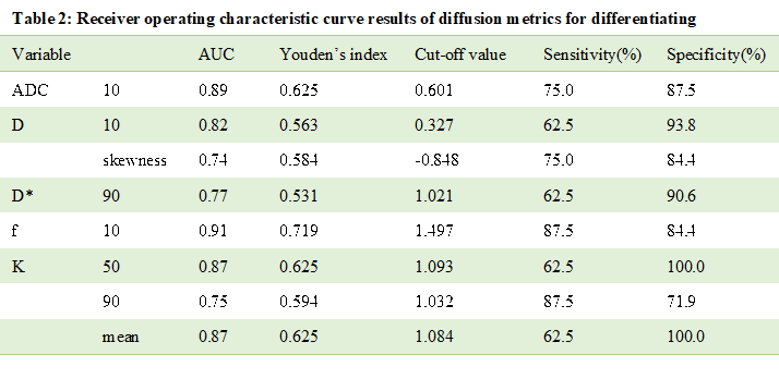
Of the 40 patients (median age, 54 years [IQR, 43–62 years], 11 men and 29 women), eight experienced recurrences after radical surgery. DWI, IVIM, and DKI parameters derived histogram metrics between the recurrent group and non-recurrent group are shown in Table 2. 10th ADC, 10th D, and 10th f of the recurrent group were significantly lower than that of the non-recurrent group, but 90th D* and mean K were opposite. No significant difference in 10th, 50th, 90th percentiles, mean and kurtosis, skewness derived from DK was found between the two groups (P >0.05). Representative cases of recurrent and non-recurrent meningioma are shown in Figure 1. Since the above-mentioned histogram metrics showed significant differences between recurrent and non-recurrent groups, ROC curve analyses were performed to evaluate the diagnostic performance (Table 3). Among the significant parameters, the highest one, 10th f, had an AUC of 0.91, with a sensitivity of 87.5% and specificity of 84.4% at the optimum threshold of 0.15×10² %. However, the diagnostic performance of DWI, IVIM, DKI parameters was similar according to the Delong test (all P >0.05, compared with each other).Discussion and Conclusion
The results of the present study elucidated that lower ADC, D, f value and higher D*, K value predicted a higher risk of postoperative recurrence of meningioma. However, there was no significant difference between the diagnostic performance of advanced diffusion techniques and that of the traditional imaging. In summary, our results preliminary indicate that advanced diffusion imaging is feasible in predicting postoperative recurrence of meningioma, although its performance is similar to traditional DWI.Acknowledgements
No acknowledgement found.References
1. Hwang W, Marciscano A, Niemierko A, et al. Imaging and extent of surgical resection predict risk of meningioma recurrence better than WHO histopathological grade. Neuro-oncology, 2016, 18(6): 863-72.
2. Ko C, Lim S, Chen T, et al. Prediction of progression in skull base meningiomas: additional benefits of apparent diffusion coefficient value. Journal of neuro-oncology, 2018, 138(1): 63-71.
3. Zhang R, Chen X, Cai J, et al. A Novel MRI-Based Risk Stratification Algorithm for Predicting Postoperative Recurrence of Meningioma: More Benefits to Patients. Frontiers in Oncology, 2021, 11: 737520-737520.
4. Nougaret S, Vargas H, Lakhman Y, et al. Intravoxel Incoherent Motion-derived Histogram Metrics for Assessment of Response after Combined Chemotherapy and Radiation Therapy in Rectal Cancer: Initial Experience and Comparison between Single-Section and Volumetric Analyses. Radiology, 2016, 280(2): 446-54.
5. Shih T, Hou H, Liu C, et al. Bone marrow angiogenesis magnetic resonance imaging in patients with acute myeloid leukemia: peak enhancement ratio is an independent predictor for overall survival. Blood 2009;113(14):3161-3167.
6. Chandarana H, Rosenkrantz A, Mussi T, et al. Histogram analysis of whole-lesion enhancement in differentiating clear cell from papillary subtype of renal cell cancer. Radiology, 2012, 265(3): 790-8.
7. She D, Lin S, Guo W, et al. Grading of Pediatric Intracranial Tumors: Are Intravoxel Incoherent Motion and Diffusional Kurtosis Imaging Superior to Conventional DWI?. AJNR. American journal of neuroradiology, 2021.
Figures