0248
Associations of Brain Iron with Cognitive and Social Emotional Performance in Children using Quantitative Susceptibility Mapping1School of Physics and Electronic Science, East China Normal University, Shanghai, China, 2MR Scientific Marketing, Siemens, Siemens Healthineers, Shanghai, China, 3Institute of Brain and Education Innovation, East China Normal University, Shanghai, China, 4Department of Radiology, Weill Medical College of Cornell University, New York, NY, United States
Synopsis
Keywords: Data Analysis, Quantitative Susceptibility mapping
Tissue iron play a critical role in cognitive functions. However, associations of basal ganglia iron concentration with cognition and emotion in children are less well understood. This study examined the correlation of susceptibility values in the bilateral basal ganglia nuclei with cognitive functions and social emotional capacity in children around the age of seven. The results highlighted that the inhibitory control, collaboration, open-mindedness showed significant association with susceptibility values in the basal ganglia. In conclusion, this QSM study indicated the potential for using brain iron content in the basal ganglia to assess cognitive and emotional performance during the children development.Introduction
Brain tissue iron is essential to healthy brain biologic functions, including neurotransmitter synthesis, generation of myelin sheets, and metabolism 1. Brain tissue showed the greatest iron concentration in basal ganglia structures, which play a critical role in a multitude of cognitive functions as part of the corticobasal ganglia-thalamo-cortical loops 2. In younger children, however, the effects of iron concentration in the basal ganglia on cognition and social emotional capacity are less well understood. Advances in quantitative susceptibility mapping (QSM) techniques have made it possible to noninvasively measure tissue iron content with high spatial resolution and high sensitivity 3. The objective of this study was to investigate the associations of iron in basal ganglia with cognitive functions and social emotional performance among normal children via QSM.Materials and Methods
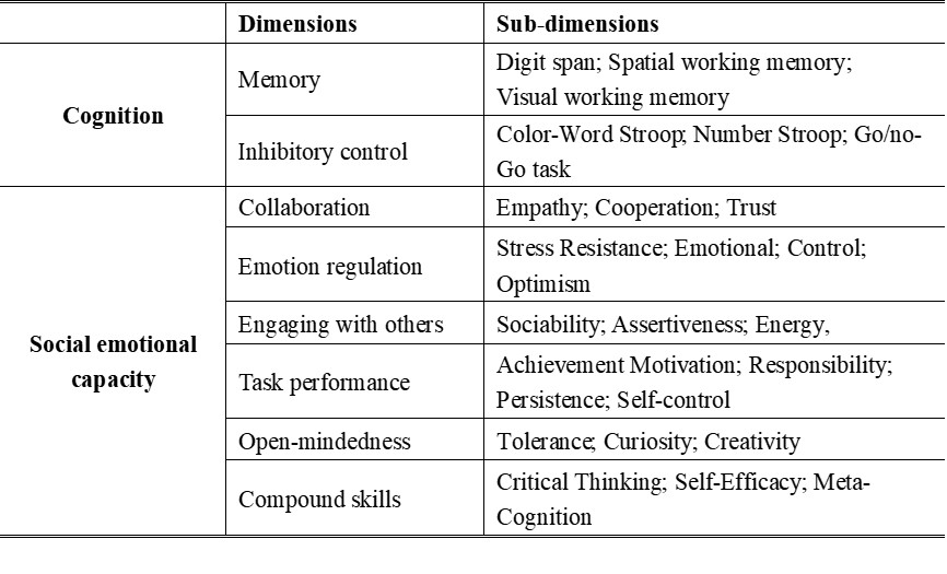
This study was approved by the local research ethics board. A total of 46 second-grade children with a mean age of 7.51 ± 0.31 years old (22 males and 24 females) were recruited in this study.The cognitive domains in this study included memory (digit span, spatial, and visual working memory) and inhibitory control contained three sub-dimensions. In the Stroop tests, the reaction time difference under inconsistent and consistent conditions reflected inhibiting ability. For social emotional capacity, including collaboration, emotion regulation, engaging with others, task performance, open-mindedness, and compound skills, the scores of each dimension reflecting emotion were calculated on the basis of sub-dimensions test scores summarized in Table 1, higher scores mean better performance.
MRI was conducted by using a clinical 3T MR imaging scanner (Prisma Fit, Siemens Healthcare, Erlangen, Germany) equipped with a 20-channel head coil. The QSM images were generated from a 3D spoiled unipolar-readout multi-echo gradient-echo sequence acquired in the axial plane with the following parameters: repetition time (TR) = 31ms, first echo time (TE1) = 3.98ms, echo spacing (ΔTE) = 4.31ms, number of echoes = 6, flip angle = 12˚, field of view (FOV) = 220*165 mm2, matrix size = 256*192, in-plane resolution = 0.83*0.83mm2, slice thickness = 0.90mm, number of slices = 176. During scanning, foam pads were placed around each subject’s head to minimize head motion.
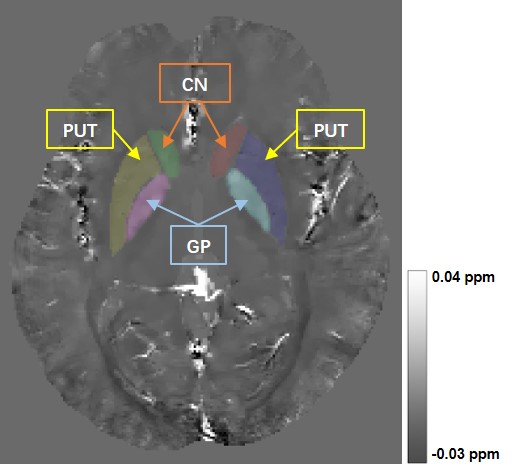
QSM maps were reconstructed using the Morphology Enabled Dipole Inversion with automatic uniform cerebrospinal fluid zero reference (MEDI+0) algorithm 4. The phase shift of even echoes induced by the gradient delay and eddy current was estimated and corrected for the data in the target domain 5. Regions of interest (ROIs), including the bilateral head of the caudate nucleus (CN), putamen (PUT), and globus pallidus (GP), were drawn manually on the QSM images using ITK-SNAP (http://www.itk-snap.org) (Figure 1). Associations of magnetic susceptibility values with cognitive and emotional scores were assessed using Pearson correlation coefficients. All statistical analyses were carried out using IBM SPSS Statistics 23.
Results
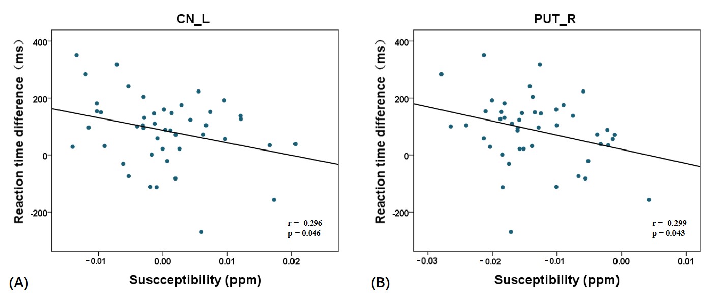
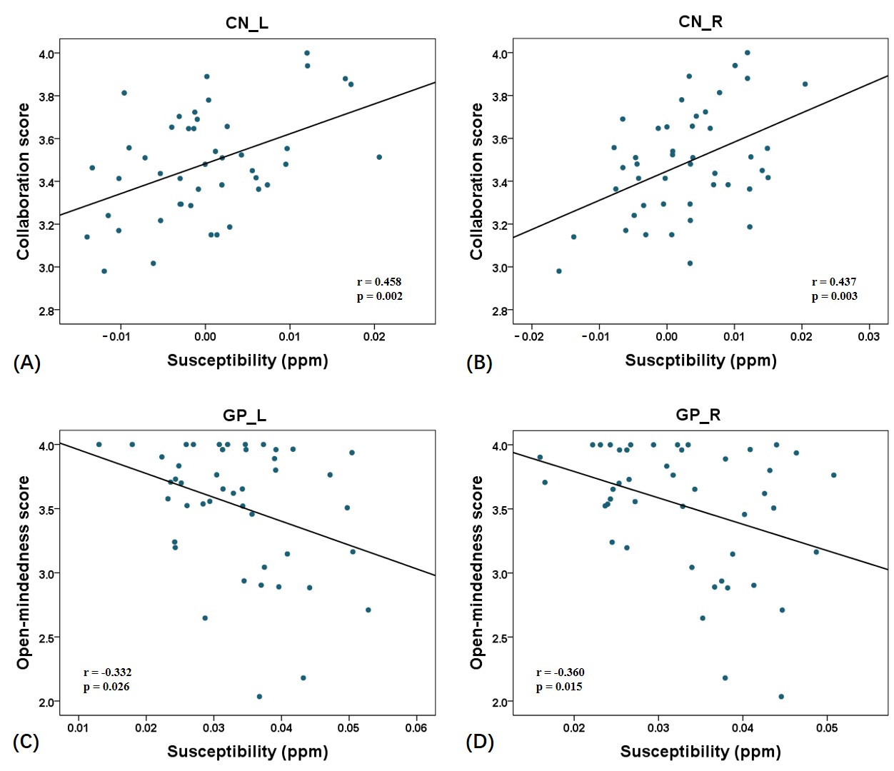
For inhibitory control of cognition, there were significant negative associations between susceptibility value and reaction time difference under inconsistent and consistent in the left CN (r = -0.296, p = 0.046) and right PUT (r = -0.299, p = 0.043) in the Color-Word Stroop test (Figure 2). The susceptibility value in the basal ganglia showed no significant association with the memory function (p > 0.05). For the social emotional capacity, significant positive correlation was found between susceptibility value and collaboration in the left of CN (r = 0.458, p = 0.002) and right of CN (r = 0.437, p = 0.003). The susceptibility values were negatively correlated with change in open-mindedness in the left of GP (r = -0.332, p = 0.026) and right of GP (r = -0.357, p = 0.016) (Figure 3).Discussion
This is the first study to evaluate the associations of iron within basal ganglia with cognitive and social emotional performance in children using QSM. Our finding showed significantly greater iron concentration in the basal ganglia associated with greater inhibiting ability in children around the age of seven. This is consistent with previous studies that iron supplementation has been found to improve cognitive performance in iron deficient adolescents 6, 7. The results also agreement with a study highlighted the transition from adolescence to adulthood as a period of dynamic maturation of tissue iron concentration in the basal ganglia that may affect individual variability in complex cognitive performance 8. Another important result of this study showed that iron content in basal ganglia correlated with some dimensions of social emotional capacity. Therefore, our results further confirmed that it is possible that efforts to enhance brain iron concentration through dietary iron supplementation may have beneficial effects on cognitive and emotional development during this critical period of development.Conclusions
In the summary, elevated brain iron was related to cognitive and emotional performance in adolescence, suggesting that QSM may be potential for measuring brain iron content in the basal ganglia region to assess cognitive and emotional performance states during the children development in the adolescence.Acknowledgements
No acknowledgement found.References
1. Ward RJ, Zucca FA, Duyn JH, Crichton RR, Zecca L. The role of iron in brain ageing and neurodegenerative disorders. Lancet Neurol 2014; 13: 1045-1060.
2. Haber SN, Knutson B. The reward circuit: linking primate anatomy and human imaging. Neuropsychopharmacology 2010; 35: 4-26.
3. Wang Y, Spincemaille P, Liu Z, et al. Clinical quantitative susceptibility mapping (QSM): Biometal imaging and its emerging roles in patient care. J Magn Reson Imaging 2017; 46: 951-971.
4. Liu Z, Spincemaille P, Yao Y, Zhang Y, Wang Y. MEDI+0: Morphology enabled dipole inversion with automatic uniform cerebrospinal fluid zero reference for quantitative susceptibility mapping. Magn Reson Med 2018; 79: 2795-2803.
5. Li J, Chang S, Liu T, et al. Phase-corrected bipolar gradients in multi-echo gradient-echo sequences for quantitative susceptibility mapping. MAGMA 2015; 28: 347-355.
6. Bruner AB, Joffe A, Duggan AK, Casella JF, Brandt J. Randomised study of cognitive effects of iron supplementation in non-anaemic iron-deficient adolescent girls. Lancet 1996; 348: 992-996.
7. Blanton C. Improvements in iron status and cognitive function in young women consuming beef or non-beef lunches. Nutrients 2013; 6: 90-110.
8. Larsen B, Bourque J, Moore TM, et al. Longitudinal development of brain iron is linked to cognition in youth. J Neurosci 2020; 40: 1810-1818.
Figures