0243
Impact of Radiomic Features Stability of Myocardial Motion on Classification of Repaired Tetralogy of Fallot Patients1Department of Biomedical Engineering and Environmental Sciences, National Tsing Hua University, Hsinchu, Taiwan, 2Department of Radiology, Kaohsiung Veterans General Hospital, Kaohsiung, Taiwan, 3Department of Pediatrics, Kaohsiung Veterans General Hospital, Kaohsiung, Taiwan, 4Department of Pediatrics, National Yang Ming Chiao Tung University, Taipei, Taiwan, 5Department of Electrical Engineering, National Taiwan University of Science and Technology, Taipei, Taiwan
Synopsis
Keywords: Radiomics, Radiomics
Myocardial motion influences the stability of radiomic features of cardiac magnetic resonance (CMR) cine images. We aimed to evaluate the impact of the stability of myocardial radiomic features on the classification performance of differentiation of repaired tetralogy of Fallot (rTOF) patients from normal volunteers. The stability of each radiomic feature during cardiac cycle was assessed by coefficient of variation (CV) of the feature value. 50 of 107 radiomic features (46.7%) in normal volunteers were stable features. The classification model established only with stable features presented the best classification performance (AUC=0.94) which was able to be improved by chi-square feature selection.Introduction
Cardiac magnetic resonance (CMR) cine imaging has been used to evaluate the cardiac function in patients with repaired tetralogy of Fallot (rTOF) 1. Radiomics refers to the extraction of quantitative features, which are unperceivable by human eyes, from radiologic images. A previous study showed that myocardial motion during the cardiac cycle influences the stability of myocardial radiomic features of CMR cine images 2. However, the impact of the stability of myocardial radiomic features on the performance of classification model has not been assessed. We aimed to evaluate the impact of the stability of myocardial radiomic features on the classification performance of differentiation of rTOF patients from normal volunteers.Methods
The stability analysis of radiomic features was conducted on 18 normal volunteers (age=21.3±0.7 years, 10 males). The classification model used to differentiate rTOF patients from normal volunteers was performed on 14 rTOF patients (age=22.3±4.2 years, 7 males) and 18 normal volunteers. All MR images were acquired in a 3T MR scanner (Skyra, Siemens, Erlangen, Germany). Cine steady-state free precession images in a short-axis view were acquired with retrospective ECG-gating and breath-hold technique and scanning parameters as follows: TR/TE=3.2/1.7 ms, flip angle=54º, voxel size=1.15×1.15×8 mm3, gap=2 mm.Fig. 1 summarizes the workflow of the present work. An institution-developed auto-segmentation tool was used to delineated the regions of interest (ROIs) of endocardial and epicardial borders of the myocardium on basal middle, and apical slices. These ROIs in normal volunteers were used for radiomic features extraction. An open-source software, PyRadiomics, was used to extract a total of 107 features and categorized as 7 categories of features, including 14 shape features, 18 first-order features, 24 Gray Level Co-occurrence Matrix (GLCM) features, 14 Gray Level Dependence Matrix (GLDM) features, 16 Gray Level Run Length Matrix (GLRLM) features, 16 Gray Level Size Zone Matrix (GLSZM) features, and 5 Neighbouring Gray Tone Difference Matrix (NGTDM) features 3. The stability of each radiomic feature in 25 cardiac frames was assessed by coefficient of variation (CV) of the feature value and categorized as four levels, as listed in Table 1.
The classification models were trained by using 7 images most close to the peak diastole and systole. The quadratic support vector machine (SVM) in a MATLAB toolbox (Classification Learner, Statistics and Machine Learning Toolbox 12.4, MATLAB 2022b) was used to train classification models. To assess the impact of performing feature selection on the classification performance, the chi-square (chi2) feature selection method was used selectively. The 6 classification models included using stable features only, unstable features only, all features, and a combination of chi2 with abovementioned 3 models.
Results
In Fig. 2, of the total 107 radiomic features in normal volunteers, 9 features (8.4%), 18 features (16.8%), and 23 features (21.5%) were with very small, small, and intermediate CV, and were all categorized as stable features; while 57 features (53.3%) were with large CV and were categorized as unstable features. All features in shape category (n=14) were stable features. All features in NGTDM category (n=5) were unstable features. The GLRLM has 10 stable features (62.5%) and was the most stable feature-group in second-order features.Fig. 3 illustrates that all models presented good performance in differentiation of rTOF patients from normal volunteers (AUC=0.85-0.95). The performance of classification model with unstable features (AUC=0.85) was substantially improved by combining chi-square feature selection (AUC=0.91). The model with stable features was mildly improved by chi-square feature selection (AUC increased from 0.94 to 0.95). The performance of classification model including all features had a lower AUC than that including only stable features (AUC=0.89 vs. 0.94).
Discussion and Conclusion
In our study, 50 of 107 radiomic features (46.7%) in normal volunteers were stable features. In 7 categories of features, all features in shape category were stable features and all features in GLSZM category belonged to unstable features. The classification model established only with stable features presented the best classification performance which was able to be improved by chi-square feature selection.Alis et al reported a large proportion of radiomic features (55.5%) with high variability and GLSZM features demonstrated the highest variability category of features in cine images 2 which is consistent with our results. Another study acquired 4D-CT images in patients with early-stage non-small-cell lung cancer and demonstrated that shape features appeared to be the most stable category of features among 841 radiomic features with regard to respiration 4. In our cine CMR images, we also found that shape features were the most stable category of features.
Using stable features to differentiate rTOF from normal volunteers had the best performance in comparison with using unstable features or all features. Therefore, a classification model established by images with motion has to consider the potential adverse impacts of unstable features. Removing unstable features from the establishment of classification model could improve classification performance. An appropriate feature selection scheme, such as chi2 in this study, could further improve classification performance.
In conclusion, each radiomic feature exhibited differential stability of myocardial motion in cine CMR images. A classification model established by stable features only had the best performance for differentiating rTOF patients from normal volunteers.
Acknowledgements
No acknowledgement found.References
1. Habert P, Bentatou Z, Aldebert P, Finas M, Bartoli A, Bal L, Lalande A, Rapacchi S, Guye M, Kober F, Bernard M, Jacquier A. Exercise stress CMR reveals reduced aortic distensibility and impaired right-ventricular adaptation to exercise in patients with repaired tetralogy of Fallot. PLoS One. 2018 Dec 31;13(12):e0208749.
2. Alis D, Yergin M, Asmakutlu O, Topel C, Karaarslan E. The influence of cardiac motion on radiomics features: radiomics features of non-enhanced CMR cine images greatly vary through the cardiac cycle. Eur Radiol. 2021 May;31(5):2706-2715.
3. van Griethuysen JJM, Fedorov A, Parmar C, Hosny A, Aucoin N, Narayan V, Beets-Tan RGH, Fillion-Robin JC, Pieper S, Aerts HJWL. Computational Radiomics System to Decode the Radiographic Phenotype. Cancer Res. 2017 Nov 1;77(21):e104-e107.
4. Du Q, Baine M, Bavitz K, McAllister J, Liang X, Yu H, Ryckman J, Yu L, Jiang H, Zhou S, Zhang C, Zheng D. Radiomic feature stability across 4D respiratory phases and its impact on lung tumor prognosis prediction. PLoS One. 2019 May 7;14(5):e0216480.
Figures
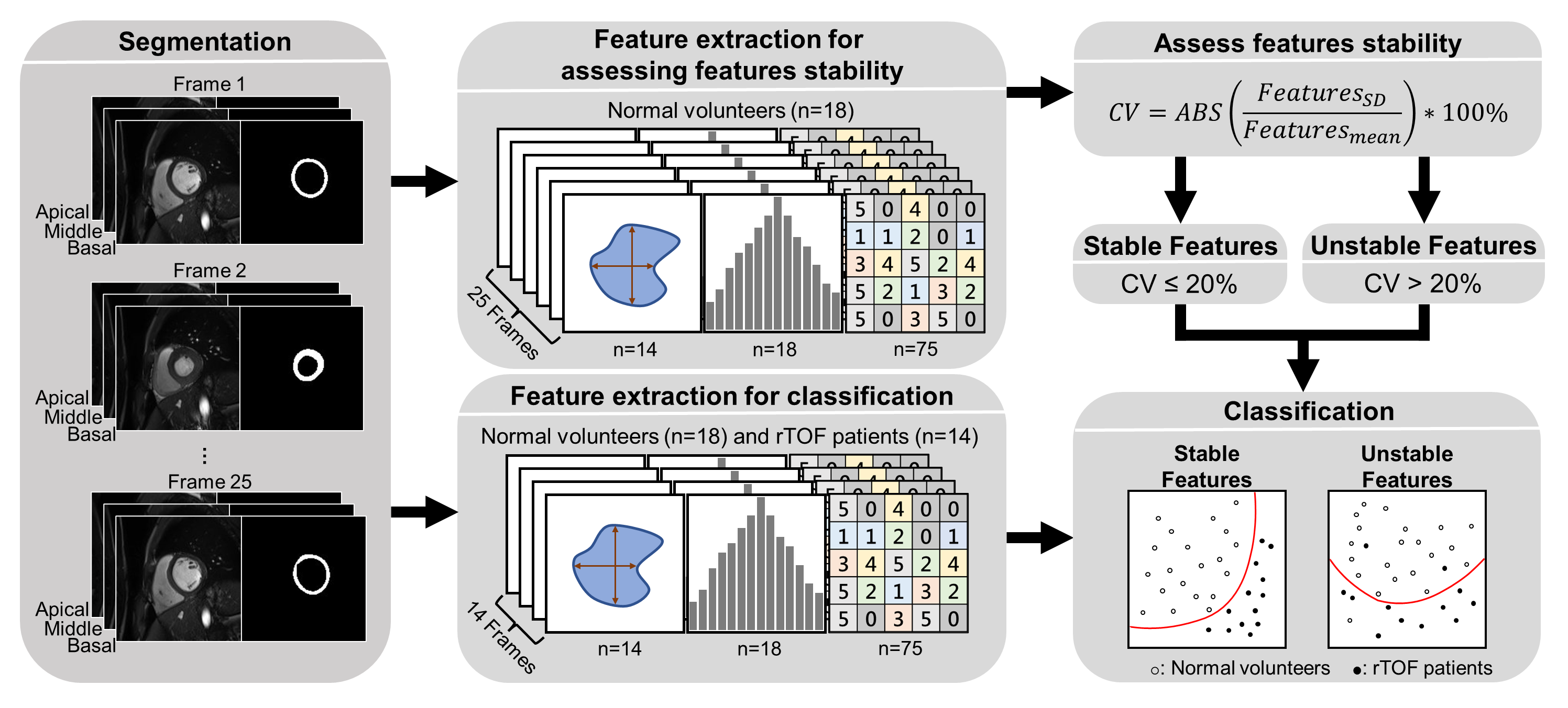
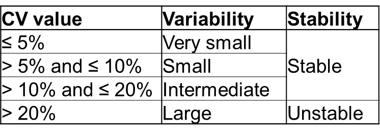
Table 1. Four variability levels of features.
CV: coefficient of variation
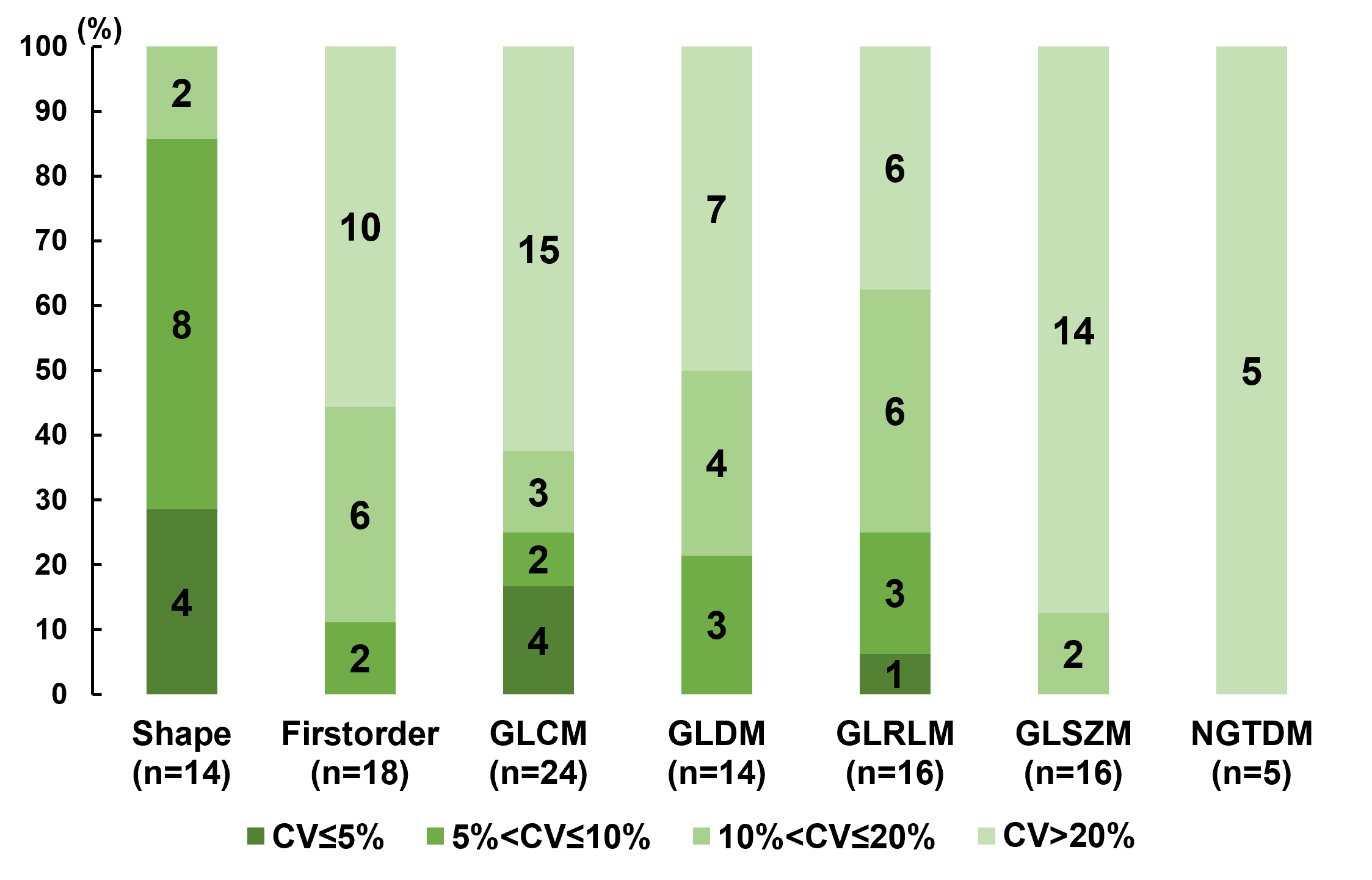
Fig. 2. The percentages of feature numbers in 7 categories of features. Different colors represent different CV levels (see Table 1). GLCM: Gray Level Co-occurrence Matrix, GLDM: Gray Level Dependence Matrix, GLRLM: Gray Level Run Length Matrix, GLSZM: Gray Level Size Zone Matrix, NGTDM: Neighbouring Gray Tone Difference Matrix.
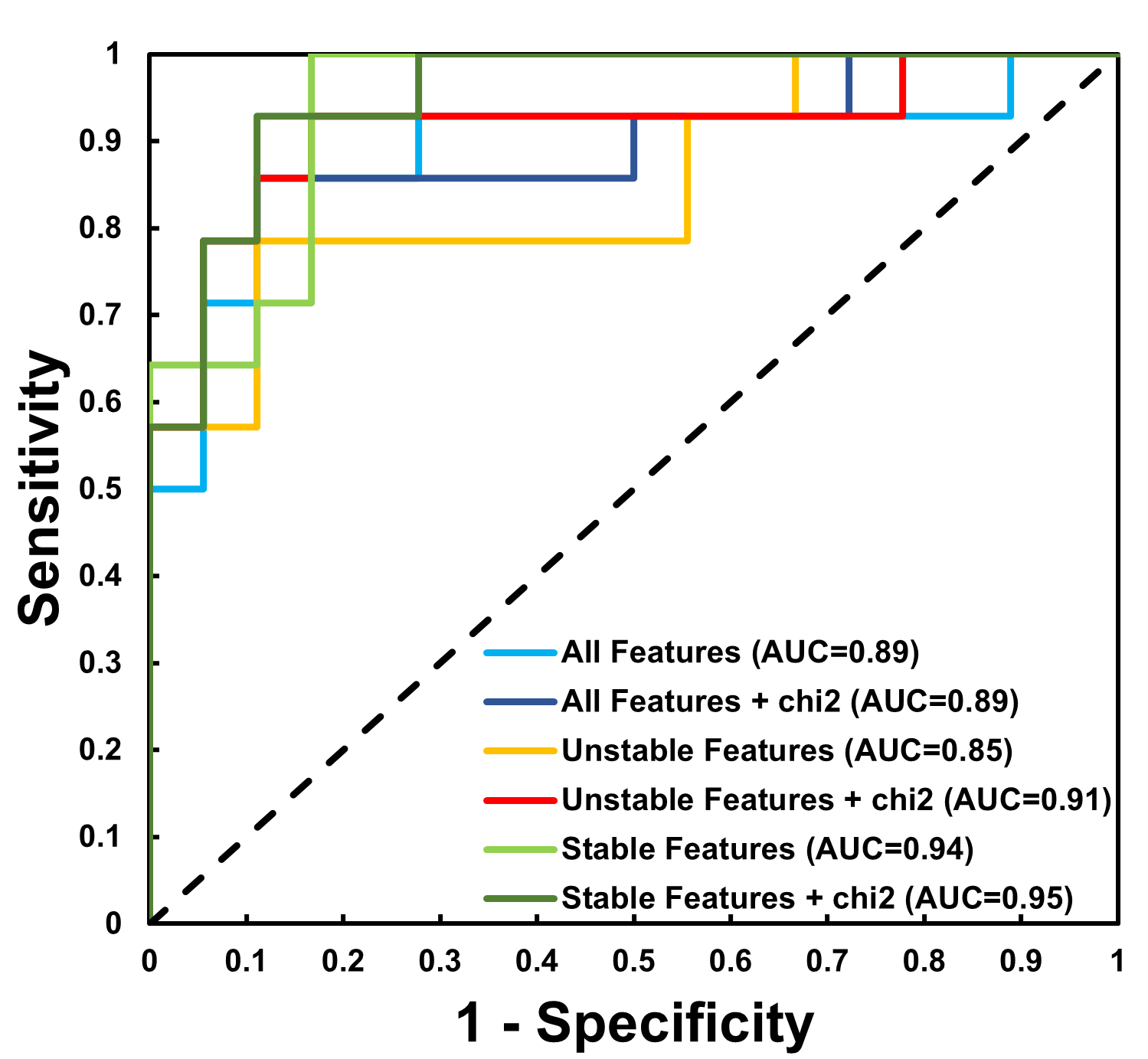
Fig. 3. The receiver operation characteristic (ROC) curve of six classification models with various combinations of features. AUC: area under curve, chi2: chi-square.
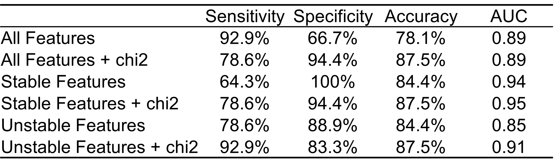
Table 2. Parameters of receiver operating characteristic curve in six classification models.
AUC: area under curve, chi2: chi-square.