0236
Longitudinal Changes of Functional Connectivity Dynamism Are Relevant for Disability Worsening in Multiple Sclerosis: A 2.5-Year Study
Paola Valsasina1, Giulia d'Amore1,2, Paolo Preziosa1,2,3, Monica Margoni1,2, Massimo Filippi1,2,3,4,5, and Maria Assunta Rocca1,2,3
1Neuroimaging Research Unit, Division of Neuroscience, IRCCS San Raffaele Scientific Institute, Milan, Italy, 2Neurology Unit, IRCCS San Raffaele Scientific Institute, Milan, Italy, 3Vita-Salute San Raffaele University, Milan, Italy, 4Neurorehabilitation Unit, IRCCS San Raffaele Scientific Institute, Milan, Italy, 5Neurophysiology Service, IRCCS San Raffaele Scientific Institute, Milan, Italy
1Neuroimaging Research Unit, Division of Neuroscience, IRCCS San Raffaele Scientific Institute, Milan, Italy, 2Neurology Unit, IRCCS San Raffaele Scientific Institute, Milan, Italy, 3Vita-Salute San Raffaele University, Milan, Italy, 4Neurorehabilitation Unit, IRCCS San Raffaele Scientific Institute, Milan, Italy, 5Neurophysiology Service, IRCCS San Raffaele Scientific Institute, Milan, Italy
Synopsis
Keywords: Brain Connectivity, Multiple Sclerosis
Here, we investigated changes in time-varying functional connectivity over 2.5 years of follow-up in 129 multiple sclerosis patients and their association with disability progression. At follow-up, 25/129 (19.3%) patients worsened clinically. At baseline, multiple sclerosis patients showed reduced time-varying functional connectivity vs controls in orbitofrontal, cerebellar, precuneal and thalamic regions. At 2.5-year follow-up, patients exhibited widespread reduction of time-varying functional connectivity over time. Such a pattern was confirmed when looking at clinically stable patients. Conversely, clinically worsened patients presented peculiar reductions of time-varying functional connectivity in default-mode network areas and in basal ganglia, this latter significant at time-by-group interaction analysis.Introduction
Time-varying functional connectivity (TVFC) is a novel analysis technique that measures temporal resting state (RS) functional connectivity (FC) fluctuations occurring during the course of functional MRI (fMRI) acquisition [1]. In multiple sclerosis (MS) patients, TVFC analysis showed abnormalities of the main functional networks, which were correlated with more severe clinical and cognitive disability [2, 3]. Preliminary evidence showed that MS-related cognitive decline was associated with progressive TVFC instability over time [4]. However, the clinical relevance of longitudinal TVFC changes has not been investigated yet. Against this background, aim of this study was to investigate changes in TVFC over 2.5 years of follow-up in MS patients and their association with disability progression.Methods
3T RS fMRI scans and clinical evaluations were obtained at baseline and at median follow-up of 2.5 years from 129 right-handed MS patients (103 relapsing-remitting [RR] and 26 progressive [P] MS) and 28 matched healthy controls (HC). At 2.5-year follow-up, MS patients were classified as clinically stable or clinically worsened according to their expanded disability status scale (EDSS) change. TVFC was quantified at voxel-wise level as the coefficient of variation (CoV) across sliding-windows of degree centrality.Results
At follow-up, 25/129 (19.3%) MS patients worsened clinically. At baseline, MS patients showed reduced TVFC vs HC in the bilateral orbitofrontal cortex (p<0.05, family-wise error corrected), bilateral cerebellum, right precuneus and left thalamus (Figure 1). At 2.5-year follow-up, a widespread reduction of TVFC over time (p<0.05, family-wise error corrected) was found in MS patients in temporal, parietal, occipital and frontal lobes, as well as in the cerebellum. Such a pattern of TVFC reduction was also found when looking at clinically stable MS patients (Figure 2). Conversely, clinically worsened MS presented peculiar TVFC reductions in areas of the default-mode network and basal ganglia (Figure 2). Reduced TVFC in the left putamen in clinically worsened vs stable MS patients was significant at time x group interaction analysis.Discussion
This is one of the first studies analyzing the longitudinal evolution of TVFC in MS patients and its association with clinical worsening. We found that longitudinal TVFC reductions differed according to patients’ clinical status. While clinically stable MS patients showed widespread, aspecific reductions of connectivity dynamism in several cortical lobes and in the cerebellum, clinically worsened MS patients presented peculiar reductions of connectivity dynamism in the default-mode network and in deep grey matter regions. Accrual of TVFC abnormalities in deep grey matter regions may represent a biological substrate of disability worsening.Conclusions
After 2.5 years of follow-up, MS patients showed widespread TVFC reduction in cortical lobes and cerebellum. A peculiar involvement of deep grey matter was found in clinically worsened MS patients.Acknowledgements
No acknowledgement found.References
[1] Calhoun VD et al,. Neuron 2014; 84: 262-74. [2] Valsasina P et al., Front Neurosci 2019; 10;13:618. [3] Hidalgo de la Cruz M et al. Brain Connect 2021;11:678-690. [4] Broeders T et al., Brain Commun 2022;4(2):fcac095.Figures
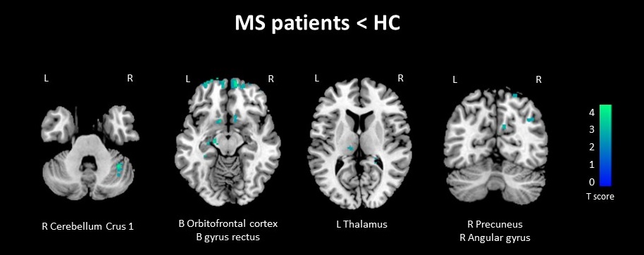
Figure 1. Abnormalities
of baseline time-varying functional connectivity (TVFC) in multiple sclerosis
(MS) patients compared to healthy controls (HC). Abbreviations: L=left,
R=right.
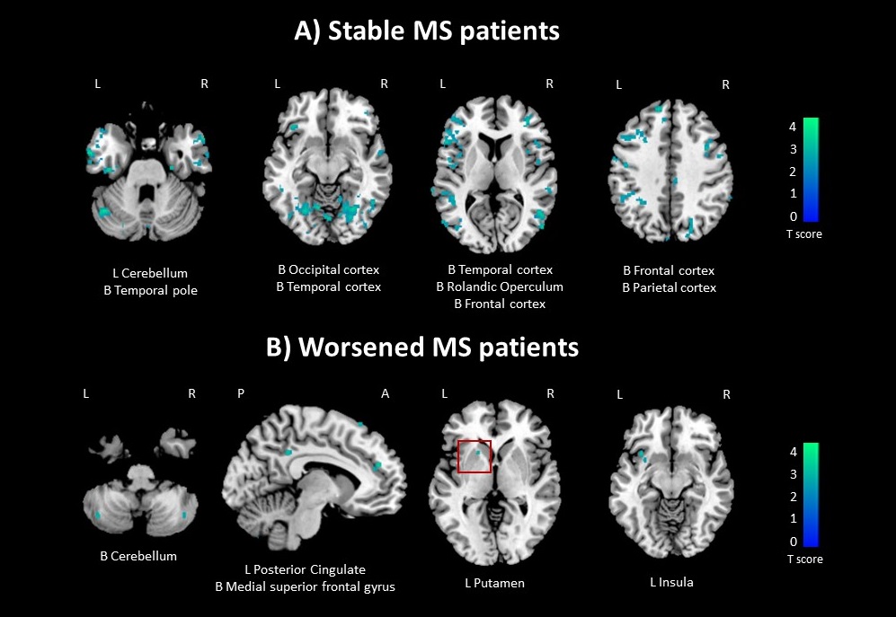
Figure 2. Changes
over 2.5-year follow-up of time-varying functional connectivity (TVFC) in
clinically stable (A) and clinically worsened (B) patients with multiple
sclerosis (MS). The red box highlights the cluster of decreased TVFC in
worsened vs stable MS significant at the time-by-group interaction
analysis. Abbreviations: L=left, R=right.
DOI: https://doi.org/10.58530/2023/0236