0233
Topological Disruption of High-Order Functional Networks in Cognitively Preserved Parkinson’s Disease1Clinical Medical College, Yangzhou University, Yangzhou, China, 2GE Healthcare, MR Research China, Beijing, China
Synopsis
Keywords: Brain Connectivity, Parkinson's Disease
Parkinson’s disease (PD) is not solely a disruption of motor-related networks but a consequence of impaired high-level cognitive processes, indicating that high-order information exchange in cognitively preserved patients with PD could be sensitively revealed in the high-order-functional connection (HOFC) networks. We thus attempted to characterize the topological alterations and classification performance of HOFC networks in normally cognitive patients with PD. Our findings identified the disrupted topology of functional interactions at high-level with extensive alterations of topological properties and improved differentiating ability in patients with PD prior to clinical symptoms of cognitive impairment, providing complementary insights into complex neurodegeneration in PD.Introduction
The aberrant topology of functional connectivity (FC) has been proved to be underlined pathological process in Parkinson’s disease (PD). However, high-level information exchange that underlies the motor disturbance in PD still needs further elucidation, which cannot be sensitively detected in traditional networks constructed by low-order FC (LOFC). In contrast to LOFC, high-order FC (HOFC) was generated by forming pairwise functional coherence on the basis of LOFC profiles between regions1. It has been validated that HOFC profiles could capture high-level modulations in several pathological conditions with improved classification performance for diagnosis2-4.Considering that high-order topological metrics, with a focus mainly on motor-related alterations, remains poorly investigated, the present study sought to explore alterations of topological metrics via a graph-theoretical approach on the basis of HOFC profiles and their relevance to clinical performance in cognitively normal patients with PD. Moreover, a machine learning approach was specifically adopted for the verification of classification performance of the fundamental HOFC between the enrolled patients and healthy controls (HCs). We hypothesized that the disrupted topology of HOFC networks might be more pronounced and achieve better discriminative ability than that of LOFC networks, providing a complementary understanding of the underlying mechanisms of motor deficits in those with PD.
Materials and Methods
SubjectsA final sample consisted of 51 (31 male and 20 female) cognitively normal patients with PD, and 60 (35 male and 25 female) matched healthy controls (HCs) were recruited. The Unified Parkinson’s Disease Rating Scale part III (UPDRS-III) and Hoehn-Yahr scale were scored for disease severity and stage of PD.
MRI experiment
MRI experiments were performed using a 3.0-tesla MRI scanner (Discovery MR750, GE, USA) with an 8-channel phased array head coil. Resting-state functional MRI data of the whole brain were acquired as follows: TR, 2,000 ms; TE, 30 ms; FA, 90°; slice thickness, 4 mm without gap; FOV, 240 × 240 mm2; matrix size, 64 × 64; voxel size, 4.0 × 4.0 × 4.0 mm3; 240 time points; total scan time, 480 s.
Data analysis
Functional data were preprocessed in SPM 12 embedded in the MATLAB 2018a platform. The networks construction (LOFC and HOFC) was performed using the GRETNA toolbox. The nodes of both networks were defined by the automated anatomic labeling atlas (2nd version) 5. Regarding edge definition, Pearson’s correlation coefficient of the mean time series (LOFC-network) or that of the LOFC profiles (HOFC-network) between any two nodes was calculated using the BrainNetClass toolbox. The topological properties for individuals were calculated at each sparsity level and were further compared between groups.
Statistical analysis
Topological properties were compared between groups using a two-sample t test in the GRETNA toolbox. The network-based statistic approach in the GRETNA toolbox was utilized for identification of intergroup differences in functional connections. The exploration of potential correlations between topological metrics and clinical scale scores in the PD group was conducted using Spearman rank correlation test with SPSS 19.0 software. The threshold for statistical significance was set to a P value < 0.05. Classification performance was analyzed using least absolute shrinkage and selection operator for feature selection and support vector machine approach for the classification.
Results
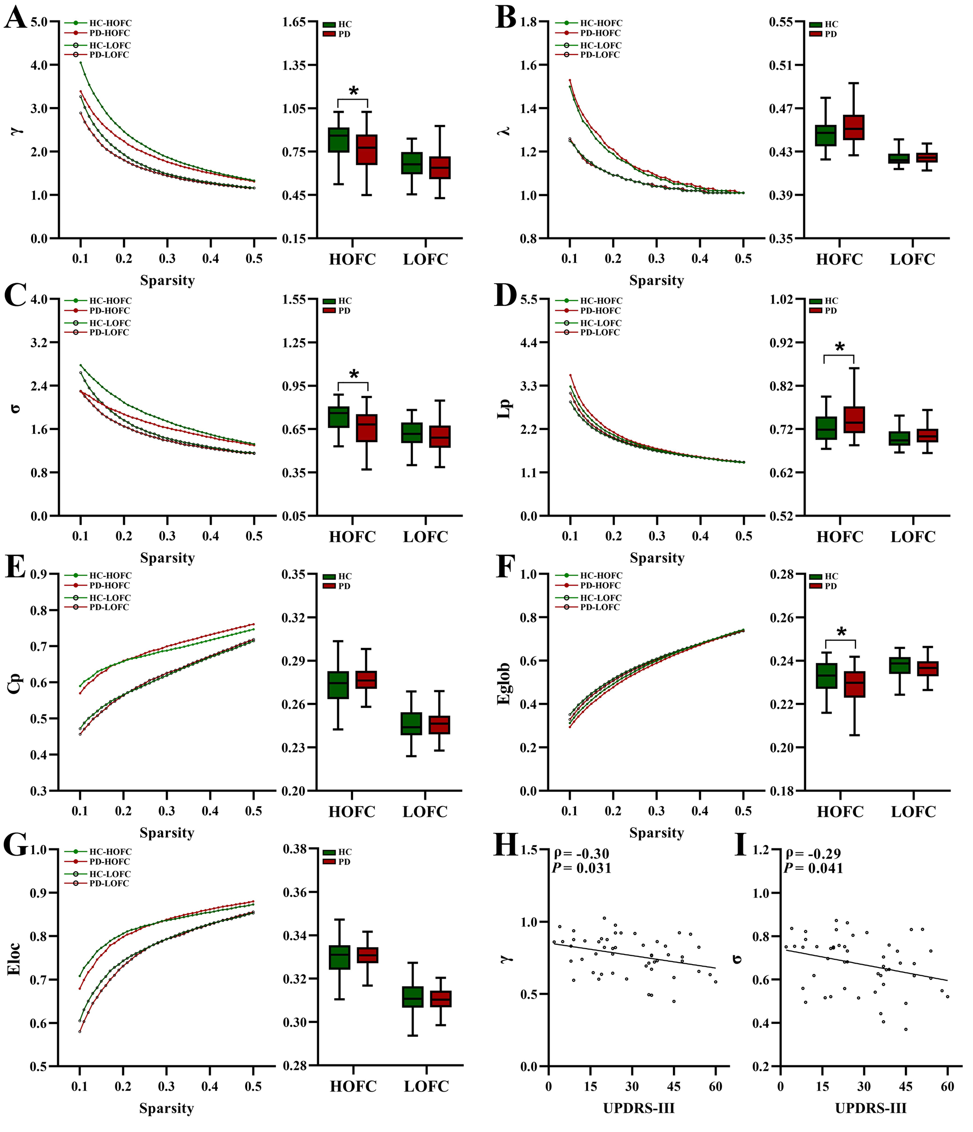
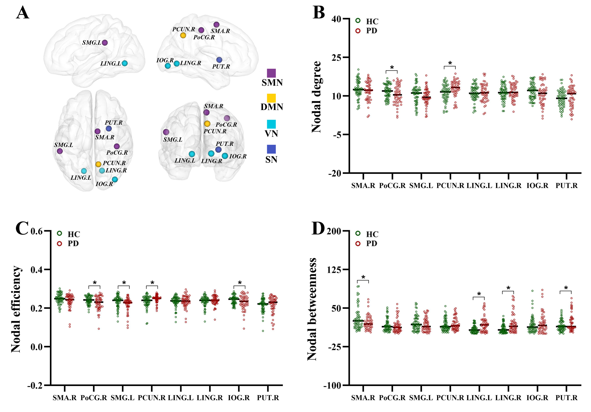
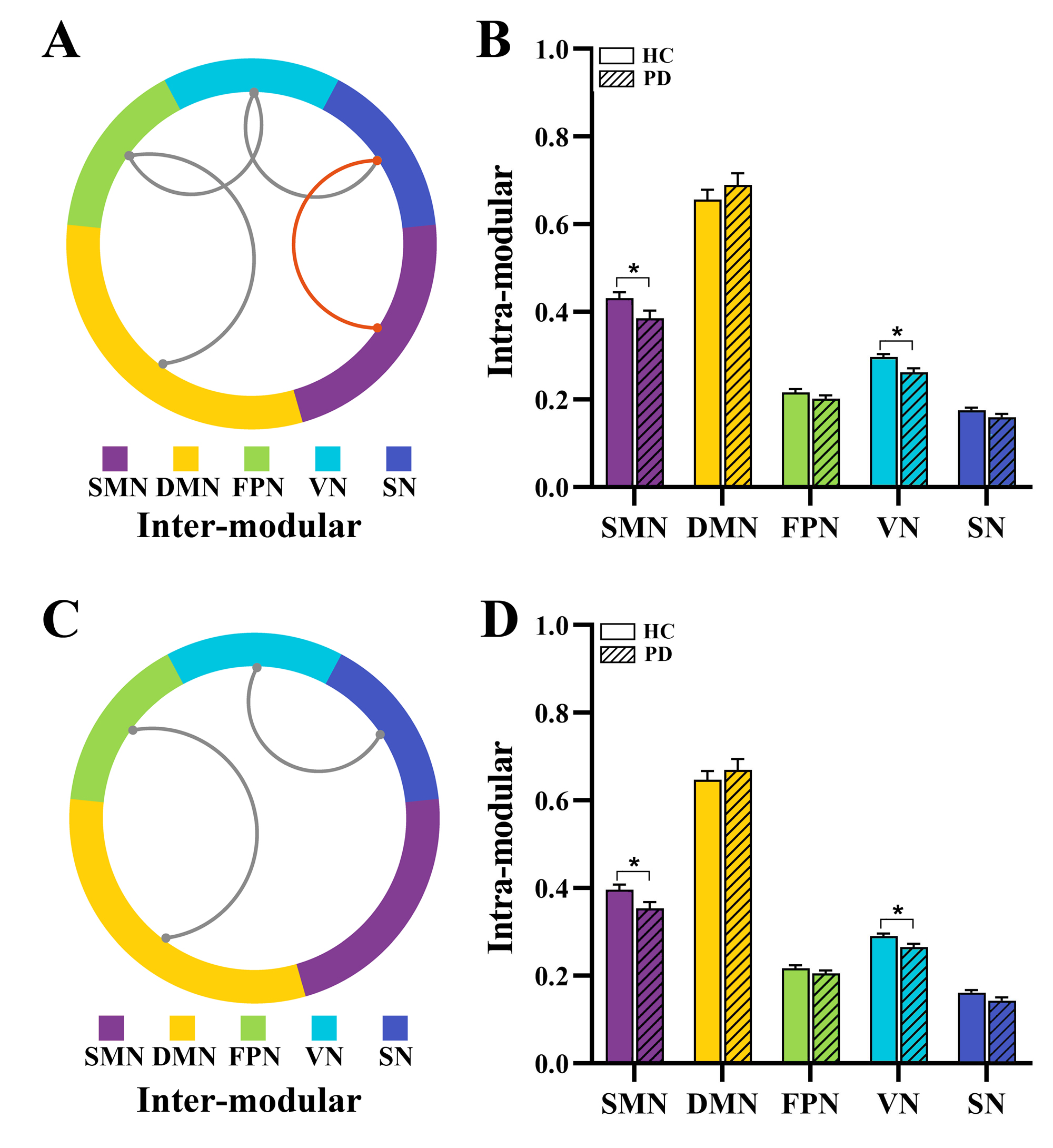
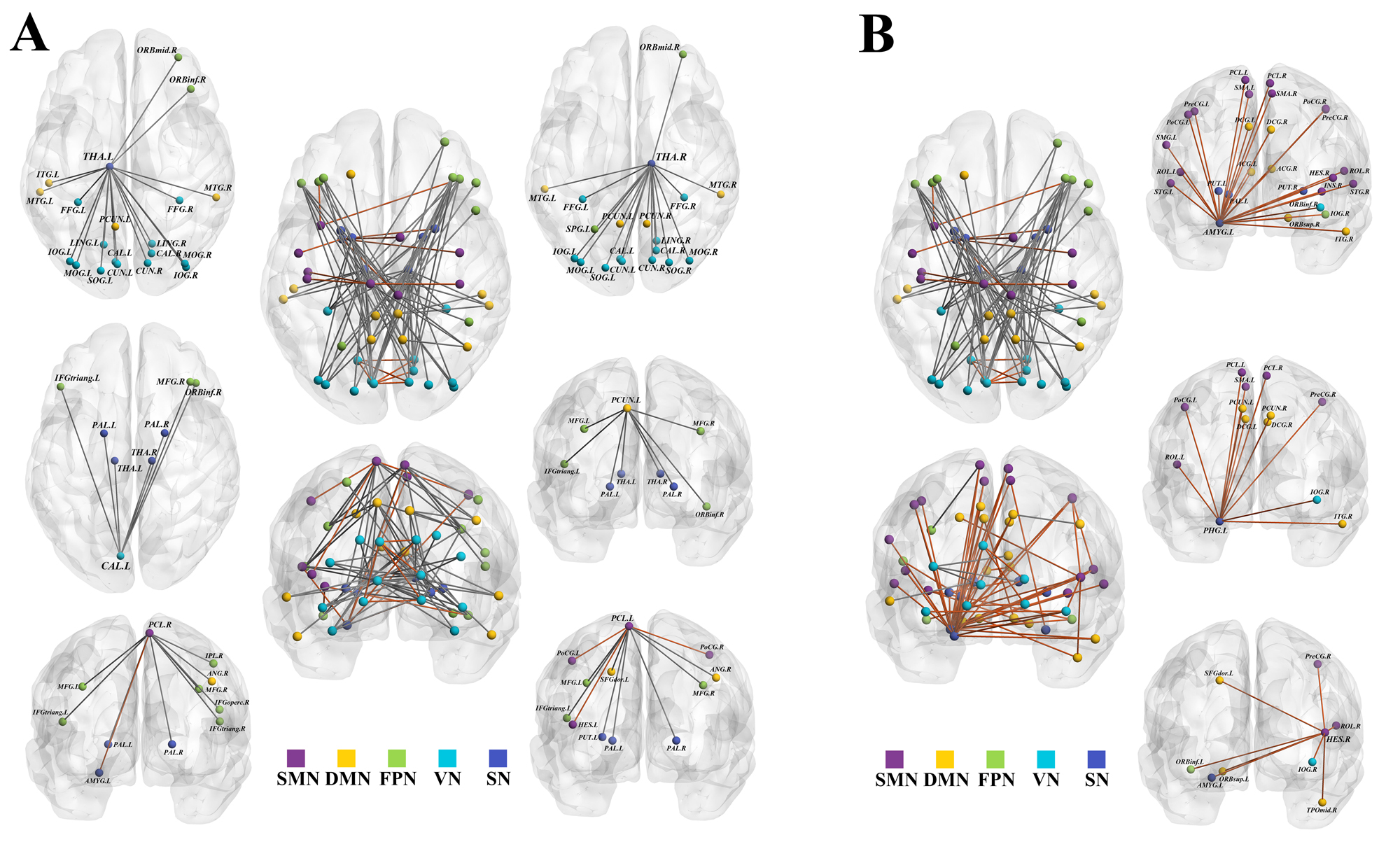
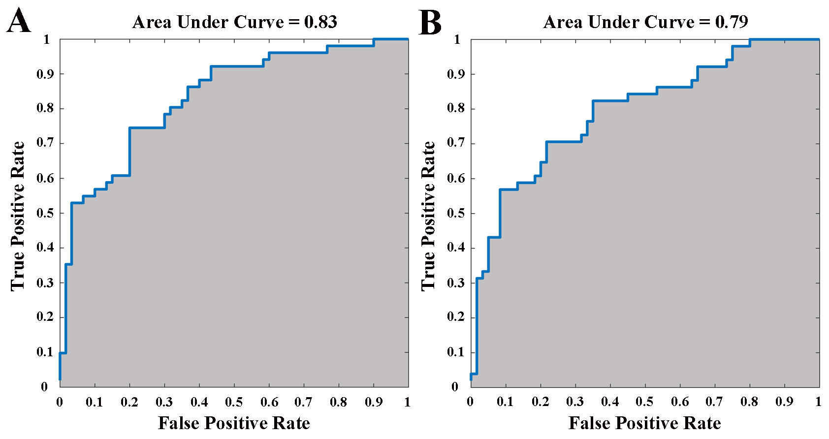
Compared to HC group, HOFC-networks in PD group showed lower values of normalized clustering coefficient (γ), small-worldness (σ) and global efficiency, but higher characteristic path length values (Fig.1). The altered nodal centralities (Fig.2) distributed in precuneus, putamen, lingual gyrus, supramarginal gyrus, supplementary motor area, postcentral gyrus and inferior occipital gyrus, as well as inter-modular FC between fronto-parietal network and visual network and between sensorimotor network and subcortical network were specific to HOFC-networks (Fig.3). Correlations between global metrics (γ and σ) and motor performance were observed in HOFC-networks. Several highly connected nodes (thalamus, paracentral lobule, calcarine fissure and precuneus) and improved classification performance were found based on HOFC profiles (Fig.4-5).Discussion and conclusion
The present study investigated the topology of high-order networks in cognitively normal patients with PD by employing HOFC construction. Relative to those of matched HC subjects, high-level information interactions in these patients were disrupted and showed altered global properties of decreased segregation (lower values of Eglob, γ, and σ),integration (higher values of Lp), and aberrant nodal metrics that were mainly distributed in the SMN, DMN, FPN, VN and SN, and impaired pairwise connections and modular architecture. Furthermore, we verified a greater sensitivity of HOFC profiles, in contrast to LOFC profiles, for the exploration of network disruptions prior to clinical evidence of cognitive impairment with more extensively altered topological properties and improved classification performance.In conclusion, the enrolled patients exhibited disrupted high-order information exchanges with lower small-worldness and extensively altered nodal centralities. The high-level functional interactions among networks featured aberrant hubs within the networks and disrupted information flow between the network modules. These findings supplied empirical evidence for comprehensive insights into the neurodegeneration associated with PD from the viewpoint of the functional connectome at a higher order, and highlighted a promising prospect of HOFC profiles for the exploration of high-level functional processes that underlie pathological status.
Acknowledgements
No acknowledgement found.References
1. Zhang H, Chen X, Shi F et al. Topographical Information-Based High-Order Functional Connectivity and Its Application in Abnormality Detection for Mild Cognitive Impairment. J Alzheimers Dis 2016;54:1095-1112.
2. Zhao F, Zhang H, Rekik I et al. Diagnosis of Autism Spectrum Disorders Using Multi-Level High-Order Functional Networks Derived From Resting-State Functional MRI. Front Hum Neurosci 2018;12:184.
3. Zheng Y, Chen X, Li D et al. Treatment-naive first episode depression classification based on high-order brain functional network. J Affect Disord 2019;256:33-41.
4. Chen X, Zhang H, Gao Y et al. High-order resting-state functional connectivity network for MCI classification. Hum Brain Mapp 2016;37:3282-3296.
5. Tzourio-Mazoyer N, Landeau B, Papathanassiou D et al. Automated anatomical labeling of activations in SPM using a macroscopic anatomical parcellation of the MNI MRI single-subject brain. Neuroimage 2002;15:273-289.
Figures