0232
Distinct subcortical and cortical functional gradient dysfunction in schizophrenia and the treatment effects after antipsychotics1Huaxi MR Research Center (HMRRC), Functional and molecular imaging Key Laboratory of Sichuan Province, Department of Radiology, West China Hospital, Sichuan University, Chengdu, China., Chengdu, China, 2College of Electronic Engineering, Chengdu University of Information Technology, Chengdu, P.R. China., Chengdu, China
Synopsis
Keywords: Psychiatric Disorders, Gradients
By characterizing the connectome gradient changes of subcortex and cortex in drug-naive first-episode schizophrenia and the treatment effect after antipsychotics, we found that the distinct fundamental functional segregation of subcortex and functional integration in cortex in patients at baseline when compared to healthy controls, and the longitudinal analyses indicated that the treatment would normalize the altered gradients. The improved subcortical gradient changes were associated with significant improvement of symptoms. This study provided the new perspective on the abnormal subcortical and cortical hierarchy organization in schizophrenia and its longitudinal subcortical gradient changes could be sensitive to reflect the antipsychotic treatment effect.Background
Identifying biomarkers indicative of treatment response in patients with schizophrenia has been a sustained area of research over the past two decades. Commonly used antipsychotics are thought to improve symptoms via the blockade of dopamine D2 receptors 1,2 which are abundant mainly in subcortical regions. Though the development of neuroimaging acquisition and analysis techniques has led to major progress in investigating the local subcortical changes including striatum in anatomy, function and chemistry before and after antipsychotic treatment, the subcortical-cortical interaction and related biological measures have yet to show consistency in relation to treatment response. Here, a novel gradient-based approach has been introduced to define a non-linear decomposition of high-dimensional resting-state functional connectivity (FC). Unlike the regional analyses, this method can comprehensively identify subcortical and cortical functional hierarchies by representing brain connectivity in a continuous, low-dimensional space. The concept of gradient focuses on connectomes where voxels with similar connectivity patterns are located close to one another along a given connectivity gradient. Leveraging this method to examine the synchronous measure of subcortical and cortical FC architecture in untreated schizophrenia patients and after treatment further in relation to symptom improvement might providing novel insight of illness- and treatment-related effects on subcortical and cortical interaction.Methods
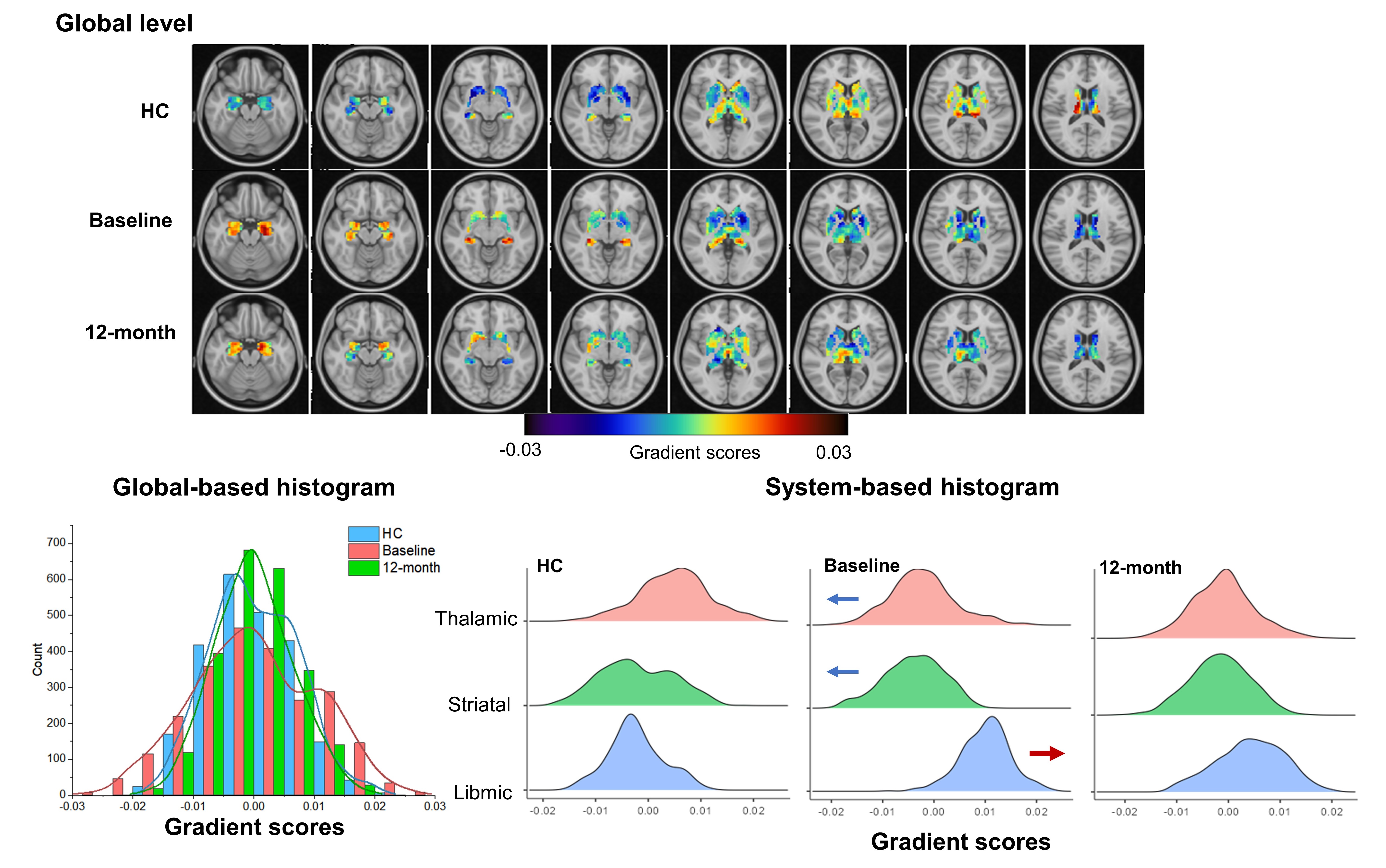
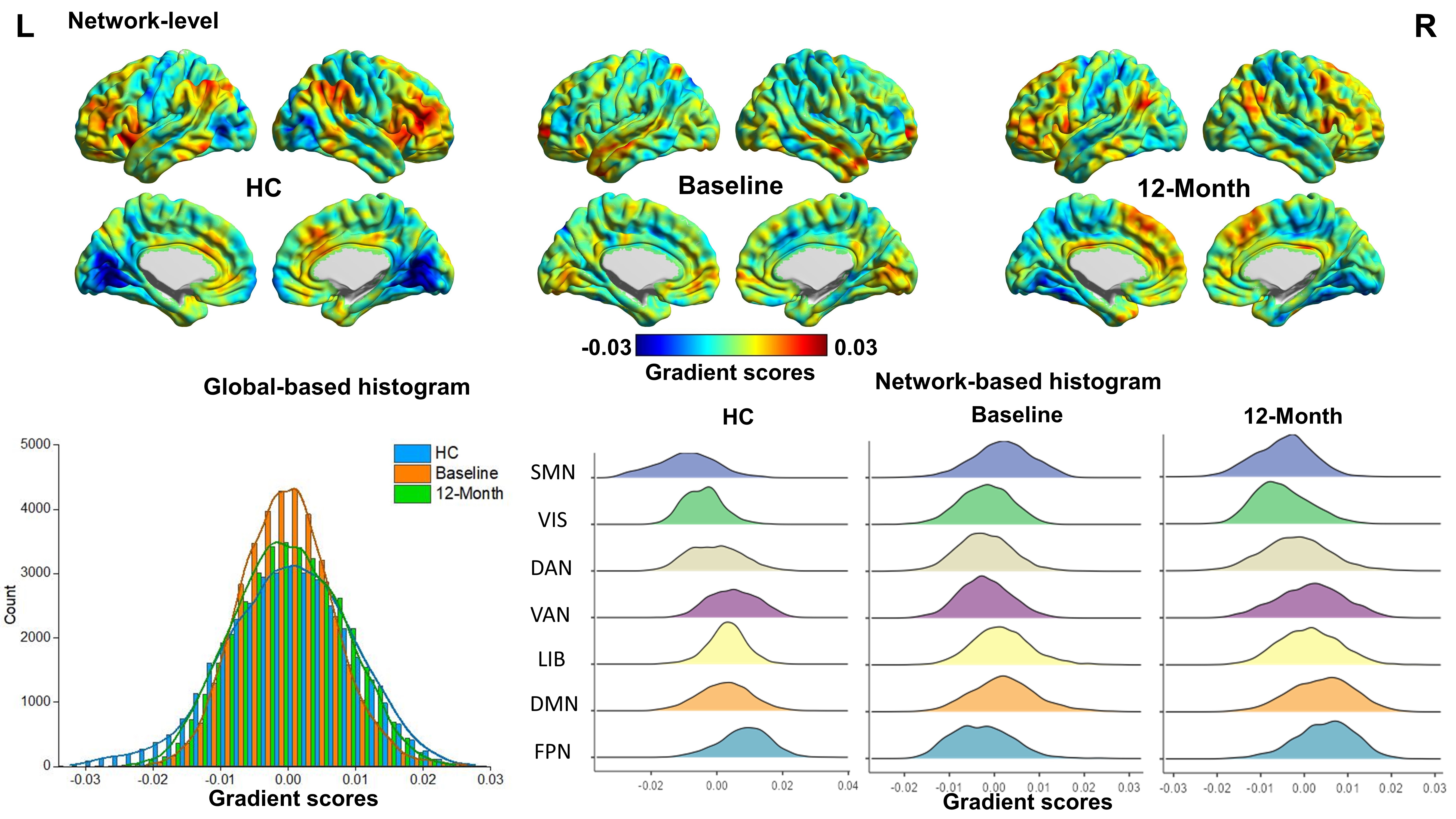
Fifty-seven patients (FES0W) and 64 healthy controls (HC) at baseline, and patients after 12-month (FES12M) treatment were recruited. The magnetic resonance imaging (MRI) scanning of participants was conducted on a GE Signa EXCITE 3.0T scanner (GE Healthcare, Milwaukee, Wisconsin) with an 8-channel phase array head coil. Resting-state functional MRI (rs-fMRI) data and high-resolution T1-weighted images (T1WI) were obtained for all participants. Functional data were preprocessed included the following steps: removal of first five dummy volumes, slice time correction, realignment, segmentation, normalization to the Montreal Neurologic Institute (MNI) space, bandpass filter (0.01-0.10 Hz) and spatial smoothing (full width at half maximum, FWHM = 4 mm). After data preprocessing, the individual subcortical-cortical/cortical-subcortical FC matrix was constructed using Pearson’s correlation between the time courses of each voxel. Gradient metrics were calculated using BrainSpace Toolbox (http://github.com/MICA-MNI/BrainSpace) 3. Voxel-based gradient values were generated and group-averaged gradient values were further extracted across all voxels (global), three systems (thalamus, limbic and striatum) in subcortex and 7 networks in cortex. The group comparisons of principal gradient alterations at global and network level were conducted separately between FES0W and HC for investigating illness effects, and between FES12M and FES0W for treatment effects. Correlational analyses were then conducted between the longitudinal gradient alterations and the improvement of clinical ratings, including the Positive and Negative Syndrome Scale (PANSS) and the Global Assessment of Functioning (GAF) scores.Results
In HC group, the gradient values were distributed along the axis from high to low in subcortex with thalamic-striatal-libmic systems and in cortex with primary to transmodal networks, while the gradient maps were consistent with previous characterizations of the spatial distribution of human 4,5. We further identified that before treatment, schizophrenia patients exhibited functional segregation in subcortical gradient with expanded global gradient scores involving increased gradient in limbic system and decreased gradient in thalamic and striatal systems compared to HC (Figure 1). While the baseline patients showed functional integration in cortical gradient with compressed global gradient scores including increased gradient in primary visual/sensorimotor networks (VIS/SMN) and decreased gradient in transmodal default mode network (DMN) (Figure 2). More importantly, these disruptions were normalized after treatment, and the longitudinal changes of subcortical gradient scores in limbic system were significantly associated with symptom improvement (negatively correlated with increase of GAF scores (r = -0.376, p = 0.018) and positively correlated with reduction of PANSS total scores (r = 0.419, p = 0.006) and subscales (disorganization scores: r = 0.416, p = 0.030 and excitement scores: r = 0.424, p = 0.030). However, there were no significant results in clinical relation to longitudinal cortical gradient alterations.Discussion
A novel functional connectome gradient algorithm calculating the spatial representation of subcortical and cortical functional hierarchy was performed by capturing the similarity of whole brain FC profiles between two voxels. The main finding was that the distinct alterations of gradient scores in subcortex and cortex in drug-naïve FES patients and were normalized after antipsychotic treatment. What’s more, the longitudinal changes of the subcortical gradients in the limbic system were highly associated with improvements in clinical symptoms. The baseline different gradient patterns of subcortex and cortex may be explained their different roles in the processing perception, motor and cognition, and the gradient-based characterization may represent a more sensitive approach to study treatment effects which reflect their interaction and normalization. The findings also highlighted the subcortical hierarchy could represent a more robust indicator of treatment response than cortical hierarchy.Conclusion
Our findings provided a novel insight into the subcortical and cortical interaction and normalization under distinct baseline functional hierarchy alterations, which were sensitive to illness and treatment effects. This might extend our understanding of the functional connectome hierarchy of subcortex and cortex in schizophrenia, and this measure in subcortex makes a promising indicator of treatment response.Acknowledgements
NoneReferences
1. Lehman AF, Lieberman JA, Dixon LB, McGlashan TH, Miller AL, Perkins DO, et al. Practice guideline for the treatment of patients with schizophrenia, second edition. Am J Psychiatry. 2004;161:1-56.
2. Kapur S, Remington G. Dopamine D(2) receptors and their role in atypical antipsychotic action: still necessary and may even be sufficient. Biol Psychiatry. 2001;50:873-883.
3. Vos de Wael R, Benkarim O, Paquola C, Lariviere S, Royer J, Tavakol S, et al. BrainSpace: a toolbox for the analysis of macroscale gradients in neuroimaging and connectomics datasets. Communications biology. 2020;3:103.
4. Tian Y, Margulies DS, Breakspear M, Zalesky A. Topographic organization of the human subcortex unveiled with functional connectivity gradients. Nat Neurosci. 2020;23:1421-1432.
5. Margulies DS, Ghosh SS, Goulas A, Falkiewicz M, Huntenburg JM, Langs G, et al. Situating the default-mode network along a principal gradient of macroscale cortical organization. Proc Natl Acad Sci U S A. 2016;113:12574-12579.
Figures