0217
A head and neck array add-on to the two-channel Nova coil using six SCC elements and Nova 32 channel receive array at 7T-MRI1Section for Magnetic Resonance, DTU Health Tech, Technical University of Denmark, Kgs Lyngby, Denmark, 2Danish Research Centre for Magnetic Resonance, Centre for Functional and Diagnostic Imaging and Research, Copenhagen University Hospital - Amager and Hvidovre, Copenhagen, Denmark, 3Department of Electrical Engineering, Technical University of Denmark, Kgs Lyngby, Denmark, 4Electrical Engineering Department, Technical University of Eindhoven, Eindhoven, Netherlands, 5Department of Neurology, Copenhagen University Hospital - Bispebjerg and Frederiksberg, Copenhagen, Denmark, 6Department of Radiology, Copenhagen University Hospital - Bispebjerg and Frederiksberg, Copenhagen, Denmark
Synopsis
Keywords: RF Arrays & Systems, RF Arrays & Systems
The advantage of low coupling ratios of shielded-coaxial-cable coils (SCCs) are used to obtain an eight-channel head-neck transmit array for 7T-MRI. The six SCCs are combined with the two-channel Nova coil by taking the advantage of low coupling between SCCs and the Nova coil. Low inter element coupling along with the independent radiation regions of the SCCs and the Nova coil made it possible to do a straightforward B1-shimming resulting in a relatively homogeneous magnetic field in both head and neck region. Structural images are acquired with large coverage down to the bifurcation while high SNR is preserved.Introduction
7T-MRI systems have higher intrinsic signal-to-noise ratios than classical systems allowing for higher resolution images, but the higher frequency has consequences for the specific absorption rate (SAR). Where the classical systems use a large birdcage coil as body coil for all transmission, 7T is restricted to use localized transmit coils like the two-channel Nova head-only-coil to stay within SAR limits [1]. This birdcage coil assures a relatively homogeneous field distribution in the cerebrum but the B1-transmit field in cerebellum and the neck region [2] is limited.Recently, shield coaxial cable (SCC) coils with high self-decoupling properties have been used for neck imaging [3,4]. SCCs are therefore great candidates to use with a volume resonator, which would satisfy high decoupling due to their different working principle.
Here, an eight-channel transmit coil have been developed as a six-channel SCC elements add-on to the two-channel Nova coil, thereby increasing the coverage the neck while keeping the Nova 32-channel receivers. We tested this eight-channel head-neck array on a human subject to show the improved coverage in structural images.
Methods
The 6-channel neck-array add-on was made with SCC elements of a loop radius of 62mm allowing 8-channel transmit/receive or 8/32-channel transmit/receive with the Nova receive coil as used in the examples. Full-wave electromagnetic simulations were performed in CST Microwave Studio (Darmstadt, Germany). Simulations were done on the Duke (homogeneous and inhomogeneous). SCCs are tuned and matched with capacitive L-networks as described in [3]. Maximum 10-gram averaged SAR is computed with the phase settings that resulted in the most homogeneous field distribution throughout the head and neck region.Experiments were performed on a Philips 7T Achieva scanner. Structural images (both 2-ch Nova and 2-ch Nova, 6-ch SCC head-neck array) were acquired. A standard magnetization prepared rapid gradient echo (MPRAGE) and a fluid attenuated inversion recovery (FLAIR) were used to show the effect of missing B1 in the lower brain and neck regions.
Results
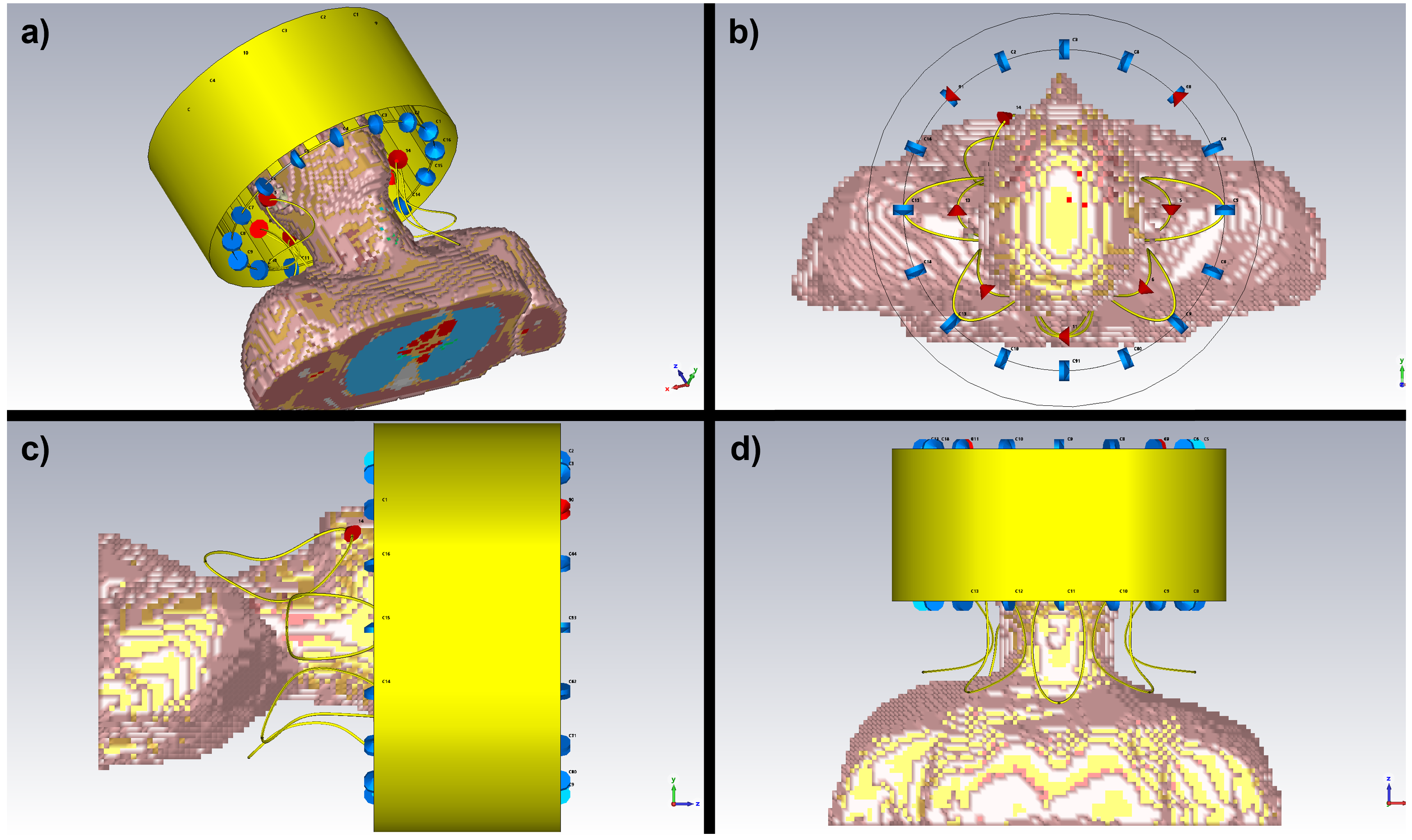
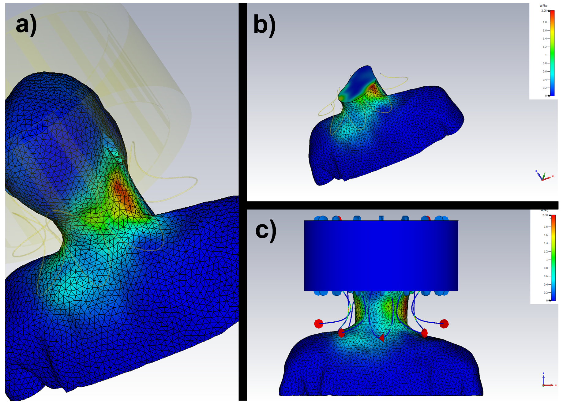
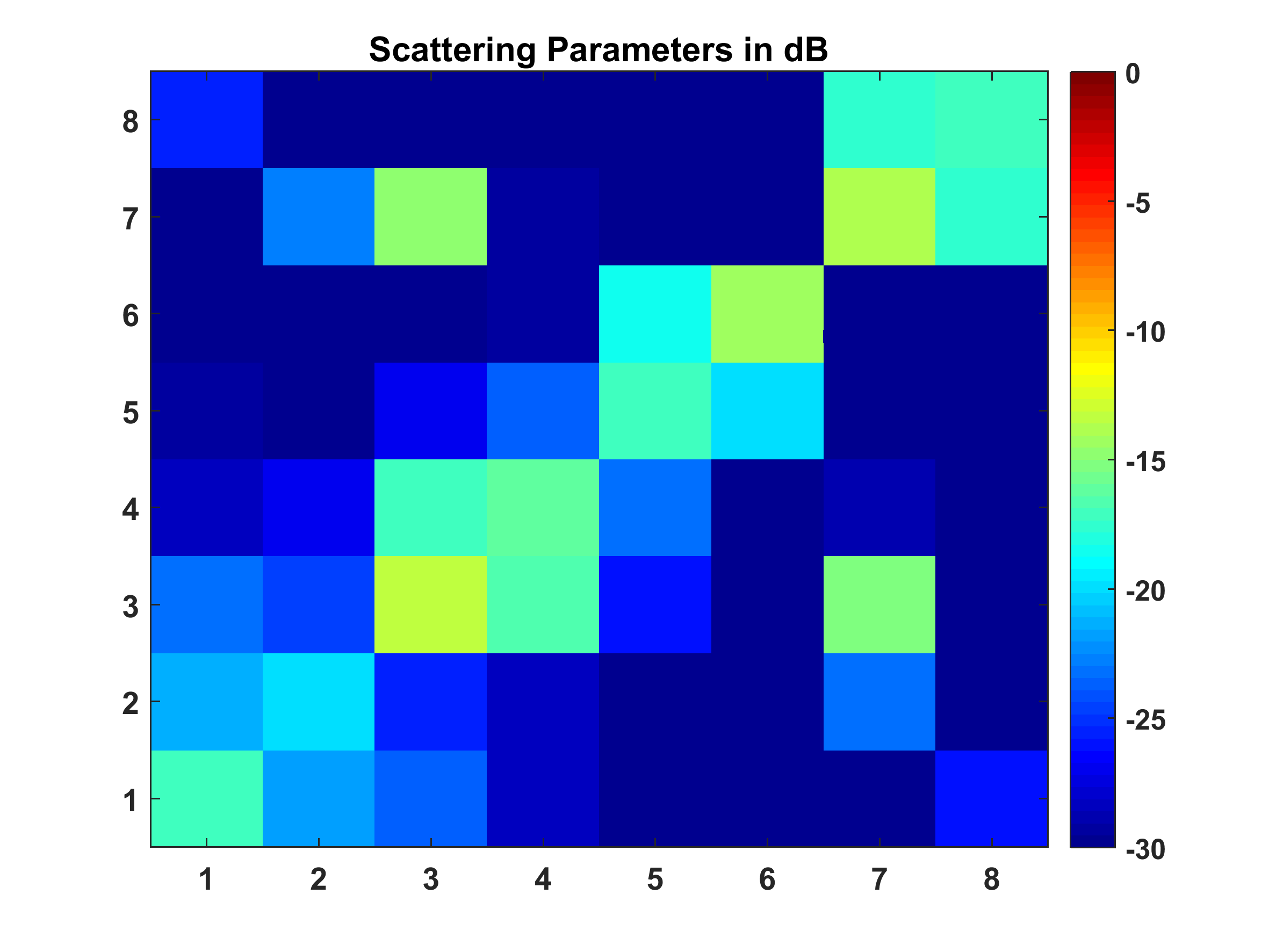
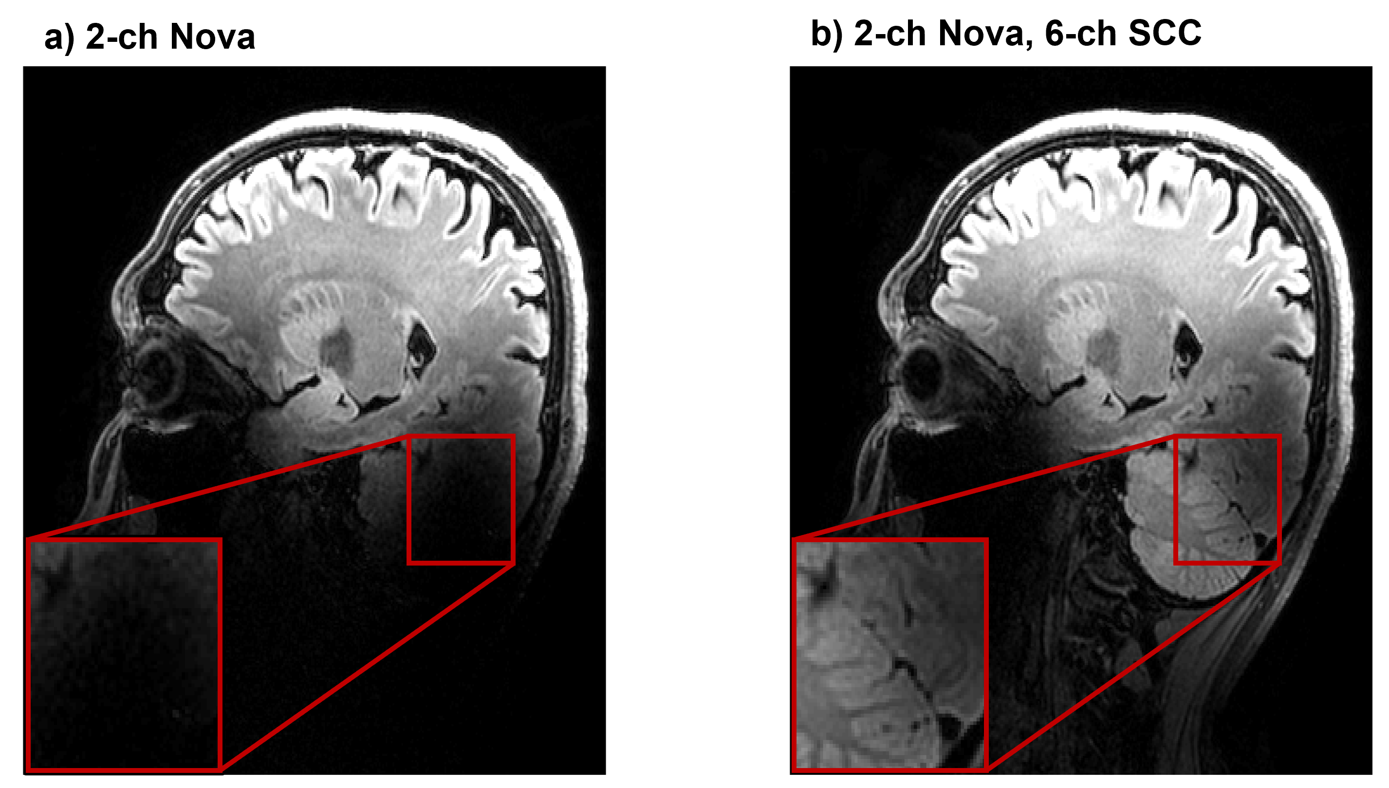
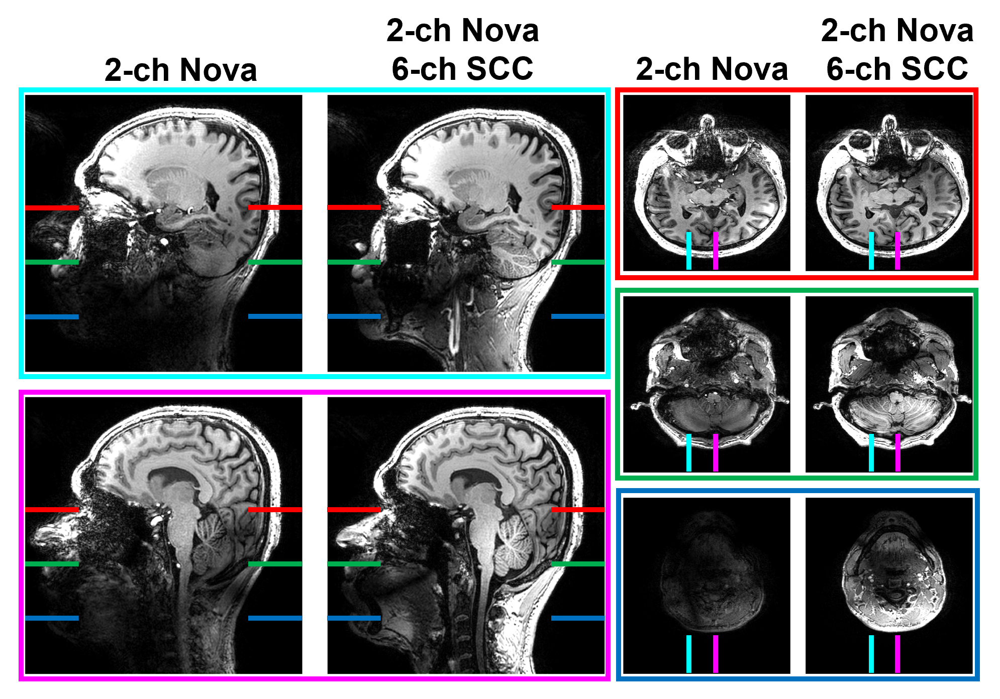
Figure 1 shows the placement of eight-channel head-neck array with inhomogeneous Duke model in the simulation environment. Figure 2 shows the SAR distribution over the homogeneous Duke model. Figure 3 presents the coupling matrix of the eight-channel head-neck array. Figure 4 shows the structural images acquired with FLAIR and finally figure 5 shows the structural images acquired with MPRAGE.Discussion
Often a loss of contrast is observed in the temporal lobe and the cerebellum depending on the sequence used using the two-channel Nova coil. The current solutions include dielectric pads, which with correct placement can improve these regions, while the neck region is out of reach. The proposed add-on transform the Nova coil into an eight-channel head-neck array, which offers a straightforward solution to the problem. Figure 3, show low coupling between coil elements and this low coupling - combined with the limited interaction of the 6 SCC coils and the two-channel nova coil makes B1 shimming straightforward. Basically, the Nova coil remains in a quadrature mode while the 6 SCC elements are shimmed to ensure a high field homogeneity in the lower brain and neck region.In figure 4, a clearly higher signal and contrast level at the lower brain and cerebellum is seen using the add-on array while the Nova coil on its own is losing contrast and signal. Moreover, figure 5 shows that higher signal levels are obtained all the way to the carotid bifurcation using the add-on array while the signal and contrast are completely lost with the Nova coil only.
A high and relative homogeneous B1 field reaching from the upper brain all the way to the carotid bifurcation, opens a large range of possibilities which so far have been limited on 7T-MRI systems. These include arterial-spin-labeling where the labeling is performed in the neck region, angiographic and vessel-wall imaging which so far has been limited to the intracranial vessels. In addition, upper c-spine imaging (C1-C4) can be done within the same scan session without changing coil.
Initial experience by comparison of simulation and experiments also suggests that a single or a limited set of fixed phase settings can be used for RF shimming which makes SAR handling easier in the multi-transmit regime used here.
Conclusion
The proposed method transforms the two-channel Nova coil into an eight-channel head-neck array which not only improves brain image quality but also extends the coverage to include the neck region. This opens up for a whole new range of studies such as arterial-spin-labeling, extracranial angiography and vessel wall as well as c-spine imaging without the need of changing coils.It is an uncomplicated and easy approach with low coupling of individual elements, independent radiation regions of the birdcage coil and SCCs, and straightforward B1-field capabilities. As the two-channel Nova coil is the standard coil on all 7T platforms, this makes it a simple task for any researcher to implement the proposed head-neck array on their multi-transmit system.
Acknowledgements
The 7T scanner was donated by the Danish Agency for Science, Technology and Innovation grant no. 0601-01370B, and The John and Birthe Meyer Foundation. The coil was sponsored by Toyotafonden j.nr.KJ/BG-9771H and Sadri Güler is paid by a DTU Alliance stipend (DTU-TU/e-DRCMR).References
[1] Ahmad SF, Kim YC, Choi IC, Kim HD. Recent Progress in Birdcage RF Coil Technology for MRI System. Diagnostics 2020;10:1017. https://doi.org/10.3390/diagnostics10121017.
[2] van Lier ALHMW, Kotte ANTJ, Raaymakers BW, Lagendijk JJW, van den Berg CAT. Radiofrequency heating induced by 7T head MRI: Thermal assessment using discrete vasculature or pennes’ bioheat equation. Journal of Magnetic Resonance Imaging 2012;35:795–803. https://doi.org/10.1002/jmri.22878.
[3] Ruytenberg T, Webb A, Zivkovic I. Shielded-coaxial-cable coils as receive and transceive array elements for 7T human MRI. Magnetic Resonance in Medicine 2020;83:1135–46. https://doi.org/10.1002/mrm.27964.
[4] Ruytenberg T, Webb A, Zivkovic I. A flexible five‐channel shielded‐coaxial‐cable (SCC) transceive neck coil for high‐resolution carotid imaging at 7T. Magn Reson Med 2020;84:1672–7. https://doi.org/10.1002/mrm.28215.
Figures