0205
RF shimming for improved B1+ in the carotid arteries using parallel transmission (pTx) head coils at 7T1Wellcome Centre for Integrative Neuroimaging, FMRIB, Nuffield Department of Clinical Neurosciences, University of Oxford, Oxford, United Kingdom
Synopsis
Keywords: Parallel Transmit & Multiband, High-Field MRI, Neurovascular
Cerebrovascular imaging methods suffer from a rapid drop in B1+ into the neck when using typical transmit head coils at 7T. Custom RF shims on regular pTx head coils could improve the B1+ magnitude in the major feeding arteries in the neck region over standard CP transmit mode. We found that this can improve the B1+ magnitude in the carotid arteries by 36% while also improving the RF homogeneity. This can be achieved using universal, phase-only RF shims, facilitating easy implementation in existing sequences and without requiring custom hardware.Introduction
Ultra-high-field MRI offers an increased signal-to-noise ratio and longer T1-relaxation time for both tissue and blood. At 7T this is expected to improve intracranial cerebral angiography and perfusion, and enhance visualization of vessel wall pathology. However, such methods are limited by an increased specific absorption rate (SAR) and by a reduced homogeneity and spatial extent of the transmit magnetic field (B1+). In particular, neurovascular imaging methods, such as arterial spin labeling1,2 and arterial blood suppression for vessel wall imaging3, suffer from a rapid drop in B1+ into the neck when using typical transmit head coils at 7T.One solution is to use specialist head-and-neck transmit coils4,5 (with or without pTx capability6) to boost the B1+ in the neck. However, this solution adds experimental complexity and expense. An alternative approach is to use standard head coils with pTx capability. For that approach, we sought to investigate by what means and by how much B1+ shimming could improve the B1+ magnitude in the major feeding arteries in the neck region over standard circular polarization (CP) transmit mode. We investigated the trade-off between improving B1+ magnitude and B1+ homogeneity in the arteries in the neck, and finally evaluated how many subjects are needed to generate a universal neck shim.
Methods
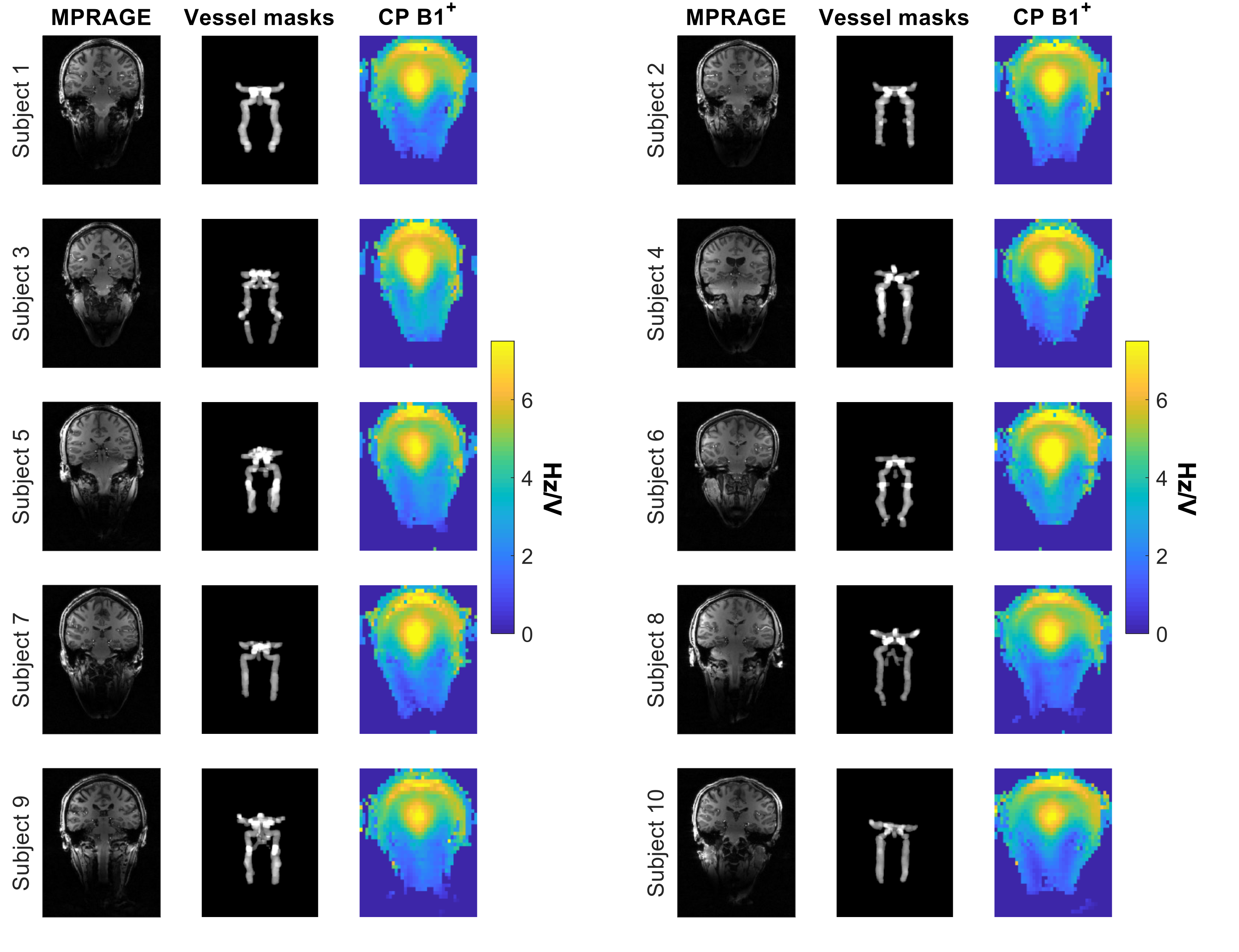
Data were acquired from 10 subjects on a Siemens Magnetom 7T scanner using a Nova Medical 8Tx/32Rx head coil under an institutional ethical agreement. Evaluations were performed using per-channel B1+ maps reconstructed from separate multi-voltage relative7 and absolute flip angle8 data sets (all acquired using a FOV of 225×225×300mm at 7.5×5.6×6.2mm resolution). Additionally, data acquired using CP-mode (45° phase increments per channel) and CP2-mode (90° phase increments per channel) were combined (B1TIAMO9), and B0 maps were acquired to account for off-resonance effects. This ensured accurate B1+ maps over a large range of transmit B1+ values and in the presence of substantial B0 inhomogeneity. The source data are provided at DOI: 10.5287/bodleian:ZB6Gk8QzN.In order to assess the theoretical upper limit for pTx to boost B1+ in the neck the maximum available B1+ was evaluated in-vivo on a voxel-by-voxel basis by summing the absolute B1+ maps across channels. For shim calculations and evaluation, hand-drawn vessel masks, comprising the internal carotid arteries and the Circle of Willis, were drawn for each subject from MPRAGE images (reconstructed as root sum-of-squares of CP-mode and CP2-mode acquisitions). These ROIs were used as masks for the RF shim calculations (see Figure 1). Both phase-and-magnitude and phase-only RF shim combinations were calculated to assess any potential benefit of the extra degrees of freedom.
Results
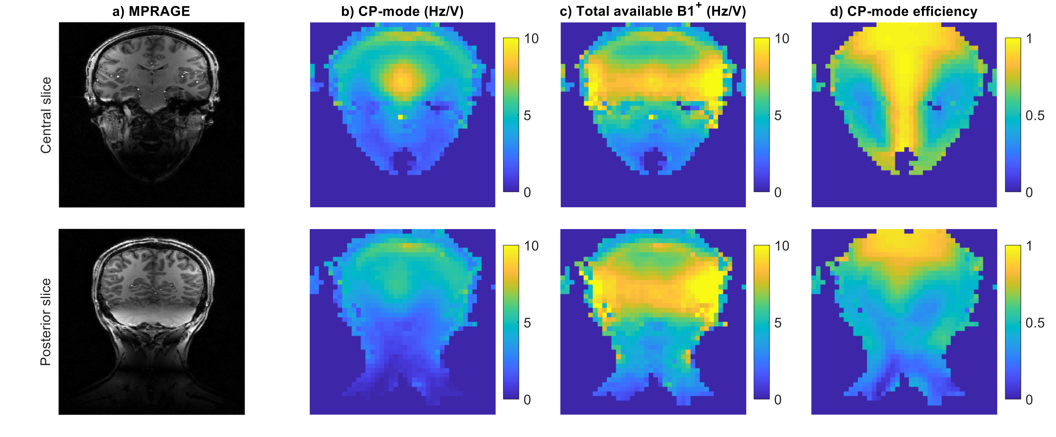
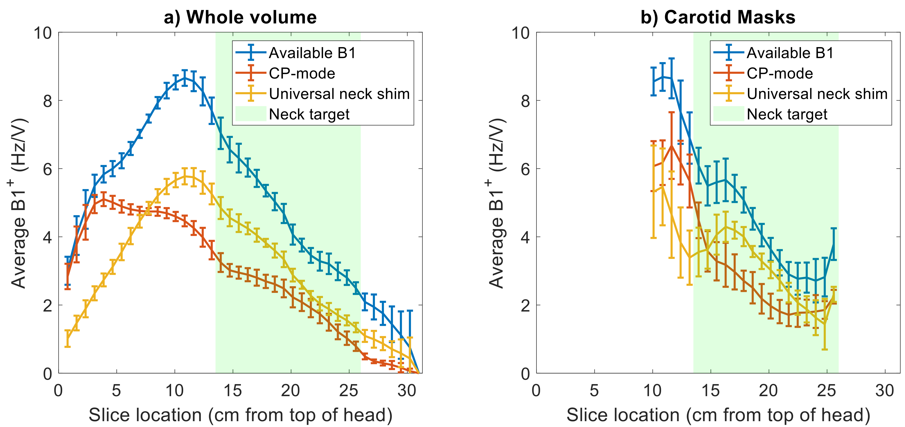
Figure 2c shows that the theoretical upper limit of B1+ in the neck is low compared to the central head region. Further, Figures 2b and 2d show that CP-mode only utilizes 57±13% of the available maximum B1+, resulting in an average B1+ magnitude in the neck for CP mode of 2.5±1.0 Hz/V, suggesting that utilising pTx should improve this very low value.Figure 3 shows that a universal shim, generated by maximizing the average B1+ in the neck, is able to outperform CP-mode, albeit not reaching the maximum theoretical voxel-by-voxel values (which are unachievable using a standard RF shimming approach). However, Fig. 3 also shows a large inferior-superior variation in the B1+ profile, since no coefficient of variation optimization was included in the calculation.
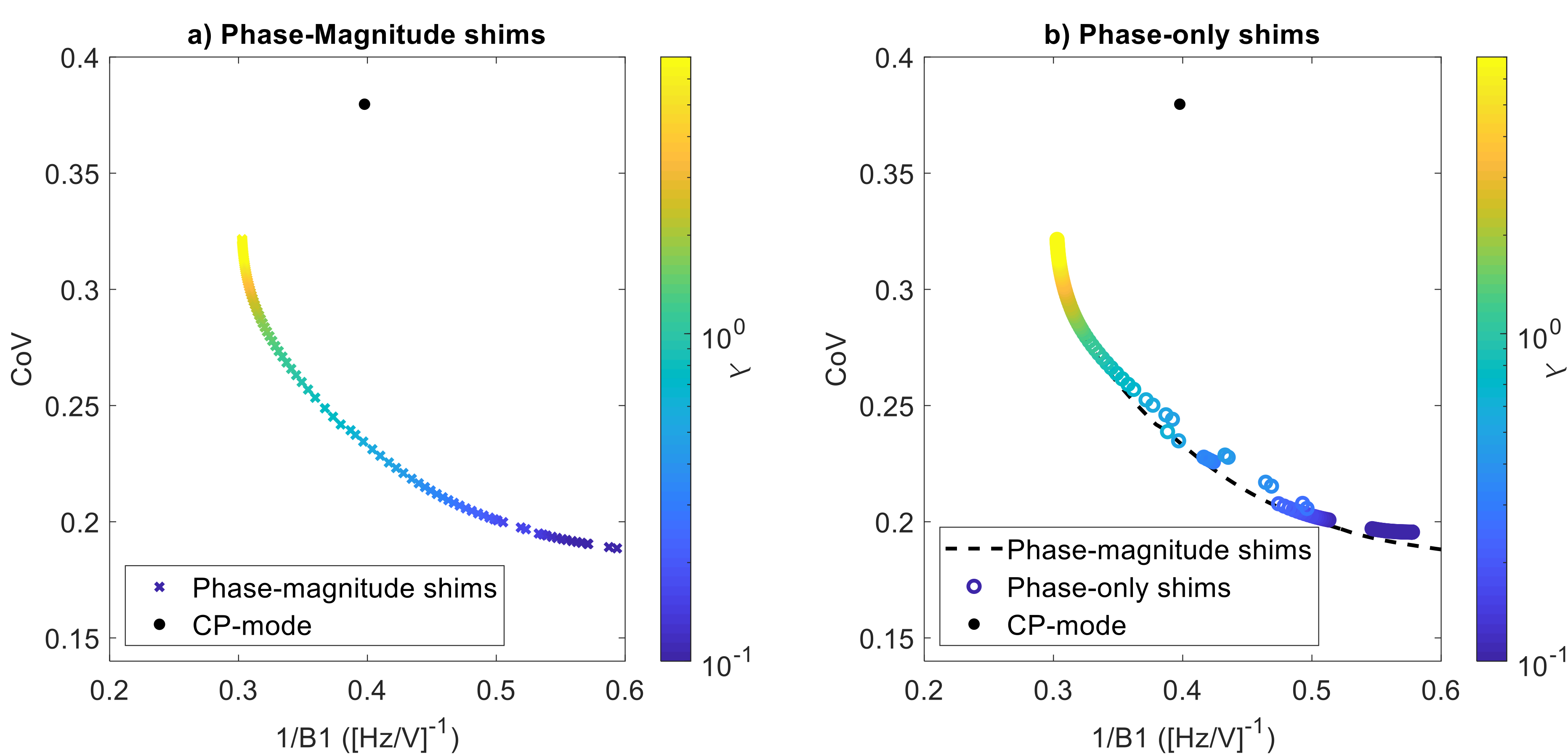
Figure 4 shows the neck shim performance within the vessel mask when CoV is included in the cost function (optimizing min[{CoV} + λ{1/B12}]). Both magnitude-and-phase, and phase-only B1+ shimming conditions were considered. We observed that phase-only shimming performed almost as well as phase-and-magnitude shimming. Based on the L-curve in Figure 4b, a regularization parameter, λ, of 1.7 is found to produce a reasonable trade-off between B1+ efficiency and minimizing the coefficient of variation.
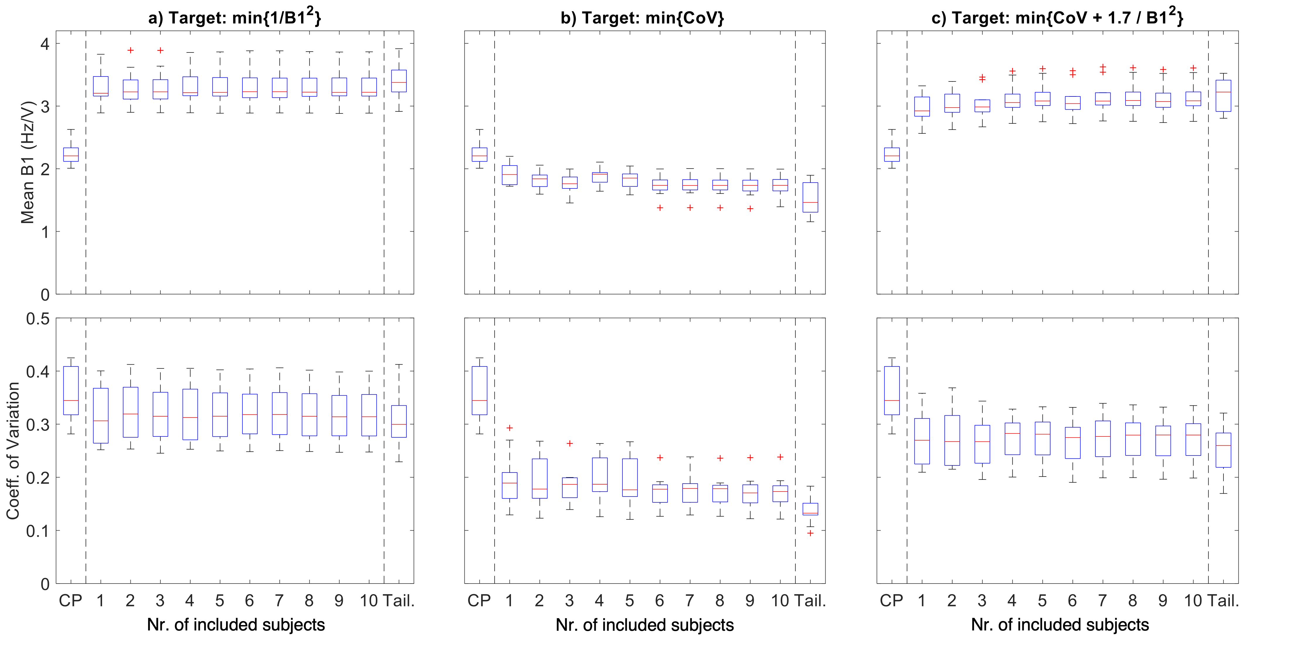
When evaluating the number of subjects needed to form a universal shim (Figure 5) it was found that only a very small number of subjects is needed to achieve convergence. In all cases, the universal shim performs only slightly worse than a fully per-subject tailored shim, and substantially better than CP-mode.
Discussion
RF shimming within a vessel mask in the neck leads to a 36% increase in B1+ magnitude, whilst reducing the coefficient of variation by 25%. This can be achieved using phase-only RF shimming and does not require phase-and-magnitude RF shimming, likely due to the inherent requirement for high B1+ utilization from all channels in order to achieve sufficient B1+ in the neck. Further, a universal shim in the neck can be easily found, and performs almost as effectively as a fully tailored per-subject shim, while consistently outperforming CP-mode in terms of both B1+ magnitude and coefficient of variation.Since a linear increase in B1+ for phase-only RF shimming implies a quadratic reduction in required SAR for a given flip angle, the reported 36% increase in B1+ magnitude corresponds to a 46% reduction in SAR.
Conclusion
The B1+ magnitude in the arteries in the neck can be increased by 36% using pTx RF shims, while also improving the homogeneity. This is possible using phase-only universal RF shims, facilitating easy implementation in existing sequences at 7T.Acknowledgements
MdB receives financial support from Siemens Healthineers and the Dunhill Medical Trust. PJ receives support from the Dunhill Medical Trust and the NIHR Oxford Biomedical Research Centre. The Wellcome Centre for Integrative Neuroimaging is supported by core funding from the Wellcome Trust (203139/Z/16/Z).References
1. Tong Y, Jezzard P, Okell TW, Clarke WT. Improving PCASL at ultra-high field using a VERSE-guided parallel transmission strategy. Magn Reson Med. 2020;84(2):777-786. doi:10.1002/mrm.28173
2. Wang K, Ma SJ, Shao X, et al. Optimization of pseudo-continuous arterial spin labeling at 7T with parallel transmission B1 shimming. Magn Reson Med. 2022;87(1):249-262. doi:10.1002/MRM.28988
3. de Buck MHS, Kent JL, Hess AT, Jezzard P. Parallel Transmit DANTE-SPACE for improved black-blood signal suppression at 7 Tesla. In: Proceedings of the 31st Annual Meeting of ISMRM; 2022:2092.
4. Adriany G, van de Moortele PF, Ritter J, et al. A geometrically adjustable 16-channel transmit/receive transmission line array for improved RF efficiency and parallel imaging performance at 7 Tesla. Magn Reson Med. 2008;59(3):590-597. doi:10.1002/mrm.21488
5. Tachrount M, Woodward B, Kopanoglu E, Italiaander M, Klomp D, Driver I. Improving RF efficiency in the brain and the neck at 7T using a novel pTx coil. In: Proceedings of the 27th Annual Meeting of ISMRM. 2019:1523.
6. Padormo F, Beqiri A, Hajnal J v., Malik SJ. Parallel transmission for ultrahigh-field imaging. NMR Biomed. 2016;29(9):1145-1161. doi:10.1002/nbm.3313
7. Padormo F, Hess AT, Aljabar P, et al. Large dynamic range relative B1+ mapping. Magn Reson Med. 2016;76(2):490-499. doi:10.1002/mrm.25884
8. Kent JL, Dragonu I, Valkovic L, Hess AT. Rapid 3D Absolute B1+ Mapping using a Sandwiched Train Pre-Saturated TurboFLASH Sequence at 7T for the Brain and Heart. Magn Reson Med. 2022;1-13. doi:10.1002/mrm.29497
9. Brunheim S, Gratz M, Johst S, et al. Fast and accurate multi-channel B1+ mapping based on the TIAMO technique for 7T UHF body MRI. Magn Reson Med. 2018;79(5):2652-2664. doi:10.1002/mrm.26925
Figures