0124

Effects of Levodopa Therapy on Neurovascular Coupling in Parkinson’s Disease Patients
Chenqing Wu1, Haoting Wu1, Cheng Zhou1, Jingwen Chen1, Jiaqi Wen1, Xiaocao Liu1, Jianmei Qin1, Zhengye Cao1, Tao Guo1, Xiaojun Guan1, Xiaojun Xu1, Baorong Zhang2, and Minming Zhang1
1Department of Radiology, The Second Affiliated Hospital, Zhejiang University School of Medicine, Hangzhou, Zhejiang, China, 2Department of Neurology, The Second Affiliated Hospital, Zhejiang University School of Medicine, Hangzhou, Zhejiang, China

Synopsis

Keywords: Parkinson's Disease, Neurodegeneration

Abnormal neurovascular coupling was found in PD patients. These impairments were partially normalized after levodopa therapy, and ALFF alterations played a primary role in this normalization effect.

Introduction

Normal brain functions rely on the coupling between cerebral blood flow (CBF) and neural activity. This neurovascular coupling was impaired in Parkinson’s Disease (PD) patients, yet how this coupling changes after levodopa therapy remains unexplored.

Objective

To investigate the effect of levodopa therapy on neurovascular coupling in PD patients.

Methods

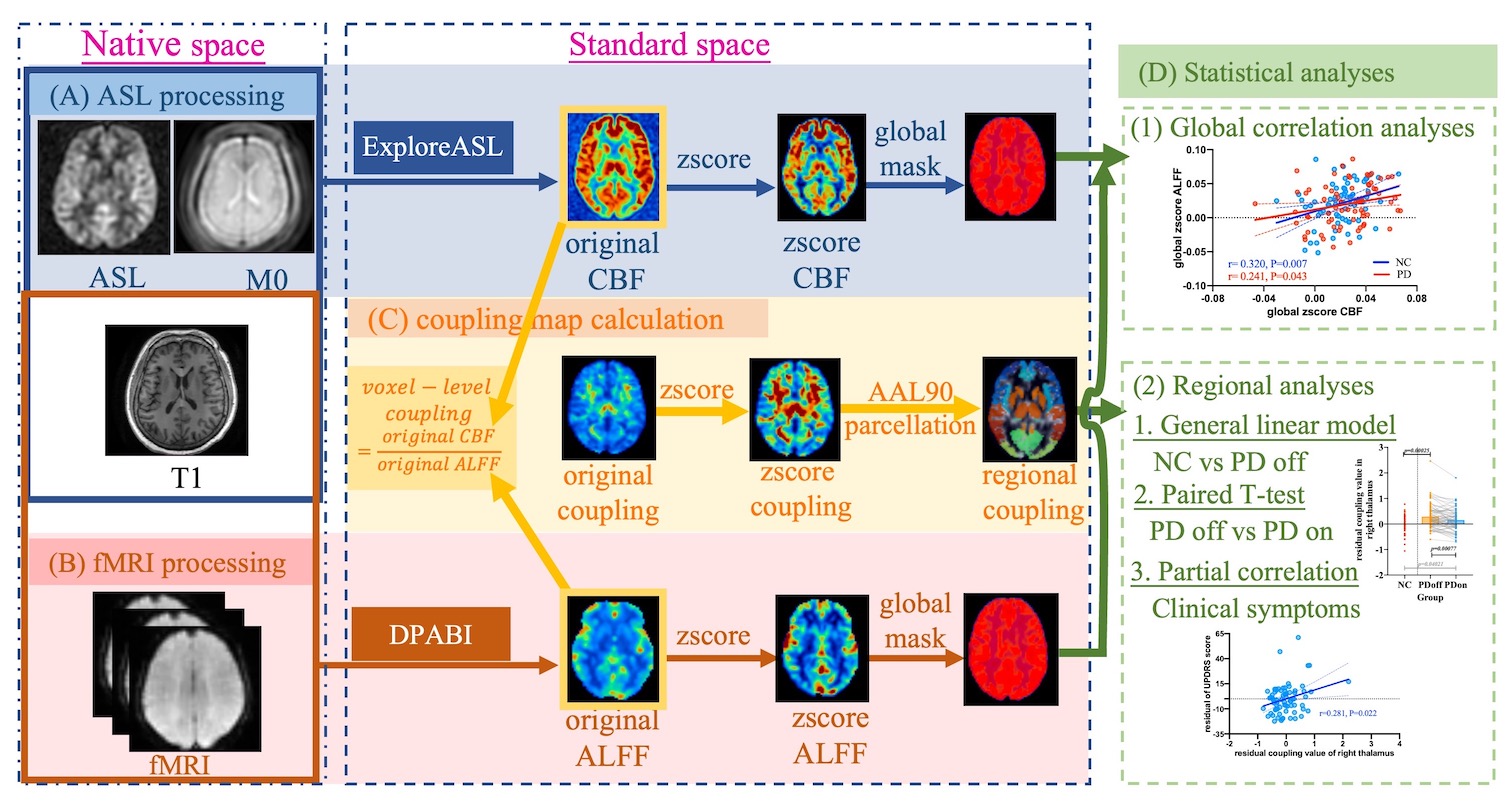
A total of 69 normal control (NC) and 71 PD patients were retrospectively enrolled. Neuropsychological assessments and MRI scanning were conducted in NC and patients before and after the levodopa therapy. CBF and neural activity were measured by arterial spin labeling (ASL) and the amplitude of low-frequency fluctuations (ALFF) from resting-state functional MRI, respectively. Global neurovascular coupling was calculated using Pearson’s correlation coefficient between CBF and ALFF. The CBF/ALFF ratio was calculated in each voxel and neurovascular coupling values were extracted in each brain region defined by Automatic Anatomical Labelling (AAL) atlas. General linear model was used for comparing regional neurovascular coupling differences between PD and NC. Paired T-test was conducted to explore regional neurovascular coupling alterations after levodopa therapy in patients. Partial correlations were calculated to investigate the relationship between regional neurovascular coupling, medication treatment, and clinical symptoms. Bonferroni correction was used for multiple comparison corrections.

Results

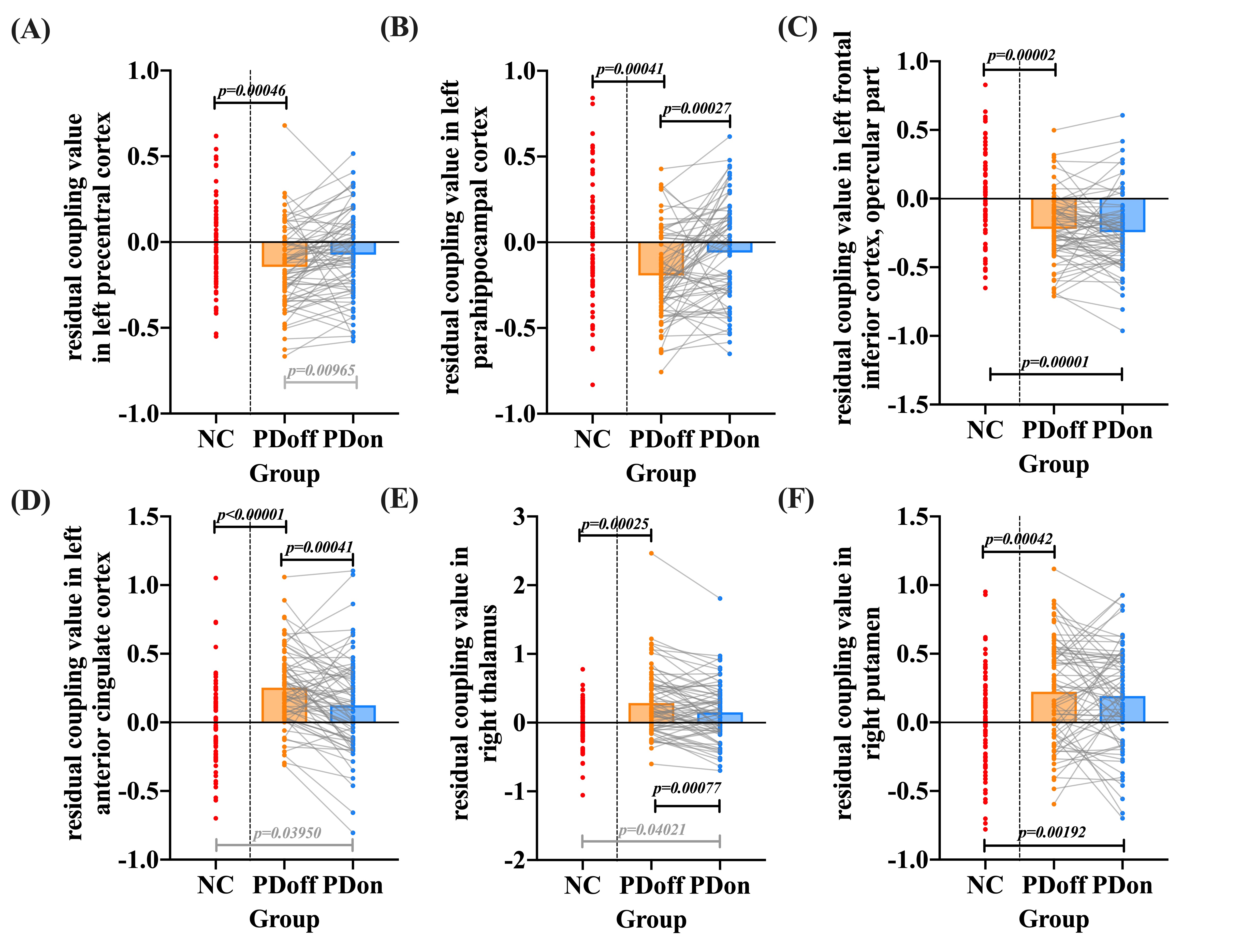
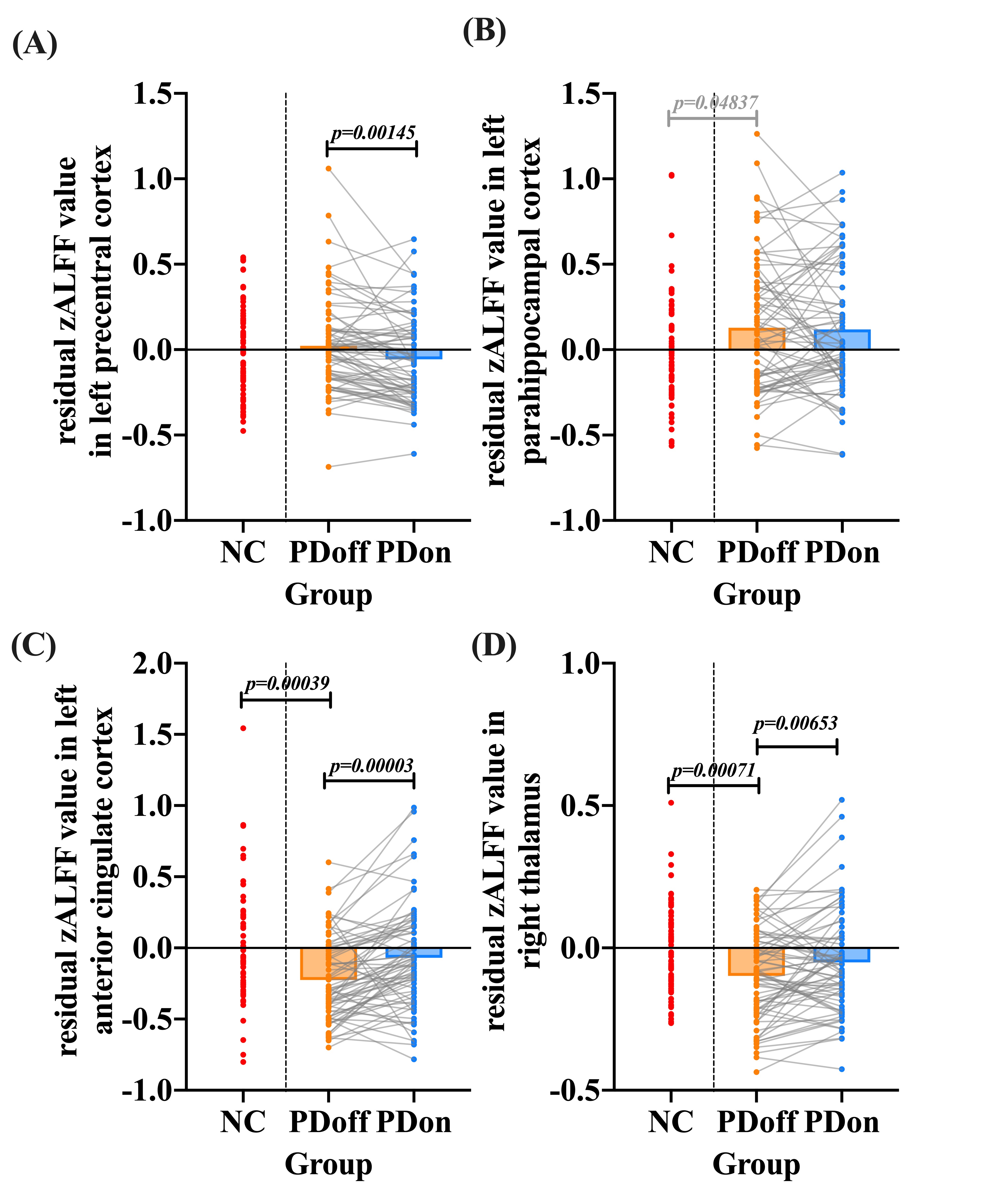
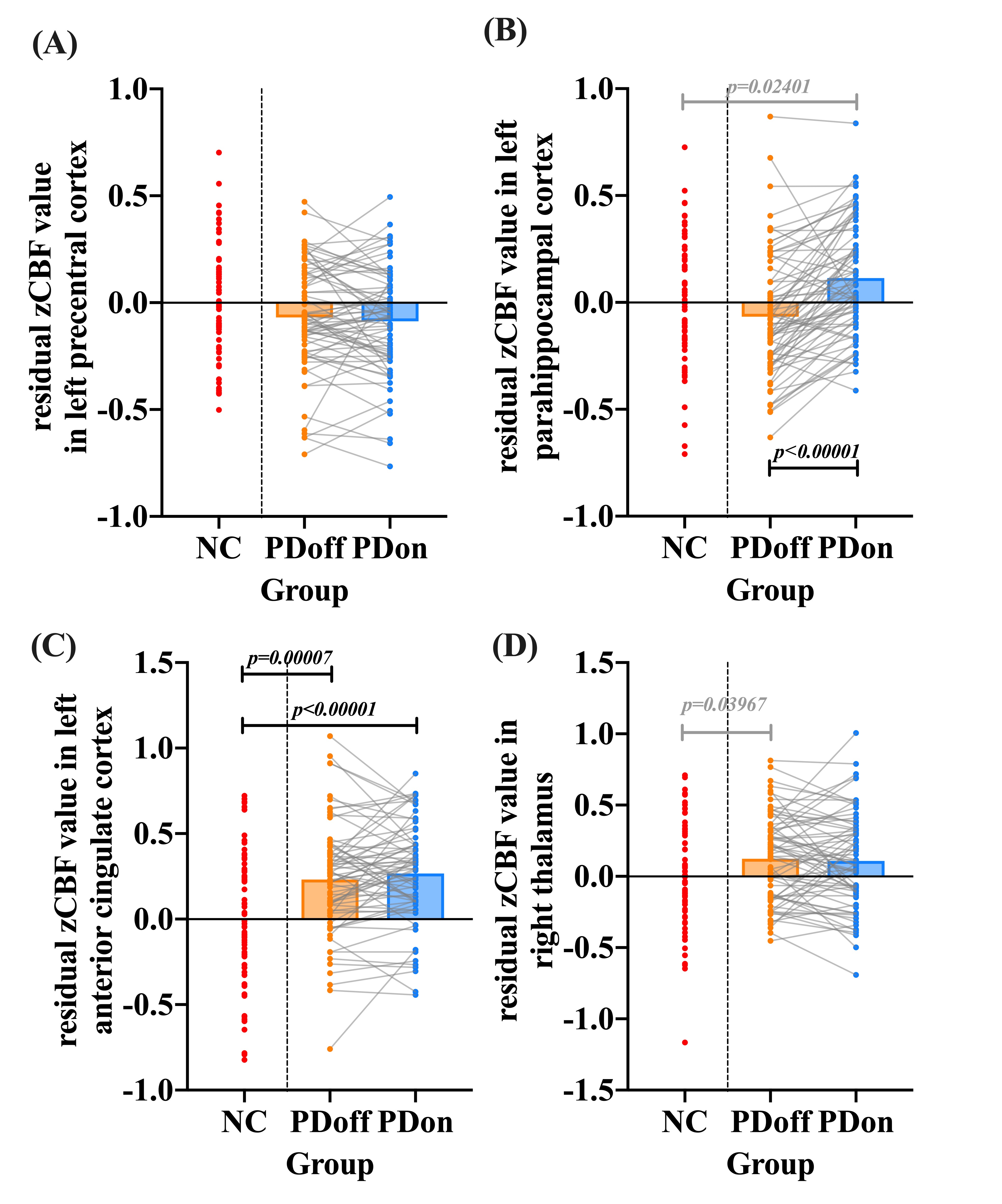
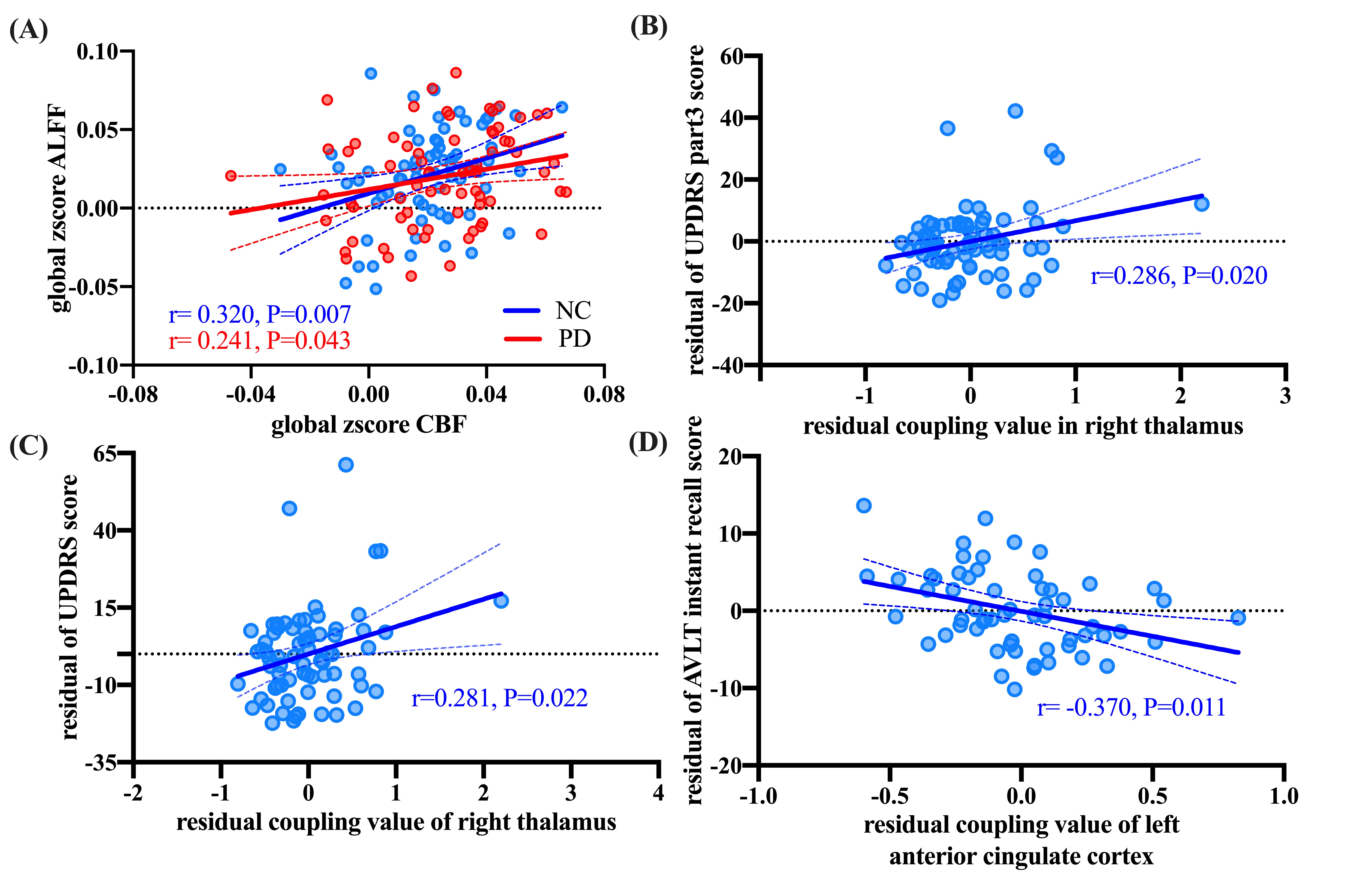
Significant positive correlations were observed between global CBF and ALFF in both NC (r= 0.320, P=0.007) and patients (r= 0.241, P=0.043). PD patients showed significantly lower regional neurovascular coupling in left precentral cortex, left frontal inferior cortex, and left parahippocampal cortex than NC (P<0.001). Meanwhile, significantly increased regional neurovascular coupling was found in left anterior cingulate cortex (ACC), right putamen, and right thalamus in patients (P<0.001). After levodopa therapy, regional neurovascular coupling in left parahippocampus, left ACC, and right thalamus was normalized in PD patients, and ALFF alterations made a major contribution to such normalization effect. The neurovascular coupling in left ACC was negatively correlated with instant memory ability (r= -0.370, P=0.011) in patients. The neurovascular coupling in right thalamus was significantly associated with the severity of motor symptoms (r=0.286, P=0.020).

Conclusion

Abnormal neurovascular coupling was found in PD patients. These impairments were partially normalized after levodopa therapy, and ALFF alterations played a primary role in this normalization effect.

Keywords

Parkinson’s disease; Levodopa therapy; fMRI (Resting State); Neurovascular coupling

Acknowledgements

This work was supported by the National Natural Science Foundation of China (Grant Nos. 82271935, 81971577, 82171888, and 82001767), the 13th Five-year Plan for National Key Research and Development Program of China (Grant No. 2016YFC1306600), the Natural Science Foundation of Zhejiang Province (Grant No. LQ21H180008 and LQ20H180012), the China Postdoctoral Science Foundation (Grant Nos. 2021T140599 and 2019M662082), the National Natural Science Foundation of China's Major Regional International Cooperation Project (Grant No. 81520108010), and the Key Research and Development Program of Zhejiang Province (Grant No. 2020C03020).

The authors have declared that no competing interest exists. We certify that we have participated sufficiently in the work to take public responsibility for the appropriateness of the experimental design and method, and the acquisition, analysis, and interpretation of the data. We also assured that all of the authors listed had sufficient contributions to the conception and design, execution, or analysis and interpretation of data. The content of this manuscript has not been published in a scientific journal, book, or other venue.

References

Reference
1. Poewe W, Seppi K, Tanner CM, Halliday GM, Brundin P, Volkmann J, et al. Parkinson disease. Nat Rev Dis Primers. 2017;3:17013.
2. Mitchell T, Lehericy S, Chiu SY, Strafella AP, Stoessl AJ, Vaillancourt DE. Emerging Neuroimaging Biomarkers Across Disease Stage in Parkinson Disease: A Review. JAMA Neurol. 2021;78(10):1262-72.
3. Lewitt PA. Levodopa for the treatment of Parkinson's disease. N Engl J Med. 2008;359(23):2468-76.
4. Pringsheim T, Day GS, Smith DB, Rae-Grant A, Licking N, Armstrong MJ, et al. Dopaminergic Therapy for Motor Symptoms in Early Parkinson Disease Practice Guideline Summary: A Report of the AAN Guideline Subcommittee. Neurology. 2021;97(20):942-57.
5. Fahn S, Oakes D, Shoulson I, Kieburtz K, Rudolph A, Lang A, et al. Levodopa and the progression of Parkinson's disease. N Engl J Med. 2004;351(24):2498-508.
6. Lang AE, Espay AJ. Disease Modification in Parkinson's Disease: Current Approaches, Challenges, and Future Considerations. Mov Disord. 2018;33(5):660-77.
7. You H, Mariani LL, Mangone G, Le Febvre de Nailly D, Charbonnier-Beaupel F, Corvol JC. Molecular basis of dopamine replacement therapy and its side effects in Parkinson's disease. Cell Tissue Res. 2018;373(1):111-35.
8. Booth S, Ramadan A, Zhang D, Lu L, Kirouac G, Jackson MF, et al. The Vasomotor Response to Dopamine Is Altered in the Rat Model of l-dopa-Induced Dyskinesia. Mov Disord. 2021;36(4):938-47.
9. Aristieta A, Ruiz-Ortega JA, Morera-Herreras T, Miguelez C, Ugedo L. Acute L-DOPA administration reverses changes in firing pattern and low frequency oscillatory activity in the entopeduncular nucleus from long term L-DOPA treated 6-OHDA-lesioned rats. Exp Neurol. 2019;322:113036.
10. Suarez LM, Solis O, Carames JM, Taravini IR, Solis JM, Murer MG, et al. L-DOPA treatment selectively restores spine density in dopamine receptor D2-expressing projection neurons in dyskinetic mice. Biol Psychiatry. 2014;75(9):711-22.
11. Schaeffer S, Iadecola C. Revisiting the neurovascular unit. Nat Neurosci. 2021;24(9):1198-209.
12. Li Z, Zhu Y, Childress AR, Detre JA, Wang Z. Relations between BOLD fMRI-derived resting brain activity and cerebral blood flow. PLoS One. 2012;7(9):e44556.
13. Baller EB, Valcarcel AM, Adebimpe A, Alexander-Bloch A, Cui Z, Gur RC, et al. Developmental coupling of cerebral blood flow and fMRI fluctuations in youth. Cell Rep. 2022;38(13):110576.
14. Zou QH, Zhu CZ, Yang Y, Zuo XN, Long XY, Cao QJ, et al. An improved approach to detection of amplitude of low-frequency fluctuation (ALFF) for resting-state fMRI: fractional ALFF. J Neurosci Methods. 2008;172(1):137-41.
15. Tahmasian M, Bettray LM, van Eimeren T, Drzezga A, Timmermann L, Eickhoff CR, et al. A systematic review on the applications of resting-state fMRI in Parkinson's disease: Does dopamine replacement therapy play a role? Cortex. 2015;73:80-105.
16. Paul G, Elabi OF. Microvascular Changes in Parkinson's Disease- Focus on the Neurovascular Unit. Front Aging Neurosci. 2022;14:853372.
17. Shang S, Zhang H, Feng Y, Wu J, Dou W, Chen YC, et al. Region-Specific Neurovascular Decoupling Associated With Cognitive Decline in Parkinson's Disease. Front Aging Neurosci. 2021;13:770528.
18. Lagana MM, Pirastru A, Pelizzari L, Rossetto F, Di Tella S, Bergsland N, et al. Multimodal Evaluation of Neurovascular Functionality in Early Parkinson's Disease. Front Neurol. 2020;11:831.
19. Iadecola C. The Neurovascular Unit Coming of Age: A Journey through Neurovascular Coupling in Health and Disease. Neuron. 2017;96(1):17-42.
20. Al-Bachari S, Vidyasagar R, Emsley HC, Parkes LM. Structural and physiological neurovascular changes in idiopathic Parkinson's disease and its clinical phenotypes. J Cereb Blood Flow Metab. 2017;37(10):3409-21.
21. Shang S, Ye J, Wu J, Zhang H, Dou W, Krishnan Muthaiah VP, et al. Early disturbance of dynamic synchronization and neurovascular coupling in cognitively normal Parkinson's disease. J Cereb Blood Flow Metab. 2022;42(9):1719-31.
22. Choi JK, Chen YI, Hamel E, Jenkins BG. Brain hemodynamic changes mediated by dopamine receptors: Role of the cerebral microvasculature in dopamine-mediated neurovascular coupling. Neuroimage. 2006;30(3):700-12.
23. Chen YI, Ren J, Wang FN, Xu H, Mandeville JB, Kim Y, et al. Inhibition of stimulated dopamine release and hemodynamic response in the brain through electrical stimulation of rat forepaw. Neurosci Lett. 2008;431(3):231-5.
24. Dirkx MF, den Ouden HE, Aarts E, Timmer MH, Bloem BR, Toni I, et al. Dopamine controls Parkinson's tremor by inhibiting the cerebellar thalamus. Brain. 2017;140(3):721-34.
25. Knutson B, Gibbs SE. Linking nucleus accumbens dopamine and blood oxygenation. Psychopharmacology (Berl). 2007;191(3):813-22.

Figures

Figure1. Workflow of image processing and statistical analyses.

Figure2. Regional neurovascular coupling differences between NC and patients, and coupling alterations after levodopa therapy in on-state patients.

Figure3. Regional Z-score ALFF differences between NC and patients, and coupling alterations after levodopa therapy in on-state patients.
Of the four regions which showed normalized neurovascular coupling after levodopa therapy, (A, C-D) significant alterations of Z-score ALFF were found in left precentral cortex, left ACC and right thalamus (P<0.001).


Figure4. Regional Z-score CBF differences between NC and patients, and coupling alterations after levodopa therapy in on-state patients.
Of the four regions which showed normalized neurovascular coupling after levodopa therapy, (B) significant alteration of Z-score CBF was only found in the left parahippocampal cortex (P<0.001).


Figure5. Partial correlation analyses.
(A) Significant positive correlations between global CBF and ALFF in both NC (r= 0.320, P=0.007) and PD patients (r= 0.241, P=0.043). (B-C) Significant positive correlations between the right thalamus coupling value and UPDRS part3 score (r= 0.286, P=0.020), and UPDRS total score (r= 0.281, P=0.022). (D). The AVLT instant recall score was negatively associated with the coupling value in left ACC (r= -0.370, P=0.011).


Proc. Intl. Soc. Mag. Reson. Med. 31 (2023)
0124
DOI: https://doi.org/10.58530/2023/0124