0099
Development and Analysis of MRI-Derived Carpal Motion Data Elements
Kevin Koch1, Andrew S Nencka1, Mohammad Zarenia2, and Rajeev Mannem1
1Radiology, Medical College of Wisconsin, Milwaukee, WI, United States, 2Radiation Oncology, Medical College of Wisconsin, Milwaukee, WI, United States
1Radiology, Medical College of Wisconsin, Milwaukee, WI, United States, 2Radiation Oncology, Medical College of Wisconsin, Milwaukee, WI, United States
Synopsis
Keywords: Joints, Joints
We present the construction and analysis of dynamic carpal data elements derived from 4D-MRI of the moving wrist. Across a clinically asymptomatic cohort of 31 subjects, dynamic data elements were constructed from capitate-normalized profiles of the scaphoid and lunate bones in a subject-specific radius-based coordinate system. Along with computing stability estimates of these measures, they are utilized to perform preliminary logistic regression modeling of radiologically-identified asymptomatic abnormalities within the study cohort. The results of this analysis suggests that the derived data elements have promising capabilities for characterizing carpal pathology and abnormalities.Introduction
Though MRI and CT arthrography can be utilized to assess carpal ligament damage, the functional impact of such damage remains elusive to current diagnostic mechanisms. To address this gap, our team has developed MRI technology for dynamic profiling of carpal bones. We hypothesize that appropriately curated dynamic carpal bone metrics could improve specificity in diagnosing damaged carpal ligaments.The acquisition and analysis paradigm utilized in our 4D-MRI approach to track carpal movements is detailed in a recent publication by Zarenia et al[1]. In the present analysis, we seek to develop and analyze a core set of data elements from these MR-derived carpal motion profiles.
Methods
Subjects: A cohort of participants (n=31) without a history of wrist injury or pain were consented into a study protocol approved by the local IRB. 19 females (aged 30.5+/-9.8 yrs) and 12 males (33.1+/9.8yrs) were enrolled and underwent an MRI examination of their dominant wrist.MRI Examination: Imaging was performed with a 3T MRI scanner using a 16-channel large flex coil. Subjects performed radial/ulnar-deviation and flexion-extension movements with no physical motion constraints. Static images were acquired using 2-point Dixon 3D-gradient echo sequence with 0.6mm isotropic voxel size. Dynamic volumes with temporal resolution of 2.3s were acquired with the same sequence at lower spatial resolution (1.6×1.6×2.5 mm3 resolution). 40 dynamic volumes were acquired for each motion, with the subject visually cued to make 3 cycles of the indicated motion during the 90 second acquisition window. Static fat-suppressed 3D FSE images (0.5 mm isotropic resolution) were also captured for identification of carpal abnormalities in the asymptomatic cohort.
Post-Processing: A 3D deep-neural network was trained using manually segmented data from a prior study to automatically segment the scaphoid, lunate, and capitate bones in the static and dynamic Dixon-reconstructed water-composition series . In addition, the head and proximal shaft of the radius was segmented using a locally-developed region-growing algorithm. Following the methods presented in Zarenia et al[1], these segmentations were utilized to derive individual rotational angles and center of mass translations of the scaphoid, lunate and capitate for each of the acquired 3D time frames.
Analysis: It is well-known that wrist motion can be referenced by the position of the capitate relative to the radius [2,3]. Using the capitate-referenced profiles of the scaphoid and lunate, quadratic polynomial coefficients were derived from each profile, along with a linear measure determined by computation of Spearman’s rho for each profile. This approach results in a total of 48 data elements across the 2 motions performed by each subject.
Coefficients of variation were computed for each derived data element. In addition, U-Tests were performed against subject gender and Spearman correlations were performed against subject age and carpal bone [scaphoid, capitate, lunate] volumes.
A radiologist with subspecialized training in musculoskeletal imaging interpreted the morphological images to identify asymptomatic abnormalities. For analysis purposes, these abnormalities were categorized as 1) triangular fibrocartilage complex (TFCC), wear/degeneration 2) carpal misalignment, 3) presence of ganglion cysts, and 4) intrinsic ligament damage. Finally, logistic regression with increasing levels of PCA-based dimensionality reduction was performed to model the derived carpal dynamic data elements against the asymptomatic finding groups. Logistic regression was performed and validated using 3-fold cross-validation and class-balancing.
Results
Figure 1 provides representative 2-point Dixon water-composition images and resulting carpal segmentations for the static (A) and dynamic (C) series.Sample dynamic metric profiles are provided in Figure 2A, with the final capitate-referenced profile representing the full-cohort plots illustrated in Figure 2B. The general low-order polynomial trends of the individual subject profiles (represented by different colors in each plot) are evident in these graphics. Sample quadratic and linear fits of these profiles are then displayed in Figure 3. Coefficients of variation for each metric across the asymptomatic cohort are provided in Figure 4.
Nineteen ( 61%) of the asymptomatic cohort had a radiologically-identified abnormality. This unexpected incidence of finding presented an opportunity to explore modeling of the derived metrics against the abnormality classes. The results of this modeling are displayed in Figure 5, along with the abnormality incidences. Two of the subjects had intrinsic ligament damage, which was too low to support the 3-fold cross-validation approach used in this preliminary small-sample sample modeling study. Surprisingly, even in this small sample, the derived dynamic elements showed relatively strong (AUC ~ 0.8) modeling performance against asymptomatic ganglion cyst and malalignment findings.
Discussion
The present study has outlined a set of simple data elements that can be derived from 4D-MRI of the moving wrist. Preliminary analysis of these metrics offers promising evidence that MRI-derived carpal dynamic profiles can be utilized to characterize pathology within the wrist.More complex multi-parametric modeling may improve the metric stability and performance presented in this work. Univariate analyses found that 13% of the data elements had significant (p< 0.05) correlations with carpal bone volume, while 11% correlated with age (only 1 element was dependent on gender). This suggests that accounting for carpal bone volume and age, using larger normative cohorts, could improve the derived metric stability.
Future work will seek to deploy the presented methods on larger normative cohorts and focused cohorts with specific classes of carpal pathology
Acknowledgements
This study was supported by NIH R21AR075327. The content is solely the responsibility of the authors and does not necessarily represent the official views of the NIH.References
[1] Zarenia, M., Arpinar, V. E., Nencka, A. S., Muftuler, L. T. & Koch, K. M. Dynamic tracking of scaphoid, lunate, and capitate carpal bones using four-dimensional MRI. Plos One 17, e0269336 (2022).
[2] de Lange A, Kauer JM, Huiskes R. Kinematic behavior of the human wrist joint: a roentgen-stereophotogrammetric analysis. J Orthop Res. 1985;3(1):56–64.Article
[3] Neu CP, Crisco JJ, Wolfe SW. In vivo kinematic behavior of the radio-capitate joint during wrist flexion-extension and radio-ulnar deviation. J Biomech. 2001;34(11):1429–38.
Figures
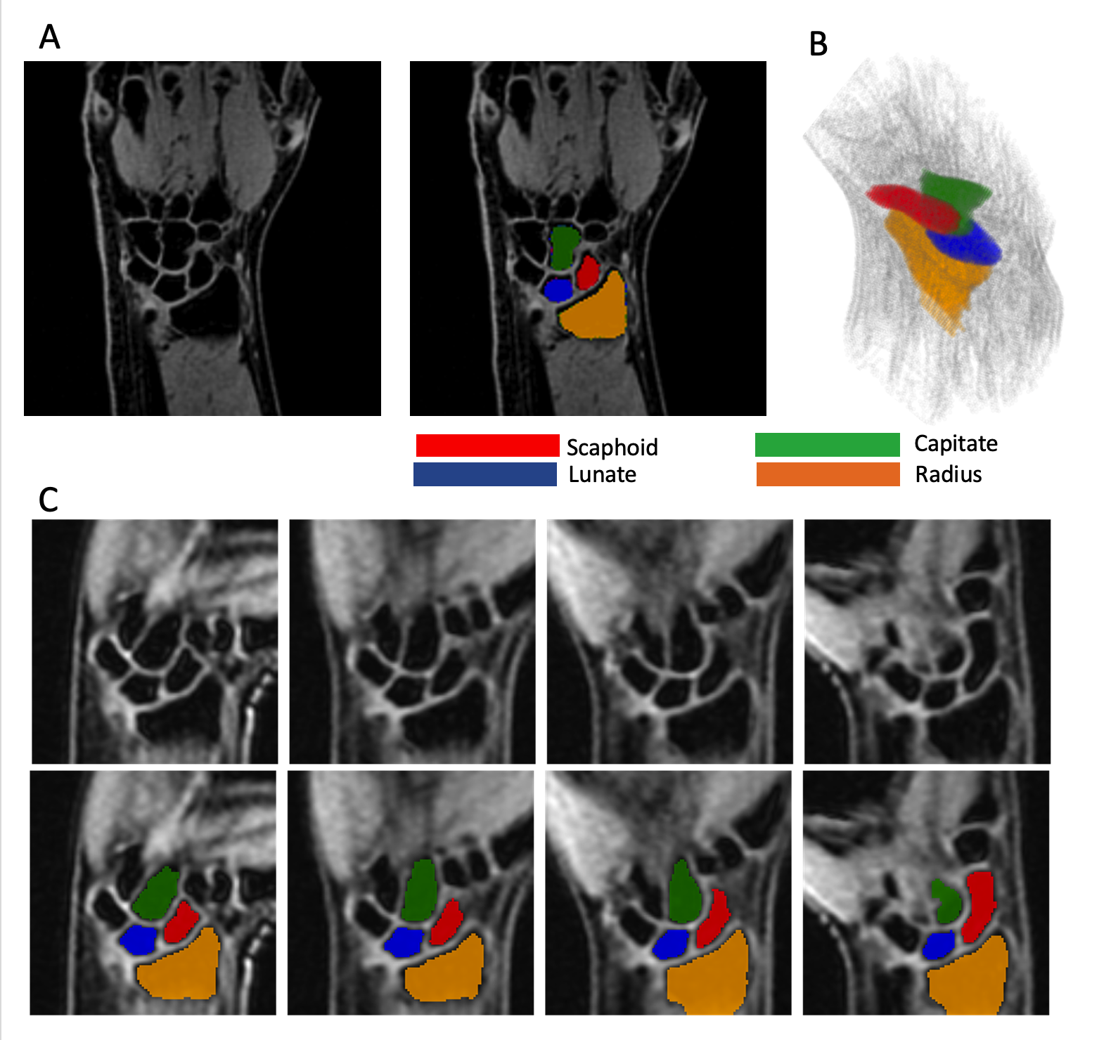
Figure 1: A) Static 2-point DIXON water-only 3D gradient-echo images provide sufficient contrast to enable automated deep-learning based 3D segmentation of analyzed carpal bones, as depicted in B). C) Slices from dynamic 3D 2-point Dixon gradient-echo images, along with auto-segmented bones in the dynamic images.
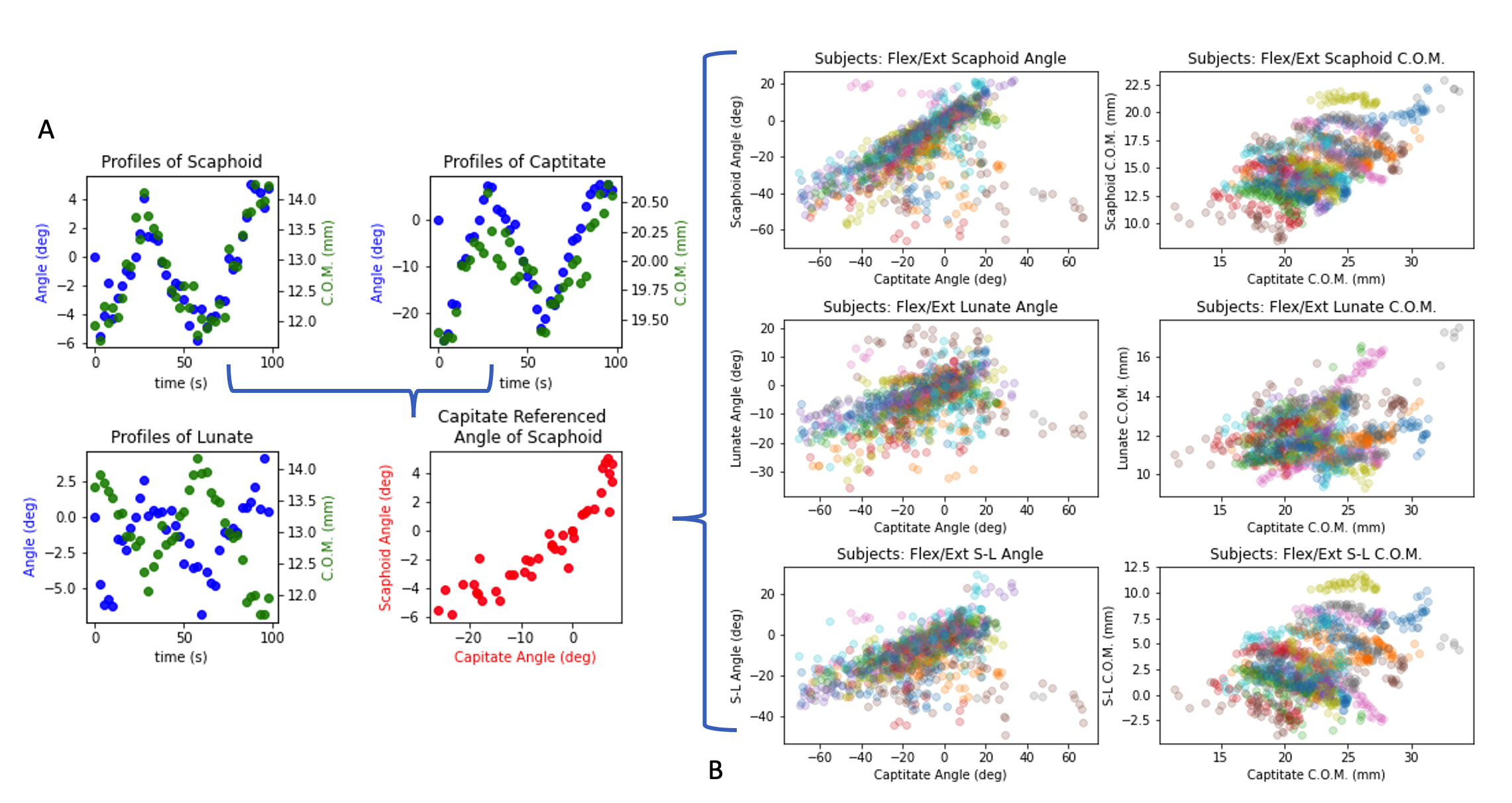
Figure 2: A) Profiles of scaphoid, capitate, and lunate angles and centers of mass (COM) during 3 motion cycles. The lower right (red) plot is the scaphoid angle relative to the capitate angle, which is the approach used to plot the full subject cohort (n=31 subjects) in B) for all 6 computed metrics. Each subject in these plots (B) is represented by a different color. The general trends of the different metrics across the subject cohort are visible in these scatter plots.
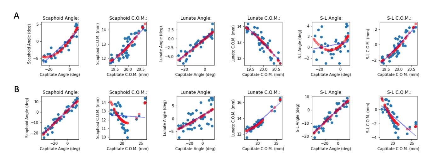
Figure 3: Example fits from one subject, illustrating the linear and quadratic regressions performed on each profile. Quadratic (red points) and linear (blue line) fits are shown for each metric profile for A) radial-ulnar deviation movements and B) flexion/extension movements. Polynomial least squares fits utilized RMS errors from point-cloud registrations at each time frame as weighting factors. S-L refers to the difference between the respective scaphoid and lunate measures (angles and COMs).
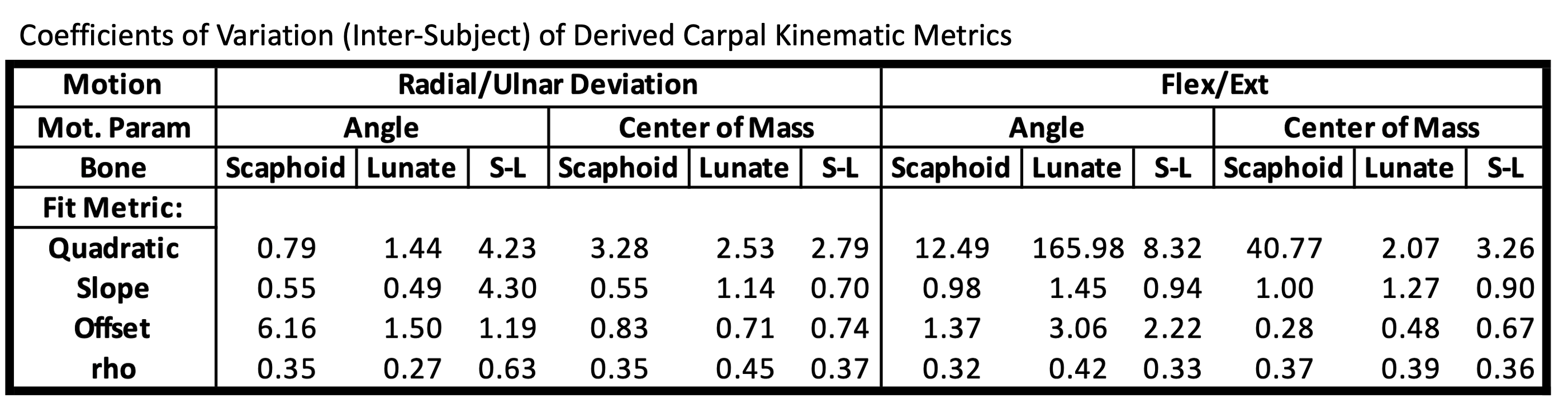
Figure 4. Coefficients of variation of the 48 derived kinematic metrics across the asymptomatic study cohort. The lowest coefficient of variation is reported for the slope and offset polynomial measures across the measures independently derived from linear and quadratic polynomial fits. The rho metric is the Spearman’s rho correlation coefficient computed for each metric. This measure of linearity of the profiles was the most stable metric derived from the study data.
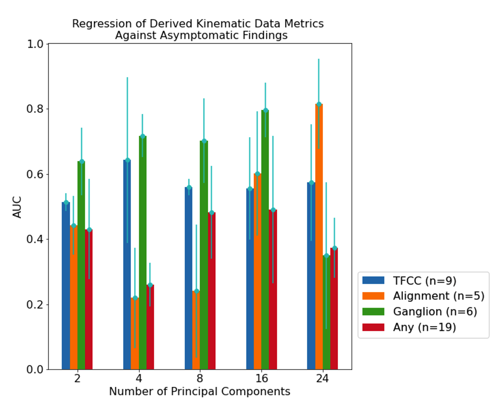
Figure 5. The mean of 3-fold cross-validated Area-Under-the Curve measures from Receiver-Operator Curves computed using derived kinematic metrics as logistic regression predictors of asymptomatic radiologically-identified pathology within the study cohort. AUC values are plotted as a function of the number of principle components utilized to reduce the dimensionality of the full metric dataset (48 metrics) . Error bars reflect the standard deviation of the computed AUC values for each model.
DOI: https://doi.org/10.58530/2023/0099