0091
Assessment of Osteoporosis in Lumbar Spine Using Ultrashort Echo Time Magnetization Transfer (UTE-MT) Imaging
Jin Liu1 and Ya-Jun Ma1
1Department of Radiology, University of California San Diego, San Diego, CA, United States
1Department of Radiology, University of California San Diego, San Diego, CA, United States
Synopsis
Keywords: Bone, Skeletal
The bone collagen matrix makes a crucial contribution to the mechanical properties of bone including elasticity and tensile strength. Its changes can be accessed by the ultrashort echo time magnetization transfer (UTE-MT) technique. This study aims to investigate the feasibility of the UTE-MT ratio (UTE-MTR) in the assessment of lumbar osteoporosis (OP). Our results demonstrated that the UTE-MTR is highly correlated with bone mineral density and Fracture Risk Assessment Tool scores and has a high ability to distinguish people with different bone masses, which indicates that the UTE-MTR has great potential in the diagnosis of the patient with OP.Introduction
Osteoporosis (OP) is a metabolic bone disease characterized by low bone mineral density (BMD) and the deterioration of bone tissue microarchitecture [1]. The bone collagen matrix plays an important role in the mechanical properties of bone by imparting elasticity and tensile strength. Ultrashort echo time magnetization transfer (UTE-MT) imaging can detect both bound and free water signals in bone [2; 3]. This study aims to investigate the feasibility of UTE-MT ratio (UTE-MTR) in assessment of lumbar trabecular bone in patients with OP.Methods
A total of 148 participants (mean age, 58 years; age range, 50-85 years) underwent MRI (Signa, Pioneer, GE Healthcare) and quantitative computed tomography (QCT) in the lumbar spine. Fracture risk was calculated for all participants using Fracture Risk Assessment Tool (FRAX). Informed consent was obtained from all participants in accordance with the Institutional Review Board. A Fermi pulse was employed to generate the MT contrast in UTE-MT sequence with duration of 8ms and bandwidth of 160Hz. The frequency offset of this MT pulse was 1500 Hz. The UTE-MT sequence was scanned twice with flip angle of 750° for MT-On and 0° for MT-Off. Other UTE-MT sequence parameters were as follows: TR = 100 ms, TE = 0.032 ms, excitation flip angle = 5°, number of spokes per-TR = 5, FOV = 28cm × 28cm, matrix = 140 × 140, slice thickness = 3.6mm, slice number = 16, bandwidth = 125 kHz, oversampling factor = 1.2, and scan time = 3min. The UTE-MTR is calculated by the signal ratio of the difference between UTE-MT-OFF and UTE-MT-ON to the UTE-MT-OFF. A product sequence, IDEAL-IQ, was used to quantify BMFF (TR = 7.3 ms, TEs = 1.2, 2.1, 3.1, 4.1, 5.0, and 6.0 ms, FA = 4°, FOV = 32×32 cm2, matrix = 160 × 160; number of slices = 12; slice thickness = 8 mm; and scan time = 16 sec). In this study, lumbar UTE-MTR and bone mineral density (BMD) values were calculated in three vertebrae (L2–L4) for each subject. Major fracture risk was also evaluated for all participants using Fracture Risk Assessment Tool (FRAX). UTE-MTR and bone marrow fat fraction (BMFF) were correlated with BMD and FRAX score for comparison. The performances of the UTE-MTR and BMFF to discriminate between three different cohorts, which included normal subjects, patients with osteopenia, and patients with osteoporosis, were also evaluated and compared using receiver operator characteristic (ROC) analysis.Results
Figure 1 shows representative UTE-MT, QCT and BMFF maps of the lumbar spines of three subjects: one from each cohort (normal, osteopenia, and OP). Lower UTE-MTR, BMD, as well as higher BMFF, were found in more osteoporotic subjects. The UTE-MTR showed strong correlation with standard BMD (r = 0.76, P < 0.001) and FRAX score (r = -0.77, P < 0.001) (Figure 2). High area under the curve (AUC) values (≥ 0.723) obtained from ROC analysis demonstrated that the UTE-MTR was capable of differentiating between the three different subject cohorts (Figure 3). Moreover, the UTE-MTR had better correlations with BMD and FRAX score than BMFF, and also performed better in cohort discrimination.Discussion and Conclusion
To our best knowledge, this is the first study to apply the UTE-MT imaging to assess lumbar trabecular bone in patients with OP. Our results showed that UTE-MTR was strongly correlated with BMD in vertebral trabecular bone, suggested that the quantities of both bone mineral and organic matrix were decreased in trabecular bone with OP. The UTE-MTR demonstrated good performance in identifying people with different bone mass, which demonstrate that the UTE-MTR may be a promising marker for assessing patient with OP.Acknowledgements
The authors acknowledge grant support from the National Natural Science Foundation of China (Grant Nos. 82172053) and the National Institutes of Health (R21AR075851 and R01AR079484).References
[1] (2001) Osteoporosis prevention, diagnosis, and therapy. Jama 285:785-795
[2] Jerban S, Ma Y, Dorthe EW et al (2019) Assessing cortical bone mechanical properties using collagen proton fraction from ultrashort echo time magnetization transfer (UTE-MT) MRI modeling. Bone Rep 11:100220
[3] Jerban S, Ma Y, Wong JH et al (2019) Ultrashort echo time magnetic resonance imaging (UTE-MRI) of cortical bone correlates well with histomorphometric assessment of bone microstructure. Bone 123:8-17
Figures
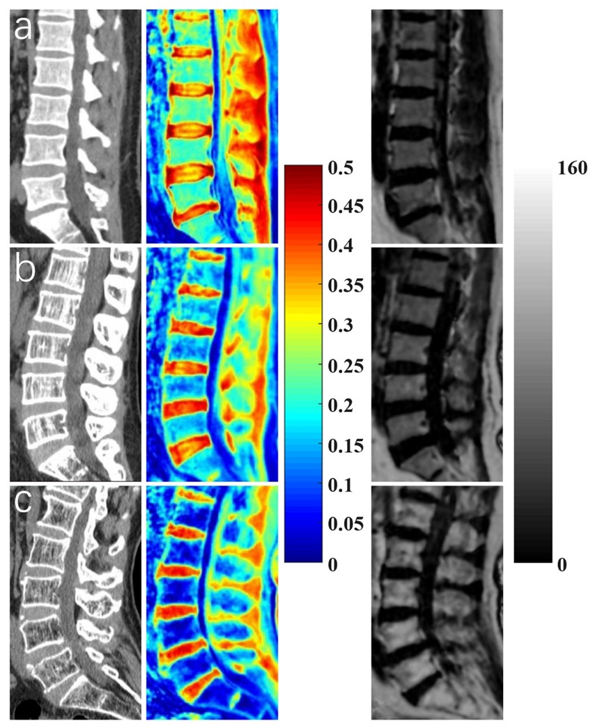
Figure
1: Representative bone
mineral density (BMD) (first column), ultrashort echo time magnetization transfer ratio (UTE-MTR)
(second column), and bone marrow fat fraction (BMFF) (third column) maps in the
lumbar spine of three subjects with normal bone mass (first row, 51-year-old
male), osteopenia (second row, 53-year-old female), and osteoporosis (last row,
72-year-old male).
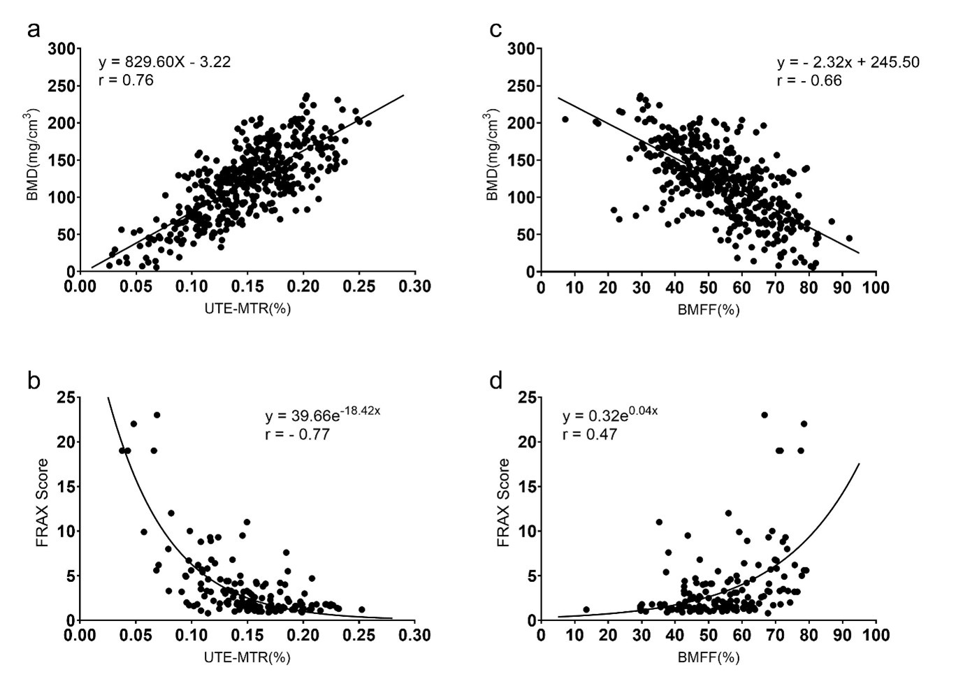
Figure 2: Correlation curves and scatter plots for the measurements between (a) ultrashort
echo time magnetization transfer ratio (UTE-MTR) and
bone mineral density (BMD), (b) UTE-MTR and Fracture Risk Assessment Tool
(FRAX) score, (c) bone marrow fat fraction (BMFF) and
BMD, and (d) BMFF and FRAX score.
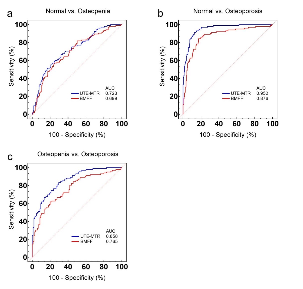
Figure 3:
Receiver operating characteristic (ROC) curves and corresponding area under
the curve (AUC) values of ultrashort echo time magnetization transfer ratio (UTE-MTR) and bone marrow fat fraction (BMFF) between (a) normal and
osteopenia, (b) normal and osteoporosis, and (c) osteopenia and osteoporosis.
DOI: https://doi.org/10.58530/2023/0091