0087
Age and gender-dependence of T1ρ-prepared PETRA sequence in knee ligaments and tendon1Center for Biomedical Imaging, NYU Langone Health, New York City, NY, United States, 2Medical College of Wisconsin, Wauwatosa, WI, United States
Synopsis
Keywords: Tendon/Ligament, Osteoarthritis
This study was performed to investigate gender-related and age-related differences in ultra-short echo-time (UTE) T1ρ parameters in the anterior cruciate ligament (ACL), posterior cruciate ligament (PCL), and patellar tendon (PT). A pointwise encoding time reduction with radial acquisition (PETRA) sequence was used to measure single-component and multi-component PETRA-T1ρ parameters in the ACL, PCL, and PT at 3.0T in 18 healthy volunteers. There were significant correlations between age and single-component and multi-component PETRA-T1p parameters in the ACL and PCL but not the PT. There were no significant differences in PETRA-T1ρ parameters in the ACL, PCL, or PT between males and females.
Introduction
Ultrashort echo time (UTE) techniques have been developed to evaluate collagen-rich short T2 musculoskeletal tissues such as ligaments and tendons. Previous studies have described the use of bi-exponential UTE decay models for evaluating the individual water components of musculoskeletal tissues but are limited by their long acquisition times1–4. The pointwise encoding time reduction with radial acquisition (PETRA) sequence is a new hybrid accelerated technique that fills the outer k-space with radial half-projections and the center with single-point Cartesian acquisitions. This study was performed to investigate gender-related and age-related differences in single-component and multi-component PETRA-T1ρ parameters in the anterior cruciate ligament (ACL), posterior cruciate ligament (PCL), and patellar tendon (PT).Methods
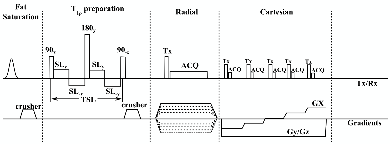
3D-T1ρ-PETRA sequenceThe 3D-T1ρ-PETRA sequence, as shown in Figure 1, is composed of a fat saturation pulse, self-compensated T1ρ preparation module, radial and Cartesian readout modules, and a longitudinal magnetization recovery delay.
The PETRA-T1ρ weighted images were acquired with a spin-lock frequency of 500Hz and TSLs: 0ms, 0.2ms, 2.6ms, 12.9ms, 12.9ms, and 30ms. These TSL values were obtained using previously described approaches5,6. Fat suppression and T1ρ preparation modules were applied at every 26 readouts to minimize acquisition time. The acquisition parameters were as follows: TR/TE: 5ms/0.07ms, FOV: 200mm×200mm, Flip angle: 6°, Slice thickness: 0.78mm, matrix size = 256×256×256, nominal voxel size: 0.78mm×0.78mm×0.78mm, receiver bandwidth = 399 Hz/px, T1 recovery delay= 500ms, number of radial spokes=8000, number of segments =350, number of readouts in each segment = 26, and number of Cartesian sample points = 1419. The total acquisition time was 23 minutes.
MRI Acquisition
PETRA-T1ρ imaging of the ACL, PCL, and PT was performed in 18 healthy volunteers (8 females, mean age: 29±8yrs, and 10 males, mean age: 35±10yrs) using a 3.0T MRI scanner (MAGNETOM Prisma, Siemens Healthcare GmbH, Germany) with a 1Tx/15Rx knee coil. Our study was approved by our Institutional Review Board (IRB). All volunteers provided written informed consent before the MRI scans.
T1ρ Relaxation Mapping
The signal decay over different spin-lock pulse durations was fitted to three different models. Mono-exponential (ME) relaxation times were calculated voxel-by-voxel by fitting the signal-intensity decay over time to:
$$ x(t)=A\exp\left(-\frac{t}{T_{1\rho,mo}}\right)+c,$$
Here, $$$A$$$ denotes the amplitude, $$$T_{1\rho, mo}$$$ the mono-exponential relaxation time, $$$t$$$ is a vector with the spin-lock pulse durations, and c is a constant to account for baseline residual noise.
Afterward, the bi-exponential (BE) relaxation components were calculated in the same manner by fitting the data to
$$x(t)=A\left(f\exp\left(-\frac{t}{T_{1\rho,s}} \right)+(1-f)\exp\left(-\frac{t}{T_{1\rho,l}}\right)\right)+c,$$
where $$$T_{1\rho,s}$$$ denotes the shorter component, $$$T_{1\rho,l}$$$ the longer component, and $$$f$$$ denotes the percentage of the total amplitude belonging to the shorter component.
Finally, the stretched-exponential (SE) relaxation components are fitted to the data according to
$$ x(t)=A\exp\left(-\left(\frac{t}{T_{1\rho,se}}\right)^\alpha\right)+c,$$
in which $$$T_{1\rho,se}$$$ denotes the characteristic relaxation time and $$$\alpha$$$ is the stretching parameter.
Statistical Analysis
Mann-Whitney tests were used to assess gender-related differences in PETRA-T1ρ parameters in the ACL, PCL, and PT. Spearman correlation coefficients were used to assess the association between age and PETRA-T1ρ parameters. Statistical significance was defined as P< 0.05.
Results
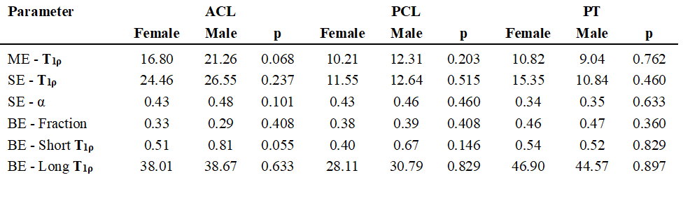
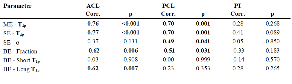
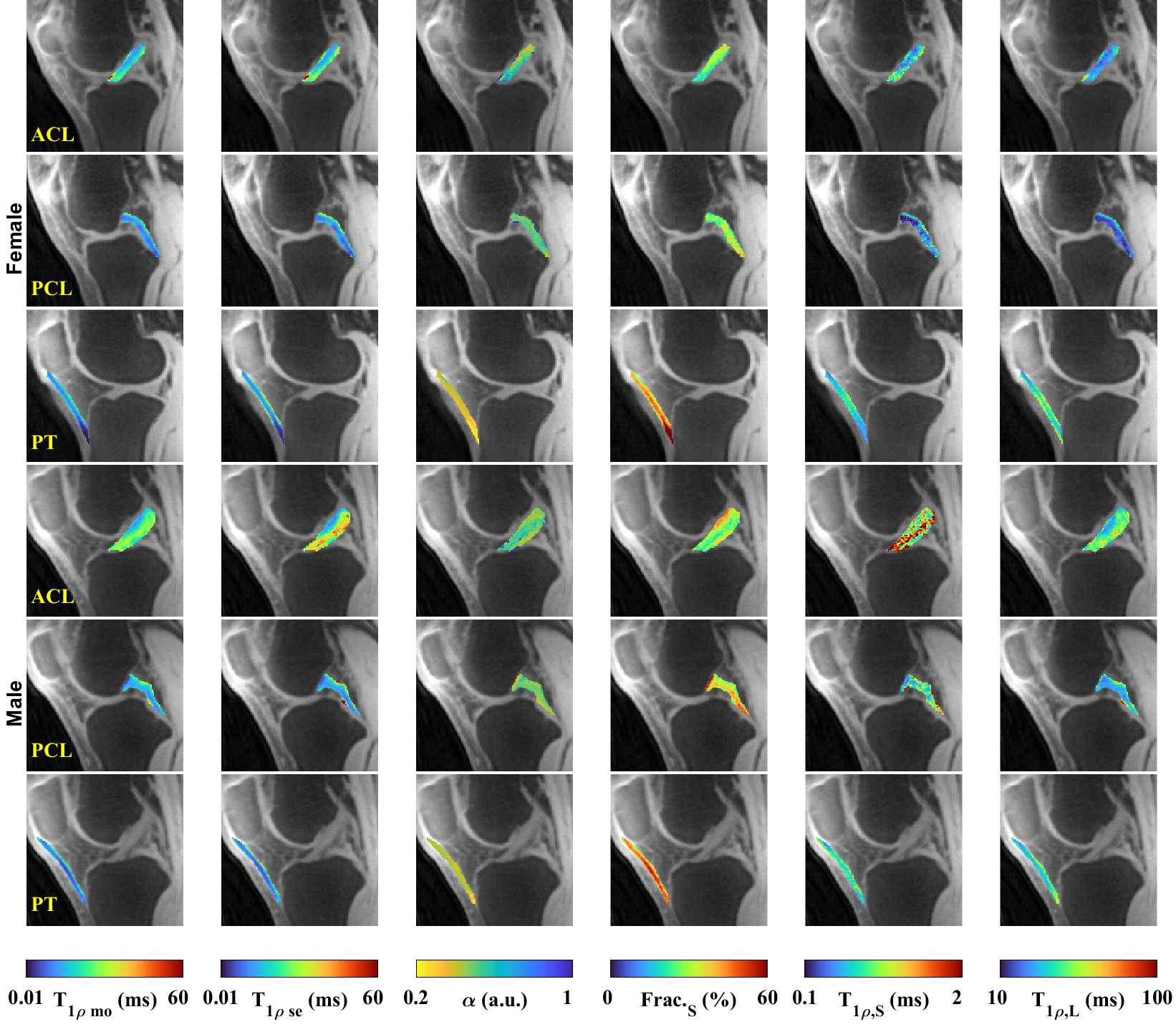
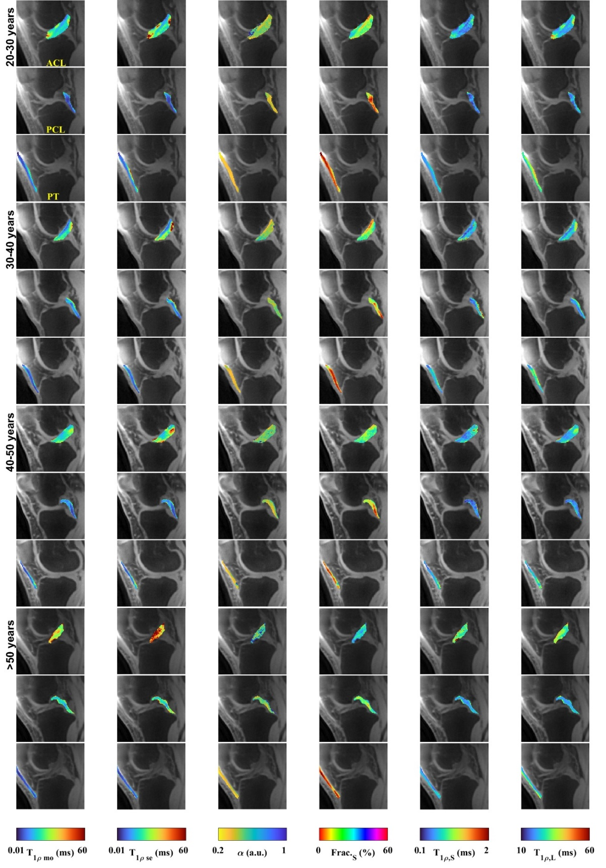
Tables 1 and 2 show gender-related and age-related differences in PETRA-T1ρ parameters in the ACL, PCL, and PT, respectively. There was no significant difference between males and females in PETRA-T1ρ parameters. There was a significant positive correlation between age and ME-T1ρ and SE-T1ρ, and a significant negative correlation between age and BE-Fraction in the ACL and PCL. There was a significant positive correlation between age and BE-Long in the ACL. There was no significant correlation between age and PETRA-T1ρ parameters in the PT. Figures 2 and 3 show representative PETRA-T1ρ parameter maps in male and female subjects and subjects of different ages, respectively.Discussion
Females have a much higher risk of ligament injuries than males7. However, the absence of gender-related differences in PETRA-T1ρ parameters in the ACL and PCL between males and females in our study suggests that the higher injury risk is likely not due to baseline differences in ligament composition and structure.Our study documented significant correlations between age and PETRA-T1ρ parameters within ligaments, with higher correlations in the ACL than the PCL, which corresponds to the much higher incidence of degeneration with aging in the ACL8. While there were significant positive correlations between age and ME-T1ρ and SE-T1ρ, the bi-component PETRA-T1ρ analysis provides better information about changes occurring within the individual water components during the aging process. The significant negative correlation between age and BE-Fraction in the ACL and PCL reflects the lower macromolecular content and higher free water content in ligaments with aging, which has been documented in histologic studies9. The significant positive correlation between age and BE-Long T1ρ but not BE-Short T1ρ in the ACL indicates greater low-frequency interactions with aging between macromolecules and loosely bound bulk water than water tightly bound to the macromolecular matrix.
The absence of age-related differences in PETRA-T1ρ parameters in the PT likely reflects the fact that the parameters were measured within the entire PT, while tissue degeneration with aging and disease typically occurs exclusively within the proximal tendon10.
Conclusion
Our study showed significant correlations between age and PETRA-T1ρ parameters in the ACL and PCL but not the PT. There were no significant differences in PETRA-T1ρ parameters between males and females.Acknowledgements
This study was supported by NIH grants, R21-AR075259-01A1, R01-AR068966, R01-AR076328-01A1, R01-AR076985-01A1, and R01-AR078308-01A1 and was performed under the rubric of the Center of Advanced Imaging Innovation and Research (CAI2R), an NIBIB Biomedical Technology Resource Center (NIH P41-EB017183).References
1. Juras, V. et al. Bi-exponential T2*analysis of healthy and diseased Achilles tendons: An in vivo preliminary magnetic resonance study and correlation with clinical score. Eur Radiol 23, 2814–2822 (2013).
2. Chang, E. Y. et al. Single- and Bi-component T2∗ analysis of tendon before and during tensile loading, using UTE sequences. Journal of Magnetic Resonance Imaging 42, 114–120 (2015).
3. Kijowski, R., Wilson, J. J. & Liu, F. Bicomponent ultrashort echo time analysis for assessment of patients with patellar tendinopathy. Journal of Magnetic Resonance Imaging 46, 1441–1447 (2017).
4. Sharafi, A., Baboli, R., Chang, G. & Regatte, R. R. 3D‐T 1ρ prepared zero echo time‐based PETRA sequence for in vivo biexponential relaxation mapping of semisolid short‐T 2 tissues at 3 T. Journal of Magnetic Resonance Imaging 50, 1207–1218 (2019).
5. Zibetti, M. V. W., Sharafi, A. & Regatte, R. R. Optimization of spin‐lock times in T 1ρ mapping of knee cartilage: Cramér‐Rao bounds versus matched sampling‐fitting. Magn Reson Med 1–17 (2021) doi:10.1002/mrm.29063.
6. de Moura, H. L., Menon, R. G., Zibetti, M. V. W. & Regatte, R. R. Optimization of spin-lock times for T1ρ mapping of human knee cartilage with bi- and stretched-exponential models. Scientific Reports 2022 12:1 12, 1–15 (2022).
7. Sutton, K. M. & Bullock, J. M. Anterior cruciate ligament rupture: Differences between males and females. Journal of the American Academy of Orthopaedic Surgeons 21, 41–50 (2013).
8. Levy, Y. D. et al. Histopathological changes in the human posterior cruciate ligament during aging and osteoarthritis: correlations with anterior cruciate ligament and cartilage changes. Ann Rheum Dis 72, 271–277 (2013).
9. McCarthy, M. M. & Hannafin, J. A. The Mature Athlete: Aging Tendon and Ligament. Sports Health 6, 41–48 (2014).
10. Johnson, D. P., Wakeley, C. J. & Watt, I. MAGNETIC RESONANCE IMAGING OF PATELLAR TENDONITIS. https://doi.org/10.1302/0301-620X.78B3.0780452 78, 452–457 (1996).
Figures