0068
4D Flow MRI Reference Values of the Portal Venous System1Department of Surgery, Section of Vascular Surgery, University of Michigan, Ann Arbor, MI, United States, 2Department of Radiology, University of Wisconsin, Madison, WI, United States, 3Department of Medical Physics, University of Wisconsin, Madison, WI, United States, 4Department of Mechanical Engineering, University of Wisconsin, Madison, WI, United States, 5Department of Biomedical Engineering, University of Wisconsin, Madison, WI, United States, 6Department of Medicine, University of Wisconsin, Madison, WI, United States, 7Department of Emergency Medicine, University of Wisconsin, Madison, WI, United States, 8Department of Radiology and Nuclear Medicine, Universität zu Lübeck, Lübeck, Germany
Synopsis
Keywords: Liver, Vessels
4D flow MRI can aid in diagnosing pathologies associated with hemodynamic changes in the portal vein, such as portal hypertension. Normal reference ranges for portal venous flow are lacking but essential for future clinical implementation. We report reference values based on radial 4D flow MRI measurements of mean flow, velocity, and effective diameter in the portal venous vasculature in 44 healthy subjects (59% female, 18-74 years). Faint helical and linear flow are physiological flow patterns observed in the portal vein. This study provides normative values for emerging clinical applications of 4D flow MRI currently under development.
Keywords: Liver, Velocity&Flow, Vessels
Introduction
4D flow MRI can help diagnose and understand pathologies that alter blood flow to the liver by providing functional and anatomical information (1). Typical pathologies in the portal venous circulation include portal hypertension, portosystemic shunts, and hypersplenism. Hitherto, 4D flow MRI studies have focused primarily on patients with pathologic hemodynamics, with healthy volunteers comprising only a small subset of studies as a reference group. However, normal reference values are needed to develop and disseminate 4D flow MRI as a standard clinical tool to evaluate portal venous flow. Therefore, the purpose of this work was to establish normal reference values for 4D flow MRI-derived flow, velocity, and vessel diameters. Further, we aimed to describe typical flow patterns in the portal venous system of healthy adult subjects.Methods
Study CohortFor this retrospective study, we screened all available 4D flow MRI exams of the upper abdomen in healthy adults acquired at our institution between 2012 and 2022. Both clinical and research studies at either 1.5T or 3.0T were included. Exclusion criteria included studies without 5 hours of fasting prior to imaging, BMI>40, or poor data quality.
Postprocessing and Analysis
Image reconstruction was performed with retrospective cardiac and respiratory gating. Background phase corrections were performed using a custom MATLAB tool followed by vascular segmentation in MIMICS (Materialise, Belgium) (2,3). Data were subsequently processed and visualized in EnSight (CEI Inc., USA). Double-oblique planes perpendicular to vessel centerlines were placed in the time-averaged complex difference angiograms (Figure 1). Manual, time-resolved segmentation of the 2D vessel contour of the splenic vein, superior mesenteric vein, and main, right, and left portal veins was then performed in a customized software package (4). Average flow, mean velocity, and effective diameter of each contour were quantified. Effective diameter was calculated as follows: Effective Diameter = 2√(Area/π).
Time-resolved pathlines emitted from the contours were used to visualize flow patterns by three readers. The portal vein was evaluated for presence and intensity of helicity (Figure 2, helical flow <360°, pronounced helical flow >360°), and whether flow reversal as a characteristic of a vortex was present.
Reference ranges for flow, velocity, and diameter for each vessel were defined as the mean ± 2 standard deviations (SD) to encompass 95% of the population (5). Conservation of mass analysis was used as a quality metric to compare inflow versus outflow at the splenomesenteric confluence and portal vein bifurcation.
Results
Study CohortWe initially identified 70 healthy liver donors and 28 research subjects with MR imaging. Fifty-three of these subjects had 4D flow MRI studies. Nine studies were excluded due to poor data quality. The resulting 44 datasets were reprocessed and reanalyzed for this study. Acquisition parameters and demographics are listed in Figure 3.
Quantitative Analysis of Hemodynamics
We reported reference values for mean flow, mean velocity, and vessel diameter in the healthy portal vein using 4D flow MRI (Figure 4). Average flow and velocity for the right portal vein were both greater than that of the left portal vein. Two subjects demonstrated trifurcation of the distal portal vein, a normal variant, instead of the typical bifurcation.
Qualitative Evaluation of Hemodynamics
Normal flow patterns in the portal vein included helical (66%) or linear flow (34%). Of the 29 helical flow cases, 4 (14%) were clockwise, and 25 (86%) were counterclockwise. Five subjects (11%) showed pronounced helical flow in the splenomesenteric confluence. No subject developed flow reversal.
Quality Control
Conservation of mass analysis demonstrated a relative error of 1.1±4.6% standard deviation (SD) at the splenomesenteric confluence and -1.4±4.1% SD at the portal bifurcation (Figure 5).
Discussion
We evaluated flow, velocity, and diameter at standardized locations in the portal venous vasculature of 44 healthy adults. For each of these parameters, we reported reference ranges encompassing two standard deviations of the mean as a basis for future 4D flow MRI studies in the portal venous system.Increased right versus left portal venous flow corresponds with studies of right and left hepatic lobular volume (6). While many of our subjects demonstrated linear flow at the splenomesenteric confluence, over half of the healthy subjects demonstrated at least faint helicity that corresponds well with the literature (7). Portal vein helices are hypothesized to impact downstream portal hemodynamics including mixing of blood from the splenic & superior mesenteric veins (7).
ll reported mean flow and velocity values from smaller studies in the literature fall within the reference ranges described in this study (8-18).
Limitations of this study include a sample size too small to evaluate age-/gender- specific characterizations and data collected from a single institution. Future studies would benefit from greater sub-population analysis and larger controlled meal challenge studies to characterize the normal postprandial hyperemic response in the portal venous circulation (14,16).
Conclusion
We have reported normal reference ranges for mean flow, velocity, and effective diameter in the portal venous vasculature based on 44 healthy subjects. Linear and helical flow are typical physiological flow patterns in the portal vein. Rarely, a pronounced helix developed at the splenomesenteric confluence. The results of this study provide normative values for emerging clinical applications of 4D flow MRI of the portal venous system currently under development.Acknowledgements
We wish to acknowledge support from GE Healthcare who provides research support to the University of Wisconsin. Dr. Oechtering receives funding from the German Research Foundation (OE 746/1-1). Dr. Reeder is the Fred Lee Sr. Endowed Chair of Radiology.References
- Oechtering TH, Roberts GS, Panagiotopoulos N, Wieben O, Reeder SB, Roldán-Alzate A. Clinical Applications of 4D Flow MRI in the Portal Venous System. Magn Reson Med Sci 2022.2.
- Liu J, Redmond MJ, Brodsky EK, et al. Generation and visualization of four-dimensional MR angiography data using an undersampled 3-D projection trajectory. IEEE Trans Med Imaging 2006;25(2):148-157.3.
- Walker PG, Cranney GB, Scheidegger MB, Waseleski G, Pohost GM, Yoganathan AP. Semiautomated method for noise reduction and background phase error correction in MR phase velocity data. J Magn Reson Imaging 1993;3(3):521-530.4.
- Stalder AF, Russe MF, Frydrychowicz A, Bock J, Hennig J, Markl M. Quantitative 2D and 3D phase contrast MRI: optimized analysis of blood flow and vessel wall parameters. Magn Reson Med 2008;60(5):1218-1231.5.
- Horn PS, Pesce AJ, Copeland BE. A robust approach to reference interval estimation and evaluation. Clin Chem 1998;44(3):622-631.6.
- Mise Y, Satou S, Shindoh J, et al. Three-dimensional volumetry in 107 normal livers reveals clinically relevant inter-segment variation in size. HPB (Oxford) 2014;16(5):439-447.7.
- Rutkowski DR, Medero R, Garcia FJ, Roldán-Alzate A. MRI-based modeling of spleno-mesenteric confluence flow. J Biomech 2019;88:95-103.8.
- Gaiani S, Bolondi L, Li Bassi S, Santi V, Zironi G, Barbara L. Effect of meal on portal hemodynamics in healthy humans and in patients with chronic liver disease. Hepatology 1989;9(6):815-819.9.
- Moriyasu F, Ban N, Nishida O, et al. Clinical application of an ultrasonic duplex system in the quantitative measurement of portal blood flow. J Clin Ultrasound 1986;14(8):579-588.10.
- Landgraf BR, Johnson KM, Roldán-Alzate A, Francois CJ, Wieben O, Reeder SB. Effect of temporal resolution on 4D flow MRI in the portal circulation. J Magn Reson Imaging 2014;39(4):819-826.11.
- Roldan-Alzate A, Frydrychowicz A, Niespodzany E, et al. In vivo validation of 4D flow MRI for assessing the hemodynamics of portal hypertension. J Magn Reson Imaging 2013;37(5):1100-1108.12.
- Brunsing RL, Brown D, Almahoud H, et al. Quantification of the Hemodynamic Changes of Cirrhosis with Free-Breathing Self-Navigated MRI. J Magn Reson Imaging 2021;53(5):1410-1421.13.
- Stankovic Z, Jung B, Collins J, et al. Reproducibility study of four-dimensional flow MRI of arterial and portal venous liver hemodynamics: influence of spatio-temporal resolution. Magn Reson Med 2014;72(2):477-484.14.
- Roldán-Alzate A, Campo CA, Mao L, Said A, Wieben O, Reeder SB. Characterization of mesenteric and portal hemodynamics using 4D flow MRI: the effects of meals and diurnal variation. Abdom Radiol (NY) 2022;47(6):2106-2114.15.
- Roberts GS, François CJ, Starekova J, Roldán-Alzate A, Wieben O. Non-invasive assessment of mesenteric hemodynamics in patients with suspected chronic mesenteric ischemia using 4D flow MRI. Abdom Radiol (NY) 2021.16.
- Roldán-Alzate A, Frydrychowicz A, Said A, et al. Impaired regulation of portal venous flow in response to a meal challenge as quantified by 4D flow MRI. J Magn Reson Imaging 2015;42(4):1009-1017.17.
- Stankovic Z, Csatari Z, Deibert P, et al. A feasibility study to evaluate splanchnic arterial and venous hemodynamics by flow-sensitive 4D MRI compared with Doppler ultrasound in patients with cirrhosis and controls. Eur J Gastroenterol Hepatol 2013;25(6):669-675.18.
- Stankovic Z, Csatari Z, Deibert P, et al. Normal and altered three-dimensional portal venous hemodynamics in patients with liver cirrhosis. Radiology 2012;262(3):862-873.
Figures
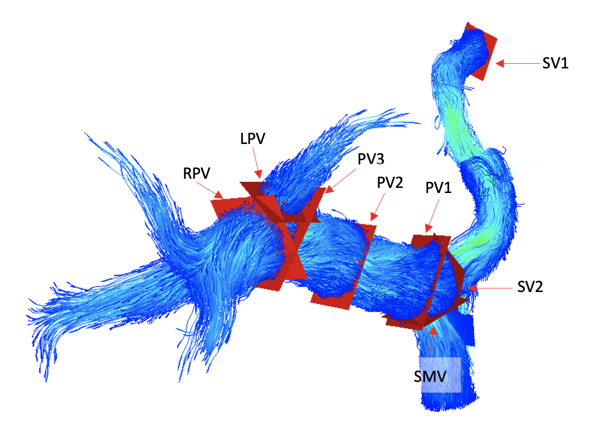
Figure 1: Standardized planes in the portal vein that were evaluated in this study:
- SV1 = Splenic Vein at the Splenic Hilum
- SV2 = Splenic Vein at the splenomesenteric confluence
- SMV = Superior Mesenteric Vein
- PV1 = Portal Vein at the splenomesenteric confluence
- PV2 = Mid Portal Vein
- PV3 = Portal Vein adjacent to the portal bifurcation
- RPV = Right Portal Vein
- LPV = Left Portal Vein
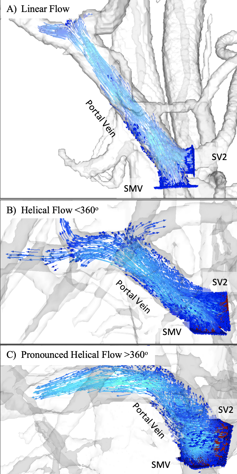
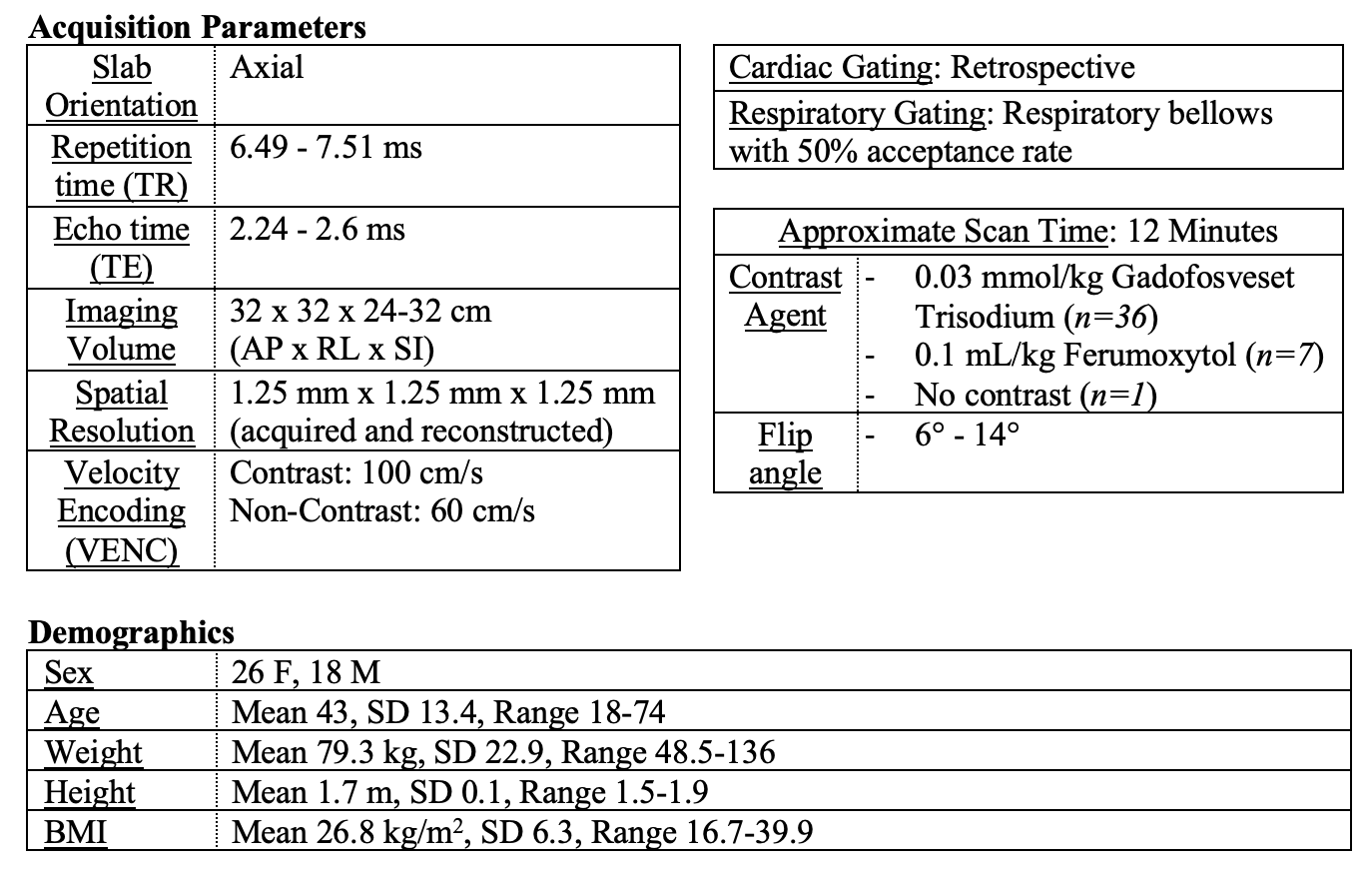
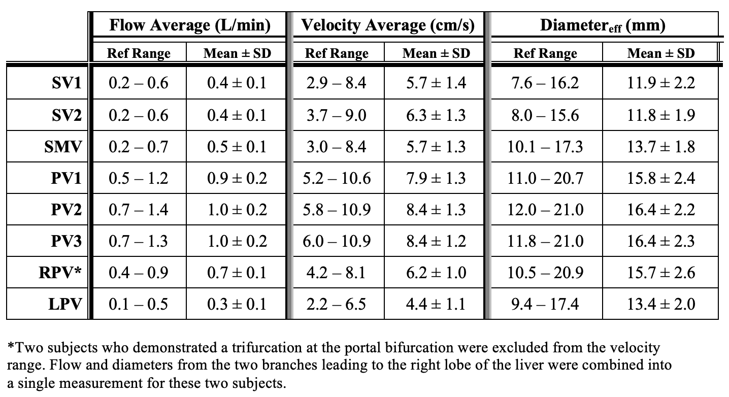
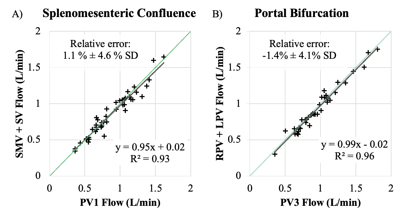
Figure 5: Quality assurance leveraged conservation of flow analysis in the splenomesenteric confluence and portal bifurcation. Green line in each chart is an equivalence line denoting 1:1 perfect conservation of flow.
A) Splenomesenteric Confluence: PV1 = Portal Vein at the splenomesenteric confluence; SMV = Superior Mesenteric Vein; SV2 = Splenic Vein at the splenomesenteric confluence
B) Portal Bifurcation: PV3 = Portal Vein at the portal bifurcation; RPV = Right Portal Vein, LPV = Left Portal Vein