5062
EPIc-reMIX, an Accelerated Routine Brain Imaging Protocol1Philips Healthcare, Gainesville, FL, United States
Synopsis
EPIMix is a multi-contrast EPI sequence developed which generates the standard brain imaging contrasts in 78 seconds. Studies using accelerated brain protocols like EPIMix and GoBrain have shown similar diagnostic performance to the routine clinical protocols. However, EPIMix suffers from EPI geometric distortions and like GoBrain is limited to 2D acquisitions. EPIc-reMIX, an accelerated brain imaging protocol, was created to leverage the speed of EPIMix and GoBrain while improving image quality and adding 3D-imaging capabilities.
Background
Recently, a new 1-minute full brain, multi-contrast EPI sequence called EPIMix was developed which generates T1-FLAIR, T2-weighted, T2-FLAIR, DWI, apparent diffusion coefficient, and T2*-weighted sequences in 78 seconds1. A prospective study of 103 patients comparing the EPIMix to the routine clinical protocol showed that the EPIMix had similar diagnostic performance (disease identification and categorization)2. Using a single scan capable of producing multiple image contrasts is also the foundation of synthetic MRI to allow for routine clinical brain exam in approximately 5-minutes3. These brain imaging approaches compliment earlier work that used an ultrafast 5-minute protocol, GoBrain, to obtain individual T1, T2, FLAIR, DWI, and T2* contrasts4. Based on these studies, extreme acceleration of brain protocols are achievable which results in comparable image quality and high diagnostic consistency4. Echo-planar imaging (EPI) used in EPIMix is a very fast imaging technique but suffers from EPI geometrical distortions and susceptibility artifacts that can reduce image quality. However, EPI correction (EPIC) provides improved image quality for fast EPI based scans by reducing image artifacts5. GoBrain sequences offer an advantage over EPIMix and synthetic MRI as it allows for acquisition of separate sequences so contrasts can be acquired in multiple imaging planes (sagittal and axial). However, all these approaches are still restricted to 2D-imaging. Newer acceleration techniques like compressed SENSE offer the potential to speed up image acquisitions even further, especially for 3D acquisitions. Here we present a combination of compressed SENSE and EPIC to produce a multi-contrast 1-minute brain protocol (figure 1).Teaching Point
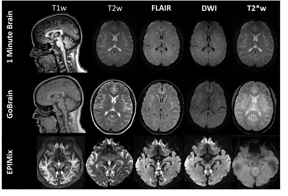
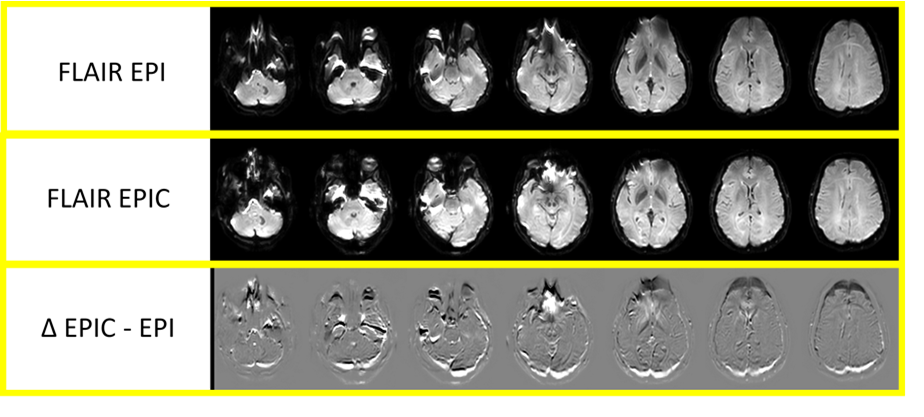
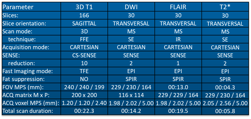
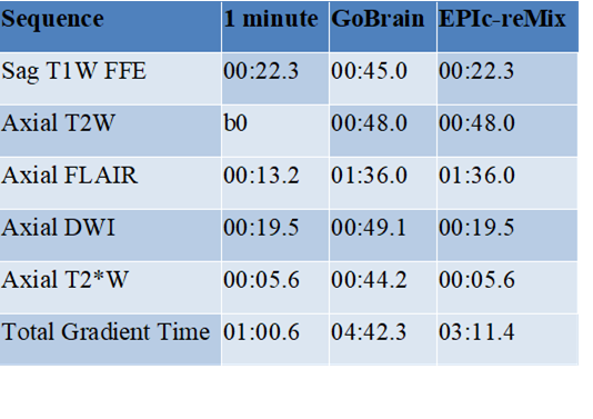
A 1-minute brain protocol was achieved by combining compressed SENSE acceleration and EPI based sequences (table 1). A magnetization-prepared 3D sagittal turbo field echo (TFE) with a compressed SENSE acceleration of 10 was used for the T1-weighted sequence. EPIC acquisition and post-processing were utilized to acquire T2-weighted, DWI, FLAIR and T2*-weighted contrasts to reduce geometric distortion. The b0 scan from the diffusion weighted sequence was used for the T2-weighted images. The GoBrain protocol was acquired to compare image quality (figure 1). Images were acquired on a Philips 3T Ingenia with 15 channel dS HeadSpine coil. EPIC also known as topup or blip-up and blip-down aims at reducing B0-induced distortions to achieve EPI scans with less geometrical distortion5. EPIC improves the geometrical match between EPI sequences and conventional brain imaging sequences like fast spin echo when compared to non-corrected EPI scans (figure 2). Through the combination of new acceleration techniques like compressed SENSE the acquisition time of 3D images can be drastically decreased while maintaining high signal to noise ratio (figure 3). Leveraging EPI based scans also allows for fast acquisitions with different imaging contrasts and image quality can be enhanced using EPIC post-processing.Summary
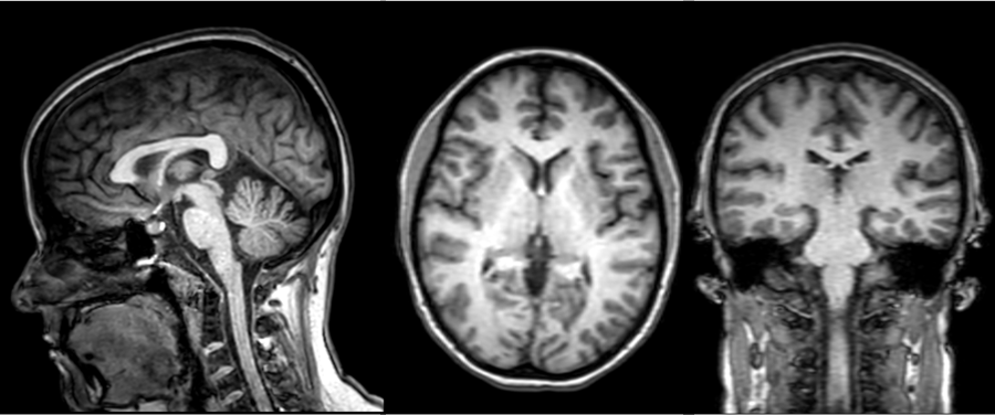
The 1-minute brain protocol generated the standard brain images with relevant image contrasts. 3D T1 TFE demonstrated better T1 contrast compared to the 2D T1 fast field echo (FFE) used in the GoBrain protocol. Moreover, the 3D T1 TFE allows for multiplanar reformats and imaging in any plane (figure 3). In the case post-contrast imaging is needed this would only require an additional 22 seconds of scan time to acquire full brain coverage. EPIC reduced geometric distortion for EPI based sequences compared to EPIMix images (figure 1). While the 1-minute protocol provided the necessary image contrasts, the dedicated axial T2-weighted sequence and FLAIR acquisitions in the GoBrain protocol demonstrated higher image quality. Based on these results we created EPIc-reMIX (table 2) which is a combination of the 1-minute and GoBrain protocols. Based on the research behind GoBrain and EPIMix, EPIc-reMix would provide a clinically viable 3-minute routine brain protocol utilizing commercially available imaging techniques. This type of accelerated protocol could also be used for certain patient populations (i.e. claustrophobic and pediatric) that cannot tolerate long examination times or as a screening protocol for time sensitive diagnoses like stroke6.Acknowledgements
No acknowledgement found.References
1. Delgado, Anna F., et al. "Diagnostic performance of a new multicontrast one‐minute full brain exam (EPIMix) in neuroradiology: A prospective study." Journal of Magnetic Resonance Imaging 50.6 (2019): 1824-1833.
2. Ryu, K. H., et al. "Clinical Experience of 1-Minute Brain MRI Using a Multicontrast EPI Sequence in a Different Scan Environment." American Journal of Neuroradiology 41.
3 (2020): 424-429. 3. Hagiwara, Akifumi, et al. "SyMRI of the brain: rapid quantification of relaxation rates and proton density, with synthetic MRI, automatic brain segmentation, and myelin measurement." Investigative radiology 52.10 (2017): 647.
4. Prakkamakul, Supada, et al. "Ultrafast brain MRI: clinical deployment and comparison to conventional brain MRI at 3T." Journal of Neuroimaging 26.5 (2016): 503-510.
5. Andersson, Jesper LR, Stefan Skare, and John Ashburner. "How to correct susceptibility distortions in spin-echo echo-planar images: application to diffusion tensor imaging." Neuroimage 20.2 (2003): 870-888.
6. Ha, Ji Young, et al. "One-Minute Ultrafast Brain MRI With Full Basic Sequences: Can It Be a Promising Way Forward for Pediatric Neuroimaging?." American Journal of Roentgenology 215.1 (2020): 198-205.
Figures