5075
Anxiety relaxation during MRI with a patient-friendly audiovisual system1Department of Radiology, Chiba University Hospital, Chiba, Japan, 2Diagnostic Radiology and Radiation Oncology, Graduate School of Medicine, Chiba University, Chiba, Japan, 3Department of Neurosurgery, National Hospital Organization Chiba Medical Center, Chiba, Japan, 4Research Center for Child Mental Development, Chiba University, Chiba, Japan
Synopsis
Many patients experience anxiety, not limited to claustrophobia, before magnetic resonance imaging (MRI) examination. Thus, to examine improvements to the environment during MRI examinations, this study employed a non-randomized controlled trial and evaluated whether a patient-friendly audiovisual system in the MR scanner room reduces patient anxiety. As a result, the patient-friendly audiovisual system compared to the traditional environment reduced anxiety in patients undergoing MRI examinations. Thus, this system can be used to provide a more patient-centered environment and may relieve the negative perceptions associated with MRI examinations.
Background
Claustrophobic reactions are a major obstacle in performing magnetic resonance imaging (MRI) examinations,as patients may be frightened and experience feelings of confinement or being enclosed [1]. Furthermore, many patients experience anxiety, not limited to claustrophobia, before MRI examination [2]. Thus, the improvement of the patient experience during MRI examination (e.g., reduction of noise and feeling of confinement) is essential [1]. A patient-friendly audiovisual (AV) system (Ambient Experience, Philips Healthcare) provides an AV imagery environment that features varying light conditions and individually selected visual wall displays with background sound effects [2]. A previous report investigated the effects of exposure to a similar ambient environment in the waiting area and patient rooms of a pediatric emergency department and demonstrated that passive distractions using ambient lighting can reduce pain and anxiety in patients [2]. However, the anxiety-reducing effects of such an ambient AV environment in MRI rooms have not yet been evaluated. We hypothesized that patient anxiety could be reduced through passive distraction using the patient-friendly AV system in the MR scanner room, and we investigated this hypothesis by performing a non-randomized controlled trial.Methods
Subjects: We randomly invited participants from outpatients who required brain MRI examinations in neurosurgery at Chiba University Hospital; these patients were told that they could choose to undergo an MRI examination using a patient-friendly AV system or the standard system. In total, 61 patients (28 women and 33 men; median age, 72 years [range, 25–86 years]) who underwent brain MRI between November 2018 and June 2020 were enrolled after obtaining verbal informed consent. To complete the MRI examination without affecting clinical practice, all patients who preferred a patient-friendly AV system were assigned to the AV group. Patients who indicated that that either system was acceptable were randomly assigned to the AV and control (using the standard system) groups. The study design is shown in Figure 1. Data acquisition: MRI was performed on a 3.0-T unit (Ingenia, Philips Healthcare) using a head-phased array coil. For all patients, the brain was scanned using the same head coil, and the scan time ranged from 15 to 20 min. The patient-friendly AV system featured a unique ambient light experience (Figure 2). The MRI room had overhead lighting that cycled through a spectrum of colors and intensities on a fixed schedule. On entering the gantry of the MRI scanner, patients could see the video projected on the light-emitting diode (LED) monitor via a prism mirror installed on the head coil. The progress of the examination was also displayed on the monitor, and the patient could confirm the end time. Furthermore, the patients could use headphones to listen to music corresponding to the projected image. In the standard system, the same MR scanner room as the patient-friendly AV system was used, with the lighting switched to white, the LED monitor turned off, and no prism mirror attached to the head coil (Figure 2). Evaluation: The claustrophobia scale (CS) has been demonstrated to have good psychometric properties and serves as a measure both in clinical practice and in other studies of fear and anxiety [3]. CS questionnaire was conducted before the MRI. In both groups, state anxiety using the State-Trait Anxiety Inventory (STAI) was assessed before and after the MRI examination (A-State-before and A-State-after MRI, respectively) [4]. Changes in A-State-before and A-State-after MRI were categorized as follows: 1) relieved high-state anxiety (A-State-before score was at the cut-off or higher and decreased to A-State-after score of under the cut-off), 2) remaining high-state anxiety (A-State-after score was at the cut-off or higher), and 3) stable easiness (Both A-State-before and A-State-after scores were under the cut-off). Statistical analyses: The Mann-Whitney U test was used to compare age, A-State-before, A-State-after, and A-Trait scores of patients between the AV and control groups. Fisher's exact test was used to compare sex, CS-positivity, high-state anxiety before MRI, and MRI experience of patients between the AV and control groups. Fisher's exact test was also used to evaluate the relationship between CS-positivity and high-state anxiety before MRI and between the A-State-before and A-State-after scores. Statistical significance was set at P < 0.05.Results
Of the 61 patients enrolled in this study, 39 were included in the AV group and 22 were included in the control group. Table 1 summarizes patient demographics. The median A-State-before score for all patients was 43 and was used as the cut-off for high-state anxiety. There was no bias between the AV and control groups in age, sex, CS-positivity, high-state anxiety before MRI, A-State-before, A-State-after, A-Trait, and MRI experience. Changes in A-State-before and A-State-after scores are presented in Figure 3, and the categorization of changes is summarized in Table 2. Significant differences were observed in the category compositions of the two groups (P = 0.048). A large proportion of patients (41.0%) in the AV group were categorized as having relieved high-state anxiety as compared to only 13.6% of patients in the control group.Conclusions
The patient-friendly AV system was shown to reduce anxiety in patients undergoing MRI examinations by providing a more patient-centered MRI examination environment. These findings may help ameliorate the negative perceptions associated with MRI examinations.Acknowledgements
The authors would like to thank the staff of the Department of Radiology at Chiba University Hospital for helping with the MRI examination.References
- Meléndez JC, McCrank E. Anxiety-related reactions associated with magnetic resonance imaging examinations. JAMA 1993;270:745–7.
- Katz RC, Wilson L, Frazer N. Anxiety and its determinants in patients undergoing magnetic resonance imaging. J Behav Ther Exp Psychiatry 1994;25:131–4.
- Robinson PS, Green J. Ambient versus traditional environment in pediatric emergency department. HERD 2015;8:71–80.
- Ost LG. The claustrophobia scale: a psychometric evaluation. Behav Res Ther 2007;45:1053–64.
- Spielberger CD, Gorsuch RL, Lushene R, Vagg PR, Jacobs GA. Manual for the State–Trait Anxiety Inventory (form Y). Palo Alto, CA: Consulting Psychologists Press; 1983.
Figures
Figure 1. Flowchart of the study design.
The diagram depicts the non-randomization and randomization procedures, patient flow, and data analysis.
AV, audiovisual; CS, claustrophobia scale; CS-Anxiety, anxiety subscale; CS-Avoidance, avoidance subscale; MRI, magnetic resonance imaging; STAI, State-Trait Anxiety Inventory; A-State-before, state anxiety before MRI; A-State-after, state anxiety after MRI; A-Trait, trait anxiety.
Figure 2. Patient-friendly AV system.
The overhead lighting was vivid and the video was projected on the wall (a). In addition, music corresponding to the video was played through the speakers in the room as well as through headphones. The same video was projected on the light-emitting diode (LED) monitor on the wall on the head side (b), and the patient could look at video through a prism mirror installed on the head coil (c). In the standard system had simple overhead lighting (d), the patient could not see the video or listen to the music (e).
Table 1. Patient demographics.
CS-positive, positive on the claustrophobia scale. Age, Score of A-State-before, A-State-after, and A-Trait were shows median [25th percentiles, 75th percentiles]. CS-positive, high-state anxiety before MRI, and MRI experience are depicted as “number (percentage)”.
*Compared using the Mann-Whitney U test.
**Compared using the Fisher's exact test.
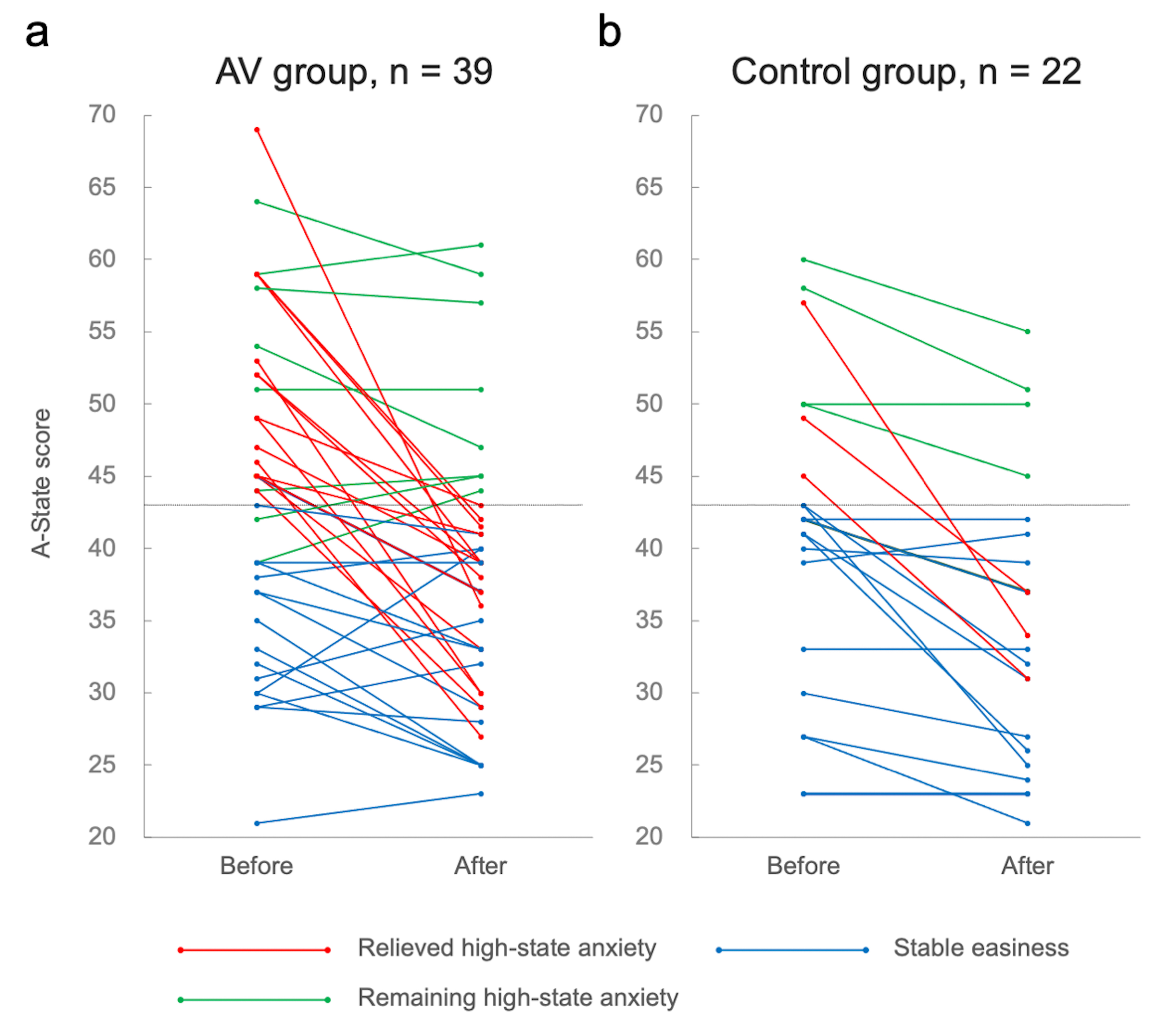
Figure 3. Changes in A-State before and after MRI in the AV group (a) and the control group (b).
The cut-off for high-state anxiety was a score of 43 (horizontal lines).