5060
Radial-Cartesian K-Space Acquisition for Consistent High-Resolution and High-Quality Knee MRI
Sajith Rajamani1, Harsh Kumar Agarwal1, Jeremy Heinlein2, and Ramesh Venkatesan1
1GE Healthcare, Bangalore, India, 2GE Healthcare, Waukesha, WI, United States
1GE Healthcare, Bangalore, India, 2GE Healthcare, Waukesha, WI, United States
Synopsis
Knee MRI is the most common Musculo-skeletal MRI [1]. It is a non-invasive tool for the evaluation of disorders such as meniscal, ligamental, soft tissue, bone and bone marrow injuries and abnormalities. Acquisition of a high-resolution and high-quality MRI is key to boost the confidence of the radiologist in diagnosis. In this manuscript we have presented how radial MRI, commercially referred to as PROPELLER (GE)/ BLADE (Siemens)/ MULTIVANE (Philips) etc. parameters, patient positioning and immobilization can be adapted for the available hardware such as receiver coil to yield high-resolution and high-quality knee MRI.
Introduction
Knee MRI is a non-invasive imaging technique for the evaluation of meniscal, ligamental, Soft tissue, bone and bone marrow injuries or abnormalities [2] For detecting specific meniscal and chondral lesions, MR Arthrography is the gold standard, but it is invasive.[3] According to the American College of Radiology (ACR) guidelines, the field of view (FOV) for standard knee sequences should be less than or equal to 16cm except for the evaluation of extended detected or suspected abnormality.[2] An optimized high resolution, high quality mri knee protocol is important to enhance the diagnostic confidence. Dedicated coils are needed for knee MRI; however, many centers may not have it and they will use medium or large surface coils. Sometime for the ease of workflow, large field of view coils such as torso coil is used for data acquisition of knee. Typically, Cartesian sampling is used as a readout method. Use of smaller field of view with respect to the size of receiver coil increases the probability of wrap-around or aliasing artifact.[4] Some of the tricks to address the issue of ghosting artifact arising from pulsation of popliteal artery as well as the aliasing is to change the phase encoding direction to superior-inferior with phase oversampling in case of sagittal plane. Slight Knee flexion, wherever tolerable or feasible inside the coil, can be used during positioning with Cartesian readout for avoiding few of the artifacts such as Annefact. But the intended clinical evaluation dictates the knee positioning during the scanning. We present data acquisition with radial k-space sampling using PROPELLER (GE) which is commercially available in both 1.5T and 3T scanners to be utilized to generate high resolution and high-quality knee MRI.Methods and Results
The study was conducted per a protocol that was reviewed and approved by an IRB for data acquisition on volunteers for experimental purposes including on commercial MRI scanner using commercially released software. The data was acquired using GE Signa HDXT 1.5T MRI scanner using HD T/R Knee coil over the right knee. We break our suggestion among two groups: High Resolution: For sagittal and coronal planes, popliteal artery pulsation forces us to choose phase encoding along the superior-inferior direction. In this case, without phase oversampling, wrap-around artifact is observed in cartesian readout but, Radial mri does not have wrap-around or ghosting artifact from popliteal artery pulsation hence, high-resolution artifact free knee data can be acquired. (Figure1) High Quality: High quality has two parts, SNR and artifacts. For SNR, radial sampling oversamples the central k-space therefore it is most optimal for SNR per unit time. For the same acquisition time when compared to Cartesian (Figure1 a & b), PROPELLER (Figure1c) has improved image quality and motion robustness as the center k-space is acquired again and again as shown in Figure 2. The repeated acquisition of central k-space is used to suppress any motion artifacts that appear due to insufficient immobilization.[5] Annefact [6] (commonly also called the cusp or star artifact), which is not present in the dedicated knee coil but can become a significant problem in the non-dedicated coils such as large surface coil and body coil. The removal of annefact can be done by several methods.[7] We observed the use of Nex=2 with RF phase cycling is the most common way to remove annefact but it is associated with doubling of the scan time which may not otherwise be required such that sufficient snr present in the image. Radial does not have this issue and can be used for this purpose. Figure 3 shows the data acquired using the extreme case large FOV coil which is the body coil. annefact is not present in PROPELLER readout.Conclusion and Discussion
We observed that Radial k-space sampling shows artifact free images without phase oversampling. Scan time of radial sampling is comparatively longer but the provision to apply parallel imaging techniques such as ARC (GE), GRAPPA (Siemens) reduces the scan time to half with acceleration factor (R) of 2. On top of that, recent advances in acceleration such as compressed sensing, which is a method of incoherent subsampling rather than uniform subsampling can be incorporated into the radial sampling with advanced deep learning-based reconstruction to produce high quality images.[8] Optimizing the knee joint protocol with radial readout can be useful for generating high-resolution, high quality knee images to enhance diagnostic confidence.Acknowledgements
No acknowledgement found.References
- Vanhoenacker F, De Vos N, Van Dyck P. Common mistakes and pitfalls in Magnetic Resonance Imaging of the knee. J Belgian Society of Radiology 2016; 100(1): 99: 1–17.
- ACR–SPR–SSR practice parameter for the performance and interpretation of magnetic resonance imaging (MRI) of the Knee.Res 6-2015
- Mathieu L, Bouchard A, Marchaland JP, et al. Knee MR-arthrography in assessment of meniscal and chondral lesions. Orthop Traumatol Surg Res 2009; 95:40–47
- Pusey E, Yoon C, Anselmo ML, Lufkin RB. Aliasing artifacts in MR imaging. Comput Med Imag Graphics 1988;12:219-224
- James G. Pipe, Wende N. Gibbs, Zhiqiang Li, et al., Revised motion estimation algorithm for PROPELLER MRI. Magnetic Resonance in Medicine. 2014;72:430–437
- Artasona LM. Annefact artifact in MRI. Blog at El Baul Radiologicó 16 Mar 2015
- Kim JK, White LM, Hinks RS, King KF. The FSE cusp artifact: a phase wrap-in artifact seen on routine clinical MR images of the knee. In: ISMRM Abstracts, Philadelphia, PA, 1999. p 1033
- Lebel, R. M. Performance characterization of a novel deep learning-based MR image reconstruction pipeline. ArXiv abs/2008.06559 (2020)
Figures
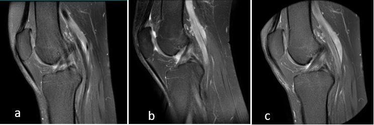
Figure 1: Sagittal Proton Density with Fatsat Cartesian readout with
Anterior-Posterior phase encoding direction showing ghosting artifact from
popliteal artery pulsation (a), Same with Superior-Inferior phase encoding
showing aliasing artifact (b), Radial readout using PROPELLER technique showing
no artifact (c)
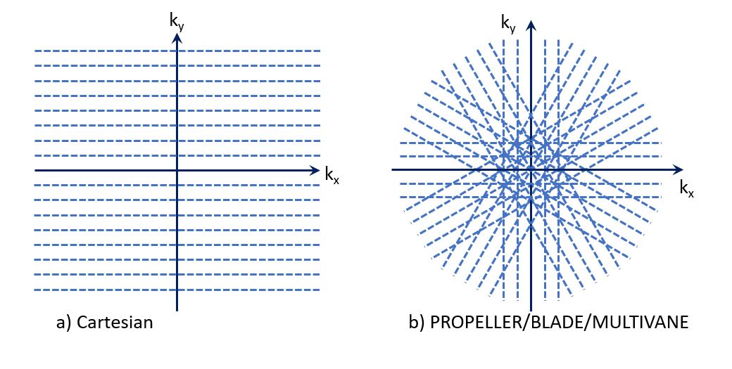
Figure 2: Cartesian k-space sampling (a), Radial k-space filling with center over-sampling
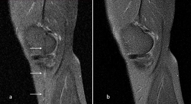
Figure 3: Sagittal PD FS Cartesian readout using Body coil showing
annefact artifact. (Arrows) (a), Radial sampling using PROPELLER doesn’t show
any annefact artifact. (b)
DOI: https://doi.org/10.58530/2022/5060