5057
Proposal for an MRI protocol for cerebrospinal flows1Radiology, CHU amiens picardie, AMIENS, France, 2CHU amiens picardie, AMIENS, France
Synopsis
Flow MRI is useful for imaging flow from the LCS and blood in the craniospinal system. Currently, at the Amiens University Hospital, we are performing these sequences in clinical routine in patients with pathologies involving potential alterations in cerebral hemohydynamics such as hydrocephalus, intracranial hypertension, Arnold Chiari's malformation. This poster presents the specifics of the MRI flow sequence applied to brain flows.
METHODS
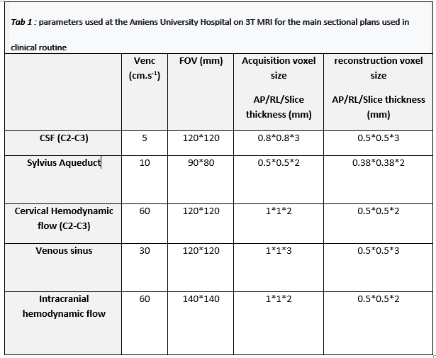
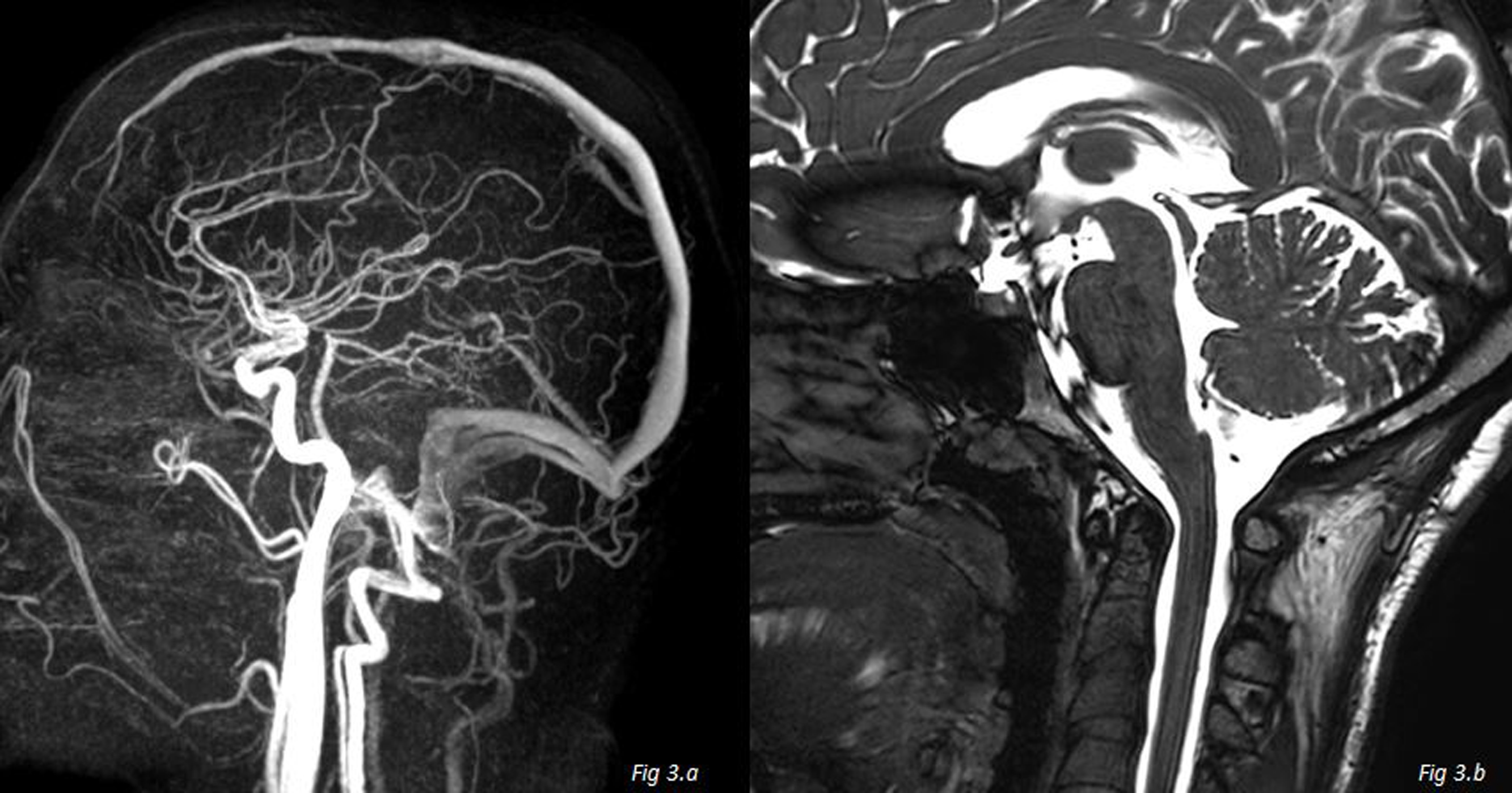
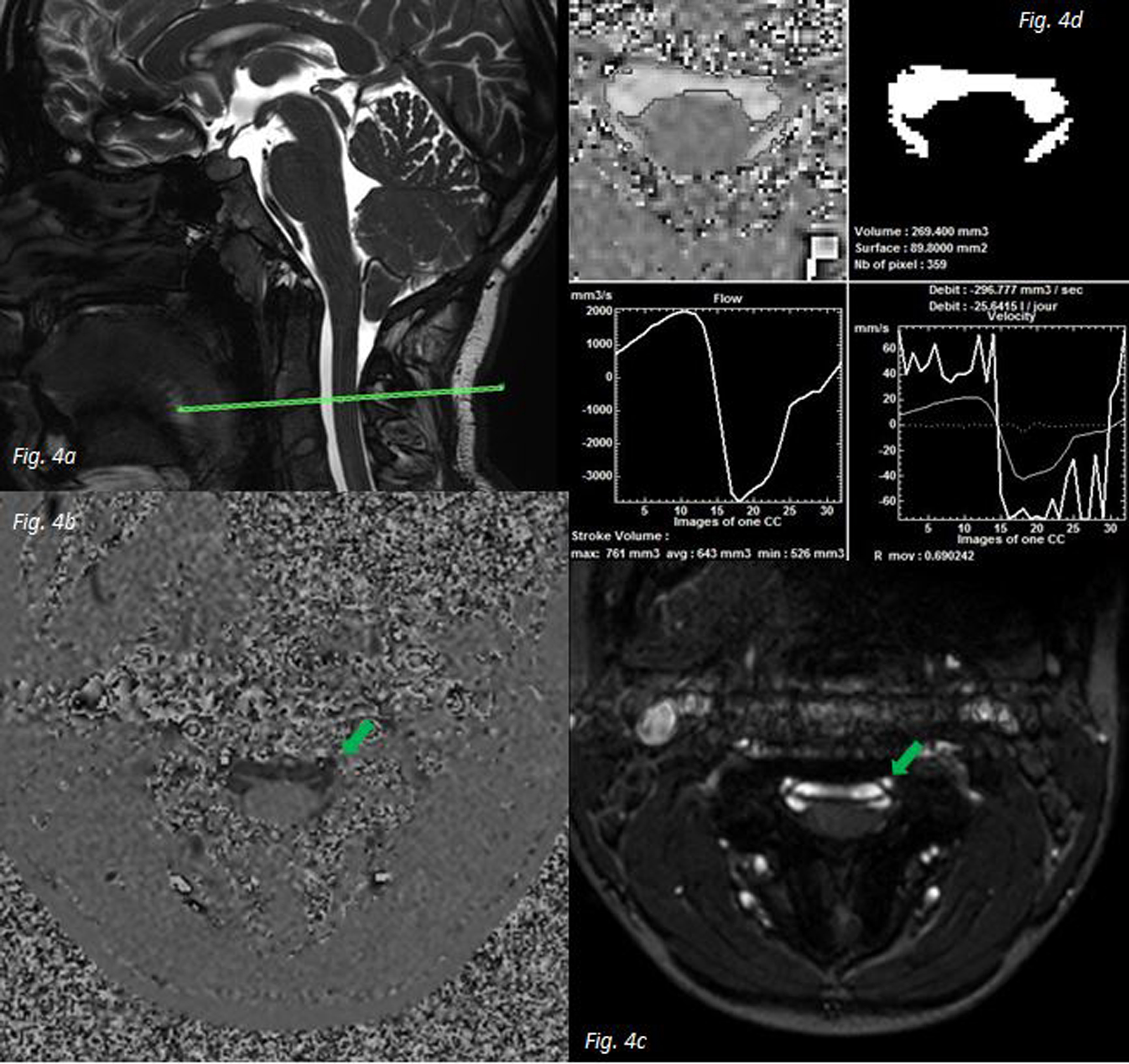
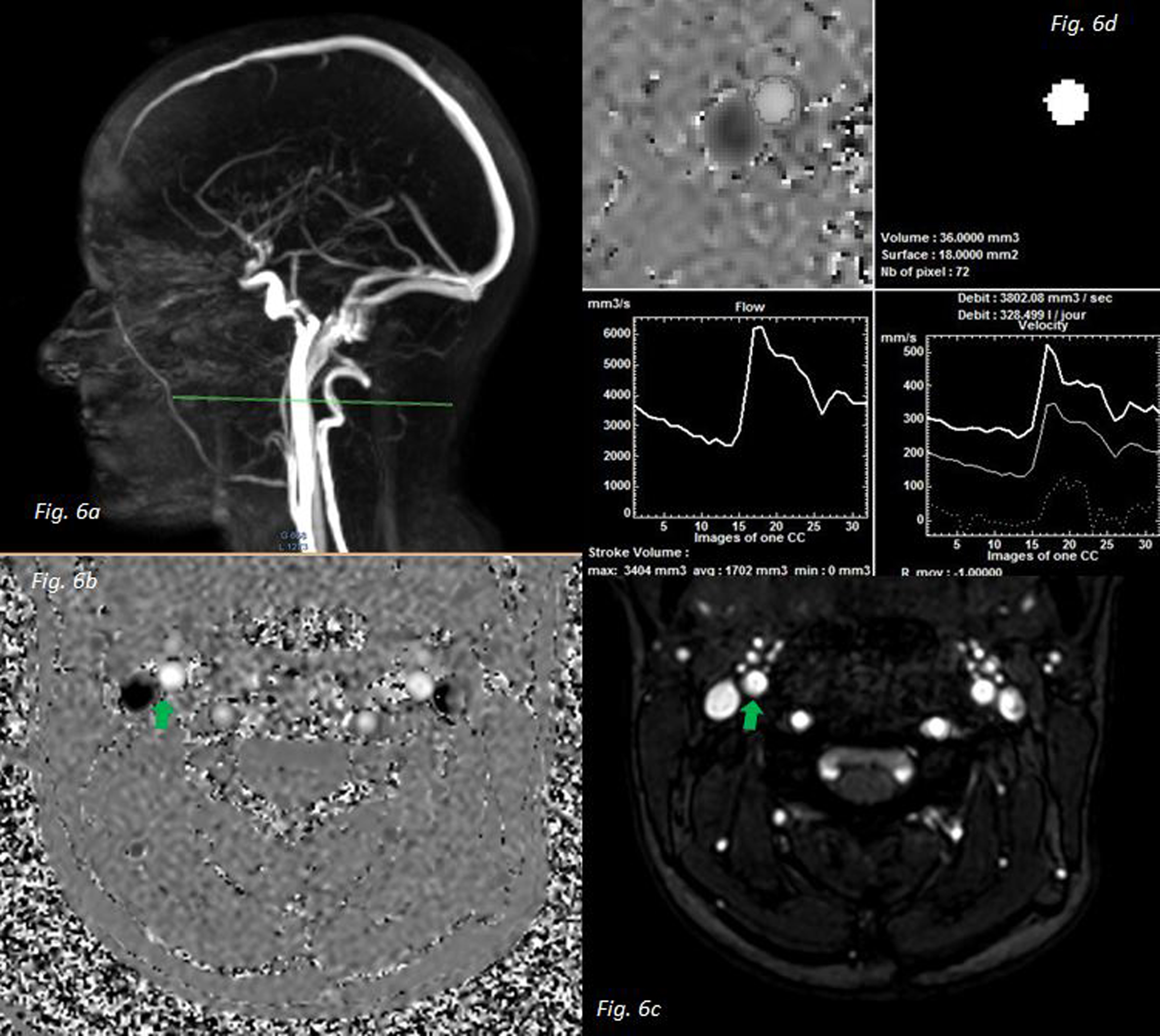
Flow MRI allows a study of the flow of a fluid or the speed of movement of a tissue in an ROI (region of interest) during a cardiac cycle. Associated with morphological images, they allow a precise and dynamic study of the CSF as well as of the intra-cerebral arteriovenous system, making it possible to orient the diagnostic and the origin of a disease due to a pressure abnormality of the cerebrospinal system. The MRI technologist must know how to make, parameterize and position the cut planes of the flow sequences correctly in order to provide the best possible information to the radiologist doctor.Patient position: The patient should be installed in the supine position with good head support to avoid movement artefacts, hearing protectors to reduce sound noise and alarm bell in the hand when needed to contact the technologist.Cardiac synchronization: ECG or PPU are possible, thirty-two cycles are reconstructed to represent an average cardiac cycle. The quality of the cardiac cycle influences the quality of the exam.Placement of sections: The section should be placed perpendicular to the vessel being studied. To facilitate placement, the 3D PCA (phase contrast angiography) and BFFE (gradient echo with balanced gradients) or Fiesta sequences are performed upstream to visualize the vascular tree and the LCS respectively, without injection of contrast product. Encoding speed (Venc): The encoding speed is the maximum speed assumed for the area of interest. It must be adapted to each vessel and is inherent to the pathology sought. The speed translates into a smooth cut section of highly contrasted black or white pixels. These pixels are used to perform measurements in the processing software. If the Venc is too low there will be an aliasing phenomenon Table 1 lists the main section plans used in clinical routine at Amiens University Hospital with the technical parameters of Philips 3T MRI. Note that for all the sequences the number of excitation is 1, the acceleration factor (SENSE) of 1.5 on the right-left, the TE and TR are at the minimum values, the flip angle at 30 and there is has 32 reconstructed heart phases. CONCLUSION Non-invasive and relatively rapid, magnetic resonance flow imaging is a real tool in the management of patients with SCL pathology. By providing information on the morphology and flow rates of the blood and the LCS, it helps guide diagnosis and therapy. This is an essential examination for these pathologies at the Amiens University Hospital.The MRI technologist plays a major role in the management by performing the MRI examination. Technical mastery of patient installation, sequence settings, the relevant choice of sequences (depending on the pathology) and image analysis are essential for a good quality examination.CONCLUSION
Non-invasive and relatively rapid, magnetic resonance flow imaging is a real tool in the management of patients with SCL pathology. By providing information on the morphology and flow rates of the blood and the LCS, it helps guide diagnosis and therapy. This is an essential examination for these pathologies at the Amiens University Hospital.The MRI technologist plays a major role in the management by performing the MRI examination. Technical mastery of patient installation, sequence settings, the relevant choice of sequences (depending on the pathology) and image analysis are essential for a good quality examination.Acknowledgements
No acknowledgement found.References
[1] Kastler B. Comprendre l’IRM : Manuel d’auto-apprentissage. 7e édition. Elsevier Masson; 2011
[1] Lokossou A. Etude de la plasticité liquidienne du système cranio-spinal : de la psychologie à la pathologie [thèse : Med]. Amiens : Université Picardie Jules Verne ; 2019
[1] Balédent O et al. Cerebrospinal fluid dynamics and relation with semiautomated cerebrospinal fluid segmentation. Invest Radiol. 2001
[1] Pagé G. Quantification et caractérisation des écoulements sanguins dans l’arborescence vasculaire de la région cervico-faciale par IRM de flux : évaluation et application [thèse : Phys]. Amiens : Université Picardie Jules Verne ; 2016 https://www.researchgate.net/publication/11848841_Cerebrospinal_Fluid_Dynamics_and_Relation_with_Blood_Flow_A_Magnetic_Resonance_Study_with_Semiautomated_Cerebrospinal_Fluid_Segmentation http://www.theses.fr/2016AMIE0041
Figures