5066
New reference ADC data for the normal brain with distortion correction in TSE- and EPI-DWI1Department of Radiological Technology, Faculty of Health and Welfare, Tokushima Bunri University, Sanuki-city, Japan, 2Department of System Control Engineering, Graduate School of Engineering, Tokushima Bunri University, Sanuki-city, Japan, 3Division of Health Sciences, Graduate School of Medical Sciences, Kanazawa University, Kanazawa, Japan, 4Otsu City Hospital, Otsu, Japan, 5Division of Clinical Radiology Service, Kyoto University Hospital, Kyoto, Japan, 6Department of Radiology, The University of Tokyo Hospital, Tokyo, Japan, 7Department of Radiology, Osaka Red Cross Hospital, Osaka, Japan, 8Department of Radiological Sciences, International University of Health and Welfare, Ohtawara, Japan, 9Division of Health Sciences, Graduate School of Medical Sciences, Kanazawa University., Kanazawa, Japan
Synopsis
We performed echo planar imaging (EPI) and turbo spin echo (TSE) diffusion-weighted imaging (DWI) using magnetic resonance imaging to obtain basic clinical data of the apparent diffusion coefficient (ADC) in 26 parts of normal brains and compared the datasets using our retrospective distortion correction technique. The ADC was significantly higher measured by EPI-DWI than by TSE-DWI. The signal-to-noise ratio of EPI-DWI was significantly higher than that of the TSE-DWI. Care must be taken when measuring ADCs near the base of the skull, such as the brain stem, where the SNR of the imaging technique is likely to decrease or distort.
Background
Diffusion-weighted imaging (DWI) in magnetic resonance imaging (MRI) is useful for acute cerebral infarction [1] and tumor characterization [2]. The apparent diffusion coefficient (ADC) is useful for distinguishing between restricted diffusion in the tissue and tissue perfusion in DWI [3, 4] for acute stroke [5], multiple sclerosis [6], and tumor assessments [7]. To evaluate ADC in the lesion area, normal values should be recognized. Therefore, normal values of ADC in each part of the brain have been reported [8]. However, echo planar imaging (EPI)-DWI tends to have artifacts (e.g., geometric distortion and susceptibility artifacts) due to inhomogeneous magnetic field and line phase discrepancies in k-space, which affects the ADC [9]. When the diffusion gradients are switched on and off, the time-varying magnetic field results in current induction (i.e., eddy currents), which lead to inhomogeneity in the magnetic field [9]. Therefore, fast spin echo (FSE)-DWI, which has advantage for the inhomogeneity in the magnetic field, is used. [10] Furthermore, comparisons of the ADCs between EPI-DWI and FSE-DWI have been reported [11-13], and the results from the different studies showed no significant difference [11], no significant difference except for the tongue in the oral cavity [12], and higher lung lesion values in TSE-DWI than in EPI-DWI [13]. Therefore, it is important to recognize the difference between the EPI- and FSE-ADC; however, a problem is that the positions of the regions of interest (ROIs) do not always match when comparing these DWIs due to image distortion. The normal data, as a reference, may be required to confirm an abnormality. Measuring ADCs in EPI- and TSE-DWI with our retrospective distortion correction technique[14] can provide reference clinical data acquisition. Therefore, ADCs in various parts of the brain were measured by EPI- and TSE-DWI and reference normal data were then extracted and compared.Methods
The study received approval from the Institutional Review Board. The normal brains of 32 patients who underwent health check were scanned on a 1.5-T MRI instrument using EPI- and TSE-DWI (Fig. 1). Distortion was corrected by (1) segmentation: the b0 images were segmented based on the plural threshold values; (2) edge detection: the edge was detected in the images obtained in step (1); (3) non-rigid image registration: non-rigid image registration using Demons algorithm was achieved between the b0 images of EPI-DWI and TSE-DWI, thereby, creating a displacement field; (4) image warp: the displacement field was applied to the b1000 image to warp. MATLAB numerical analysis software (version 2018a; MathWorks, Inc., Massachusetts, USA) was used for the distortion correction. Twenty-six parts of the brain (Pons, Middle Cerebellar Peduncle [MCP], Dentate Nucleus [DN], Pedunculus Cerebri [PC], Hippocampus, Temporal Lobes [TL], Tegmentum of Midbrain [TM], Genu of the Corpus Callosum [GCC], Putamen, Globus Pallidus [GP], Thalami, Occipital Lobes [OL], Splenium of the Corpus Callosum [SCC], Frontal Deep White Matter [FDWM], Parietal Deep white Matter [PDWM]) were measured from the images of b0 and b1000. ROIs were set by two radiological technologists with 18 and 20 years of experience in MRI. Based on the consensus of both technologists, the ROIs were carefully set while referring to b0. The ROIs of EPI- and TSE-DWI were carefully measured at the same location of the same size so as not to contaminate other tissues, such as cerebrospinal fluid (CSF) (Fig. 2) The ROI volumes ranged from approximately 14 to 52 mm^2 (average, 29.8 ± 11.8 mm^2) The software used was ImageJ v.1.52 (National Institute of Health, Bethesda, MD, USA) for signal intensity (SI) measurement. ADCs were calculated according to the following formula: ADC = –ln (S1000/S0) / (b1000–b0), where S1000 is the signal value when the b-value is 1000 s/mm2 (b1000), and S0 is the signal value when the b-value is 0 s/mm2 (b0). The signal-to-noise ratio (SNR) of the cerebrospinal fluid was measured to identify the cause of the difference between the two sequences. These were compared using Wilcoxon paired rank test (P = 0.05). EZR software (version 1.42; Jichi Medical University Saitama Medical Center, Saitama, Japan) was used for statistical analysis.Results
There was a significant difference in the ADC results, especially in the brain stem, between the two imaging sequences, with the ADC from EPI-DWI significantly higher than the ADC from TSE-DWI (P < 0.05). ADC showed high TSE-DWI in some parts of the brain, such as in globus pallidus and hippocampus; however, the difference was slight. (Fig. 2 and 3) The median (interquartile range [IQR]) values for SNRs were as follows: EPI-DWI Right, 35.06 (21.49); Left, 40.19 (28.85); FSE-DWI Right, 25.85 (18.30); and Left, 31.46 (17.32). The Wilcoxon signed-rank test showed that the SNR was significantly higher for EPI-DWI than for TSE-DWI (P < 0.01)Conclusion
ADCs in various parts of the brain were measured by EPI- and TSE-DWI, and basic normal data were extracted and compared. Care is needed when measuring ADCs near the base of the skull, such as the brain stem, where the SNR is likely to decrease or distort.Acknowledgements
Nothing in particular.References
[1] Van Everdingen KJ, van der Grond J, Kappelle LJ, Ramos LM, Mali WP, Diffusion-weighted magnetic resonance imaging in acute stroke, Stroke. 29 (1998) 1783–1790.
[2] Barajas RF, Cha S, Metastasis in Adult Brain Tumors, Neuroimaging Clin. 26 (2016) 601–620.
[3] Le Bihan D, Breton E, Lallemand D, Grenier P, Cabanis E, Laval-Jeantet M, MR imaging of intravoxel incoherent motions: application to diffusion and perfusion in neurologic disorders, Radiology. 161 (1986) 401–407.
[4] Le Bihan D, Breton E, Lallemand D, Aubin ML, Vignaud J, Laval-Jeantet M, Separation of diffusion and perfusion in intravoxel incoherent motion MR imaging, Radiology. 168 (1988) 497–505.
[5] Warach S, Chien D, Li W, Ronthal M, Edelman R, Fast magnetic resonance diffusion-weighted imaging of acute human stroke, Neurology. 42 (1992) 1717–1723.
[6] Roychowdhury S, Maldjian JA, Grossman RI, Multiple sclerosis: comparison of trace apparent diffusion coefficients with MR enhancement pattern of lesions, Am J Neuroradiol. 21 (2000) 869–874.
[7] Hayashida Y1, Hirai T, Morishita S, Kitajima M, Murakami R, Korogi Y, Makino K, Nakamura H, Ikushima I, Yamura M, Kochi M, Kuratsu JI, Yamashita Y, Diffusion-weighted imaging of metastatic brain tumors. comparison with histologic type and tumor cellularity, Am J Neuroradiol. 27 (2006) 1419–1425.
[8] Naganawa S, Sato K, Katagiri T, Mimura T, Ishigaki T, Regional ADC values of the normal brain: differences due to age, gender, and laterality, Eur Radiol. 13 (2003) 6–11.
[9] Le Bihan D, Poupon C, Amadon A, Lethimonnier F. Artifacts and pitfalls in diffusion MRI. J Magn Reson Imaging. 24 (2006) 478–488.
[10] Baltzer PA, Renz DM, Herrmann KH, Dietzel M, Krumbein I, Gajda M, Camara O, Reichenbach JR, Kaiser WA, Diffusion-weighted imaging (DWI) in MR mammography (MRM): clinical comparison of echo planar imaging (EPI) and half-Fourier single-shot turbo spin echo (HASTE) diffusion techniques, Eur Radiol. 19 (2009) 1612–1620.
[11] Mikayama R, Yabuuchi H, Sonoda S, Kobayashi K, Nagatomo K, Kimura M, Kawanami S, Kamitani T, Kumazawa S, Honda H, Comparison of intravoxel incoherent motion diffusion-weighted imaging between turbo spin-echo and echo-planar imaging of the head and neck, Eur Radiol. 28 (2018) 316–324.
[12] Hirata K, Nakaura T, Okuaki T, Kidoh M, Oda S, Utsunomiya D, Namimoto T, Kitajima M, Nakayama H, Yamashita Y, Comparison of the image quality of turbo spin echo- and echo-planar diffusion-weighted images of the oral cavity, Medicine (Baltimore). 97 (2018) e0447.
[13] Wan Q, Lei Q, Wang P, Hu J, Zhang T, Yu D, Li X, Liang C, Intravoxel incoherent motion diffusion-weighted imaging of lung cancer: comparison between turbo spin-echo and echo-planar imaging, J Comput Assist Tomogr. 44 (2020) 334–340.
[14] Takatsu Y, Sagawa H, Nakamura M, Suzuki Y, Miyati T, Novel distortion correction method for diffusion-weighted imaging based on non-rigid image registration between low b value image and anatomical image, Magn Reson Imaging. 57 (2019) 277–284.
Figures
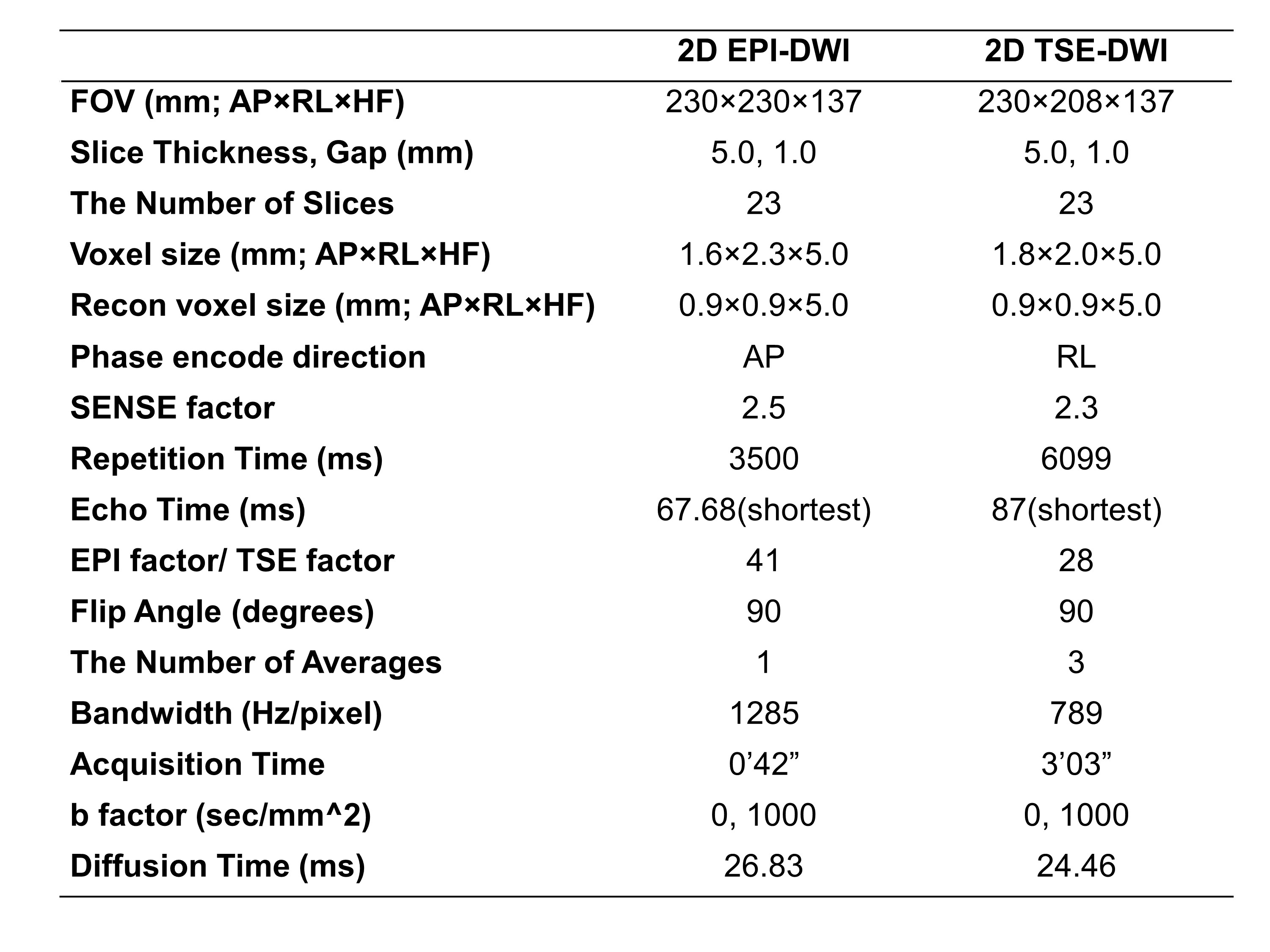
Sequence parameters;
AP, anterior-posterior direction; DWI, diffusion weighted image; EPI, echo planar imaging; HF, head-foot direction; SENSE, sensitivity encoding; TSE, turbo spin echo
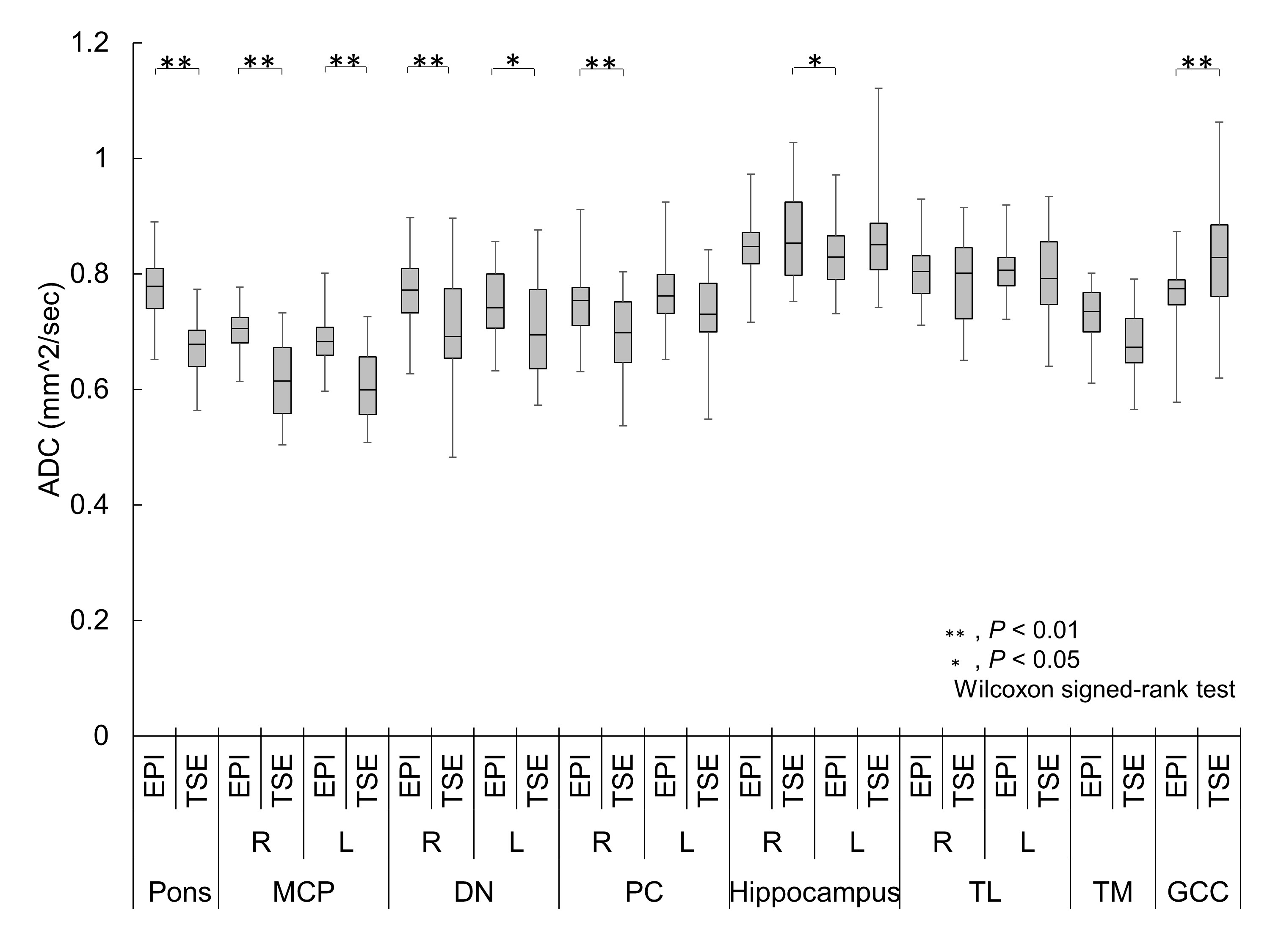
ADC measurement results 1;
DN, Dentate Nucleus; GCC, Genu of the Corpus Callosum; MCP, Middle Cerebellar Peduncle; PC, Pedunculus Cerebri; TL, Temporal Lobes, TM, Tegmentum of Midbrain
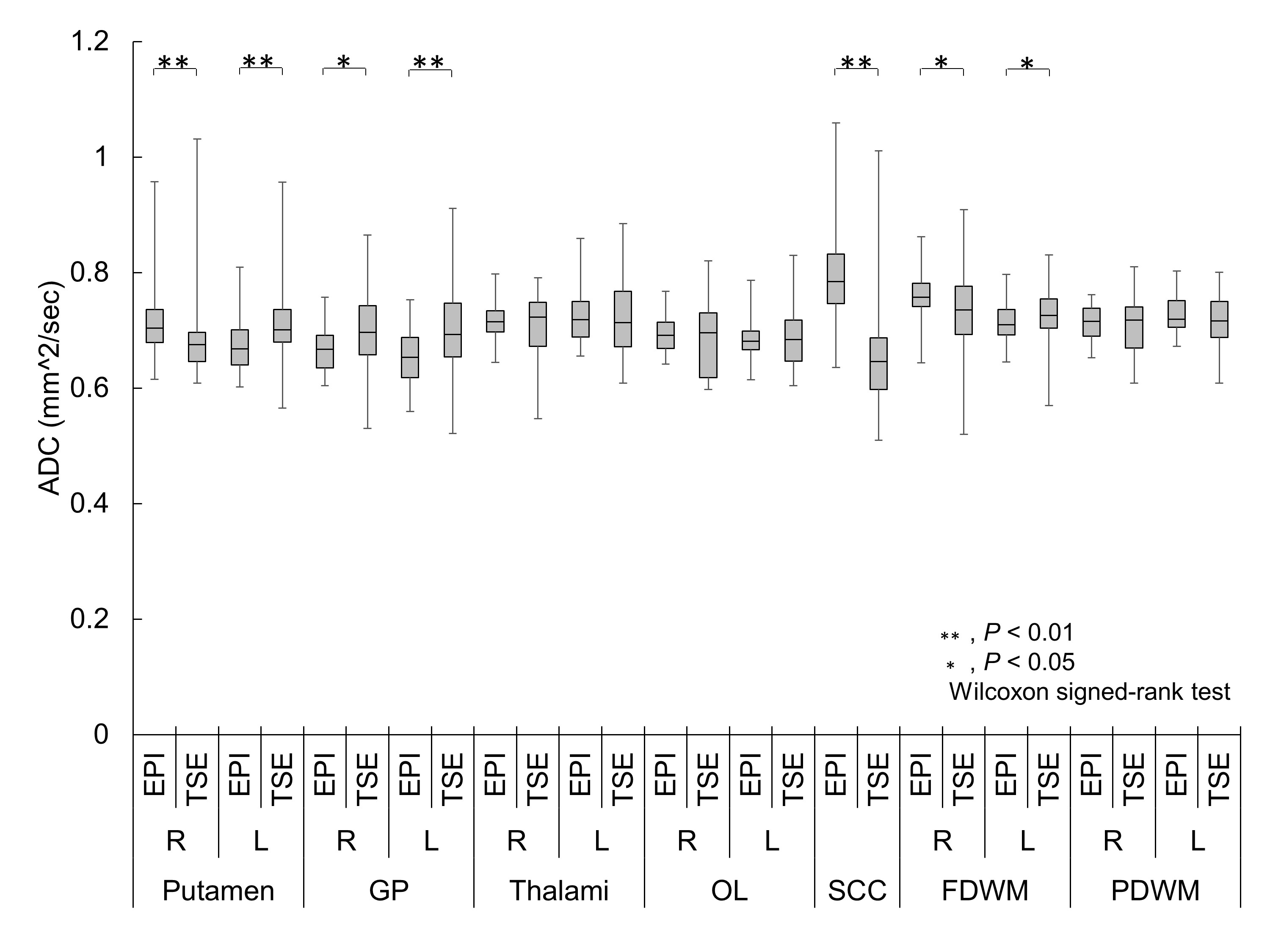
ADC measurement results 2;
FDWM, Frontal Deep White Matter; GP, Globus Pallidus; OL, Occipital Lobes; PDWM, Parietal Deep white Matter; SCC, Splenium of the Corpus Callosum