Preclinical Imaging of Myocardial Viability
1University College London, London, United Kingdom
Synopsis
Cardiac imaging has revolutionized our ability to diagnose heart disease, quantify mechanisms of pathology, and determine optimal therapy in patients. Current techniques can interrogate structure, contractility and metabolism but struggle to directly quantify one of the most important determinants of patient morbidity, myocardial viability. This presentation will provide an overview of current clinical and preclinical methods for evaluating cardiac function and viability in acute myocardial infarction, then discuss some emerging technologies that could improve our ability to monitor disease progression and response to experimental therapies.
Summary
Early measurements of tissue viability after myocardial infarction are essential for accurate diagnosis and treatment planning but are challenging to obtain. The presentation will cover the advantages, disadvantages and practical aspects of a range of established preclinical MR and non-MR imaging methods which can evaluate cardiac function, including· Myocardial contractility
· Dobutamine stress imaging
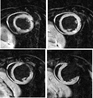
· Late gadolinium-enhanced MRI
· T1 mapping and extracellular volume fraction measurements
· 18F fluorodeoxyglucose positron emission tomography (FDG-PET)
· 99mTc MIBI and 201TI single-photon emission computed tomography (SPECT)
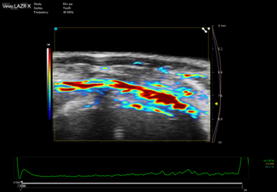
Recent developments in myocardial viability imaging using manganese-enhanced MRI and photoacoustic tomography will also be presented and the potential clinical translation of these novel approaches will be discussed.
Acknowledgements
D.J.S. was supported by British Heart Foundation Intermediate and Senior Basic Science Research Fellowships (FS/15/33/31608, FS/SBSRF/21/31020), the BHF Centre for Regenerative Medicine RM/17/1/33377, the MRC MR/R026416/1, and the Wellcome Trust 212937/Z/18/Z.References
Jasmin NH, Thin MZ, Johnson RD, Jackson LH, Roberts TA, David AL, Lythgoe MF, Yang PC, Davidson SM, Camelliti P and Stuckey DJ. Myocardial Viability Imaging using Manganese-Enhanced MRI in the First Hours after Myocardial Infarction. Adv Sci. 2021;8:e2003987
Chow A, Stuckey DJ, Kidher E, Rocco M, Jabbour RJ, Mansfield CA, Darzi A, Harding SE, Stevens MM and Athanasiou T. Human Induced Pluripotent Stem Cell-Derived Cardiomyocyte Encapsulating Bioactive Hydrogels Improve Rat Heart Function Post Myocardial Infarction. Stem Cell Reports. 2017;9:1415
Stuckey DJ, McSweeney S, Thin MZ, Habib J, Price AN, Fiedler L, Gsell W, Prasad SK, Schneider MD. T1 Mapping Detects Pharmacological Retardation of Diffuse Cardiac Fibrosis in Mouse Pressure-overload Hypertrophy. 2014, Circulation: Cardiovascular Imaging 7:240-9
Stuckey DJ, Carr CA, Tyler DJ, Aasum E, and Clarke K. Novel MRI method to detect altered left ventricular ejection and filling patterns in rodent models of disease. 2008, Magn Reson Med 60: 582-587.
Stuckey DJ, Carr CA, Tyler DJ, and Clarke K. Cine-MRI versus two-dimensional echocardiography to measure in vivo left ventricular function in rat heart. 2008, NMR Biomed 21: 765-772.

