Exercise Physiology & Dynamic MRI
Benjamin Marty1
1Association Institut de Myologie, Paris, France
1Association Institut de Myologie, Paris, France
Synopsis
Dynamic magnetic resonance spectroscopy (MRS) and imaging (MRI) techniques are perfectly suited to investigate the physiological adaptation of the skeletal muscle during exercise. The consumption and resynthesis of adenosine triphosphate (ATP), which is the principal high-energy phosphate molecule that enables muscle contraction can be precisely followed using dynamic 31P MRS. Oxygen delivery and extraction, which limit the rate of ATP resynthesis during aerobic exercise can be estimated from the Fick law, using perfusion MRI coupled with susceptometry and/or deoxy-myoglobin 1H MRS. All these valuable data can be recorded at once during the same exercise bout using multinuclear interleaved sequences.
 Slide #1
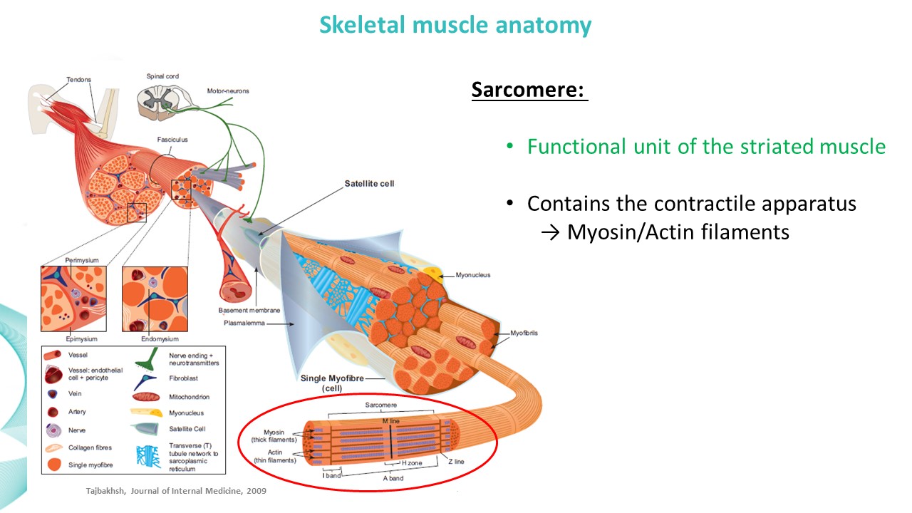 Slide #2
 Slide #3
 Slide #4
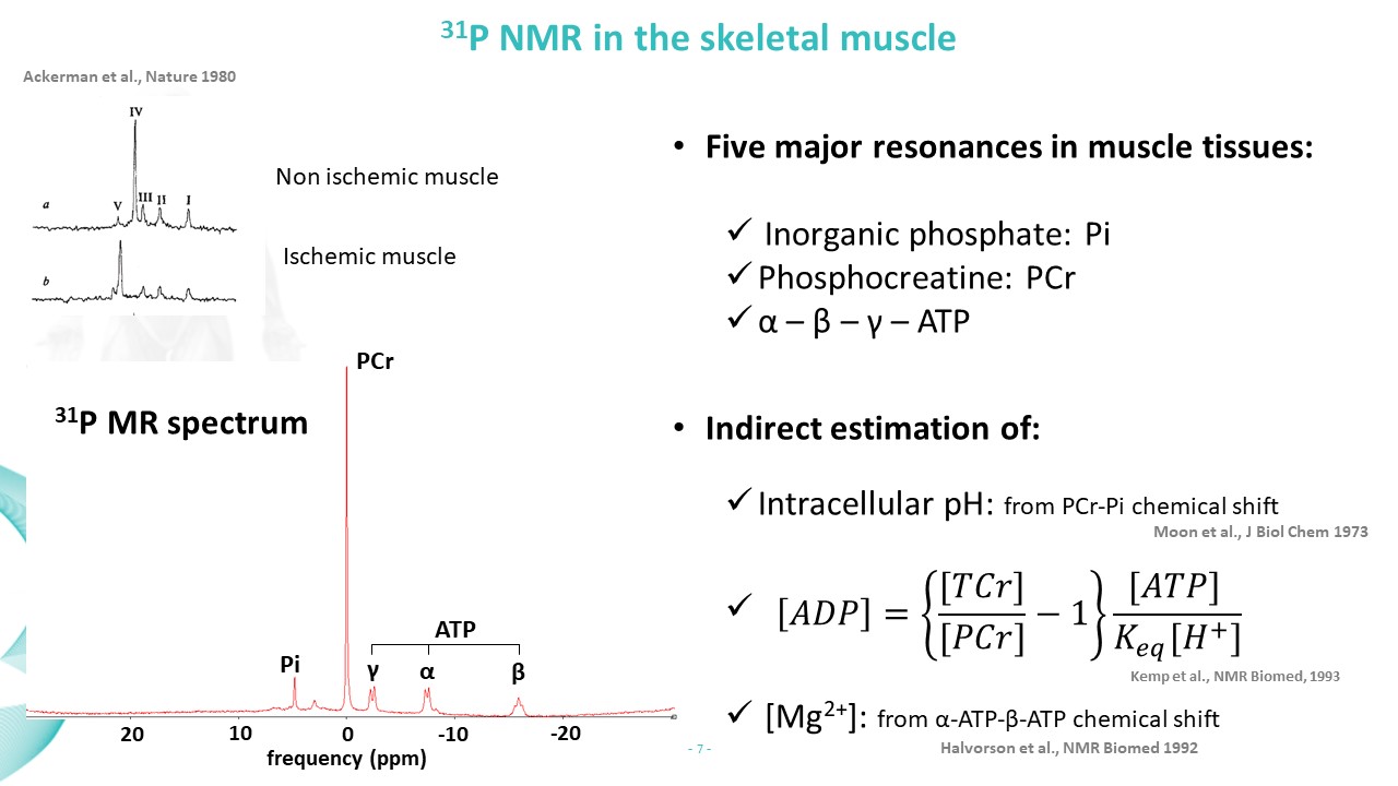 Slide #5
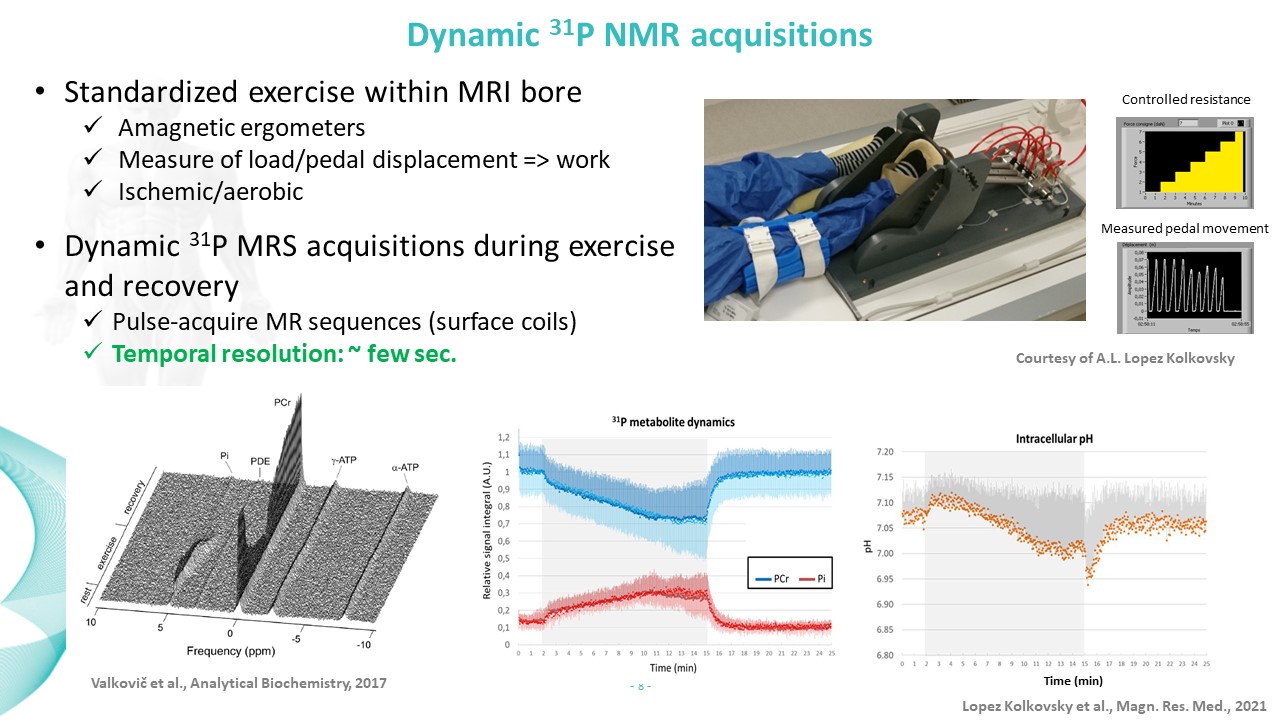 Slide #6
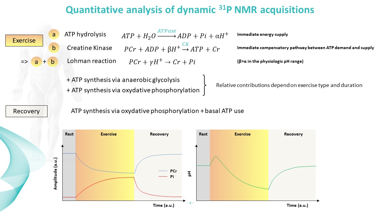 Slide #7
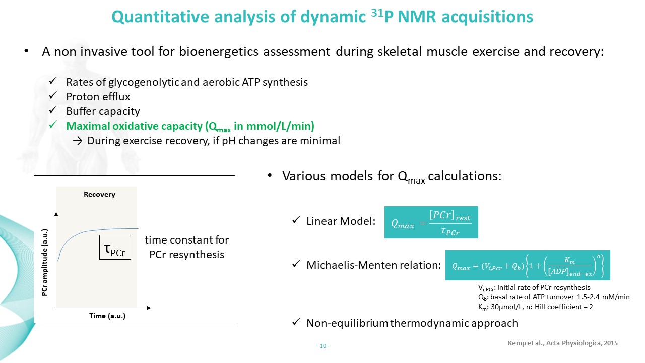 Slide #8
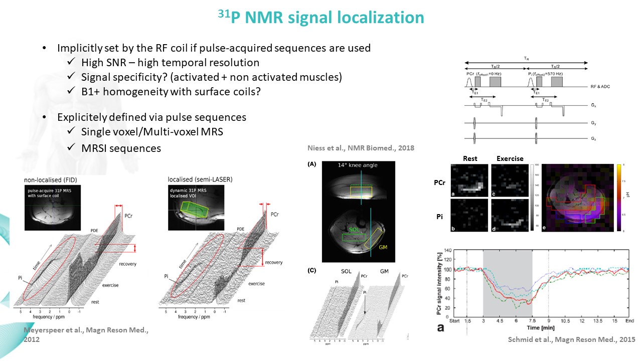 Slide #9
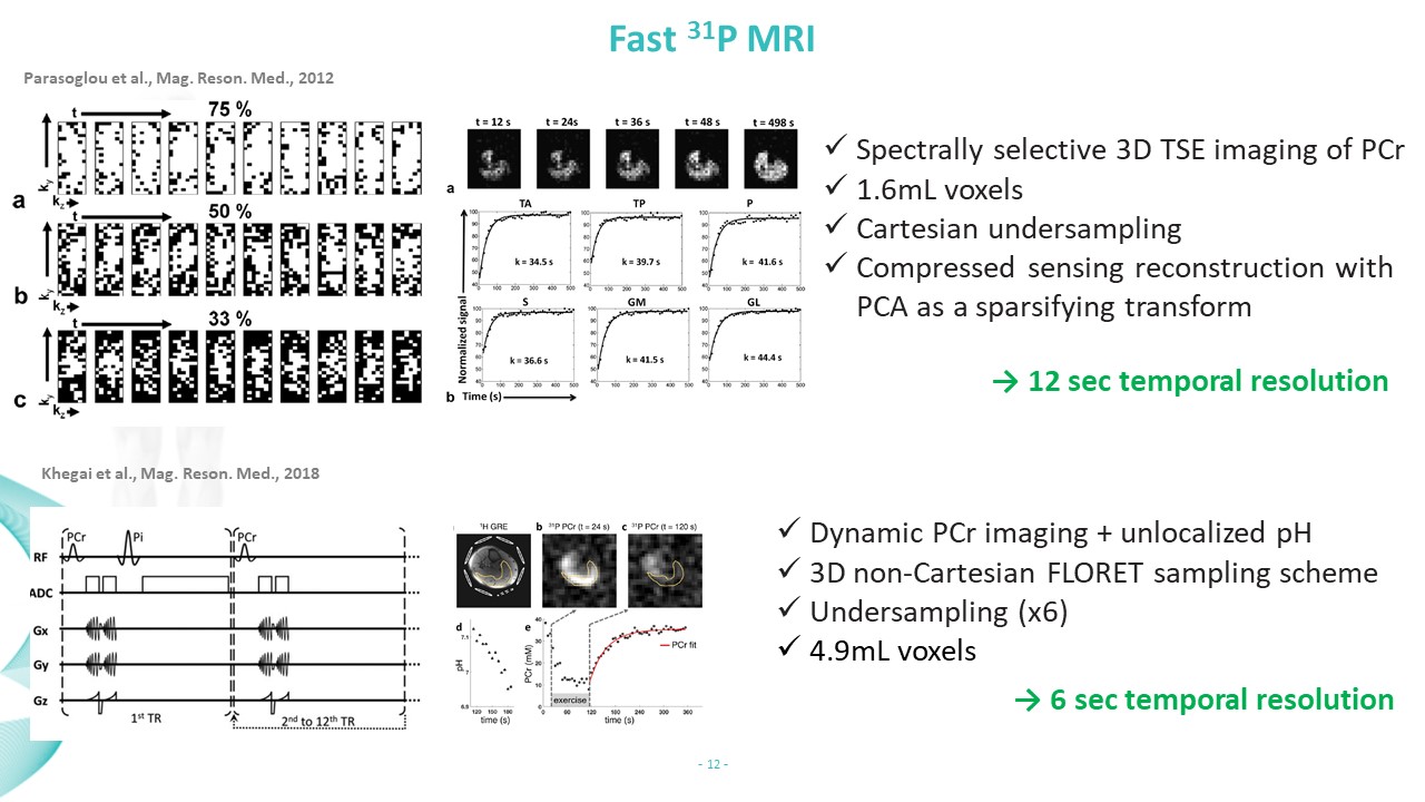 Slide #10
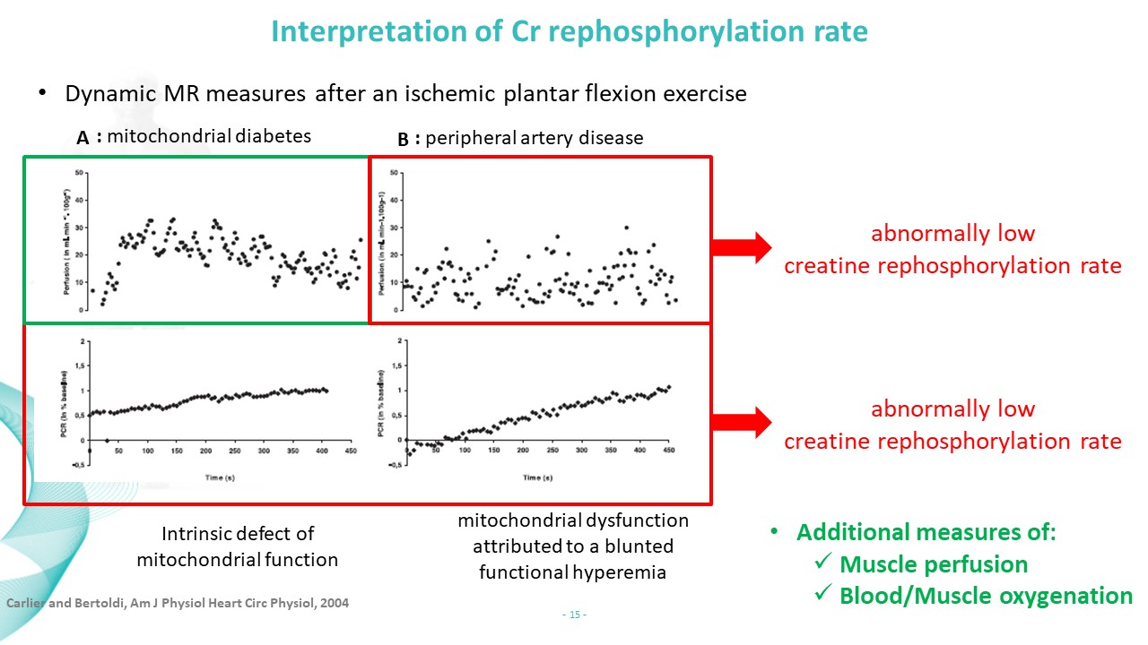 Slide #11
 Slide #12
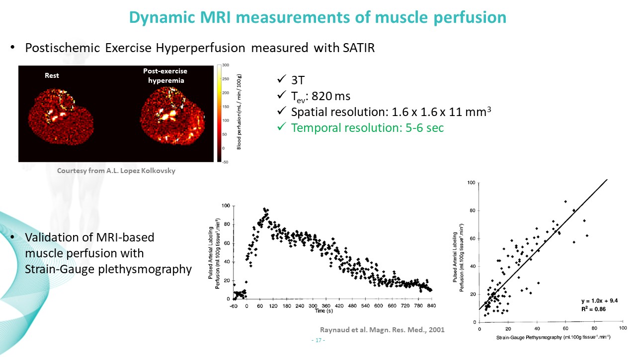 Slide #13
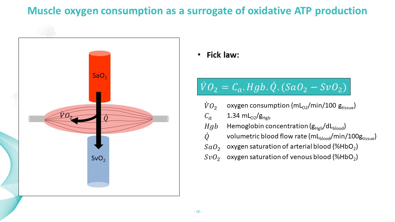 Slide #14
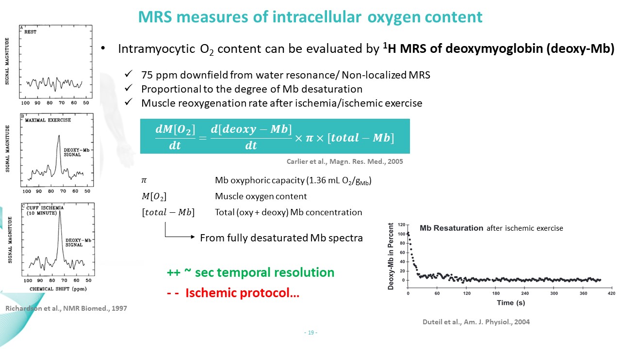 Slide #15
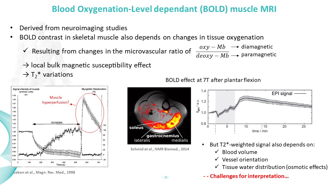 Slide #16
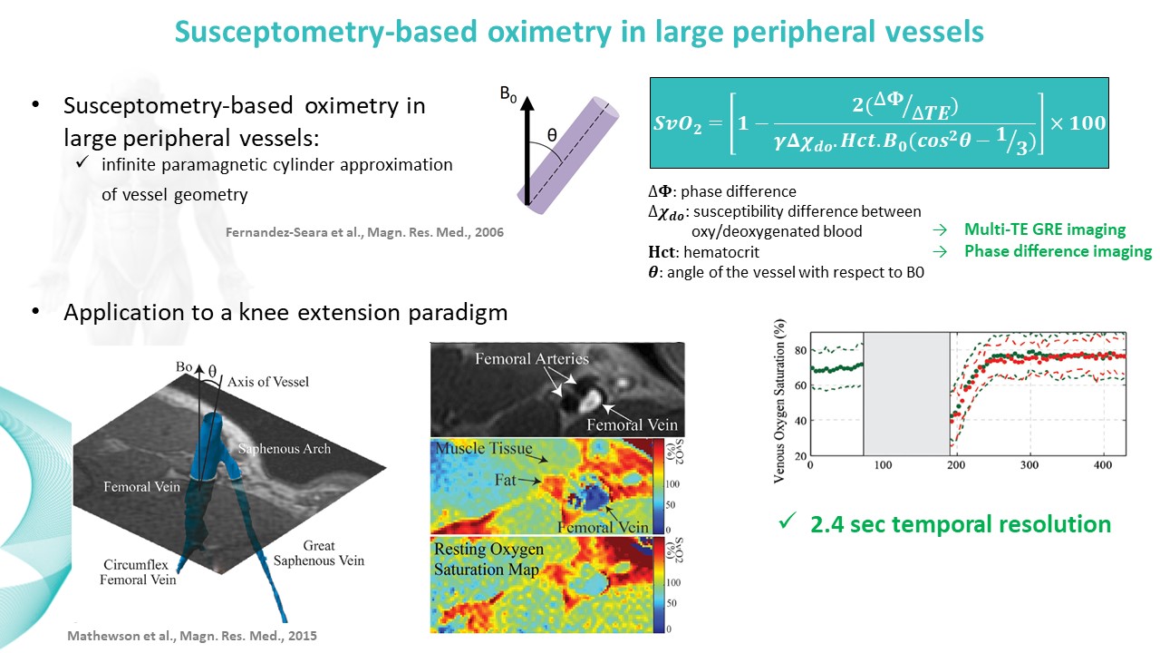 Slide #17
 Slide #18
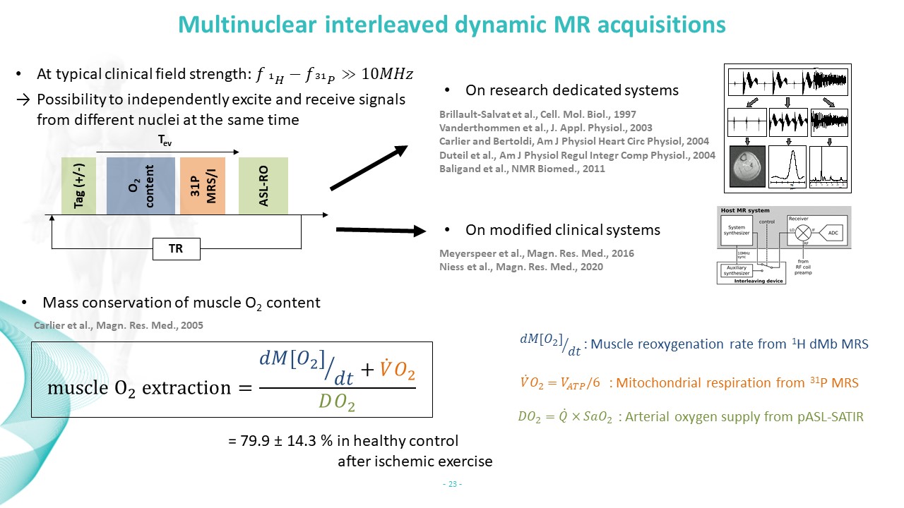 Slide #19
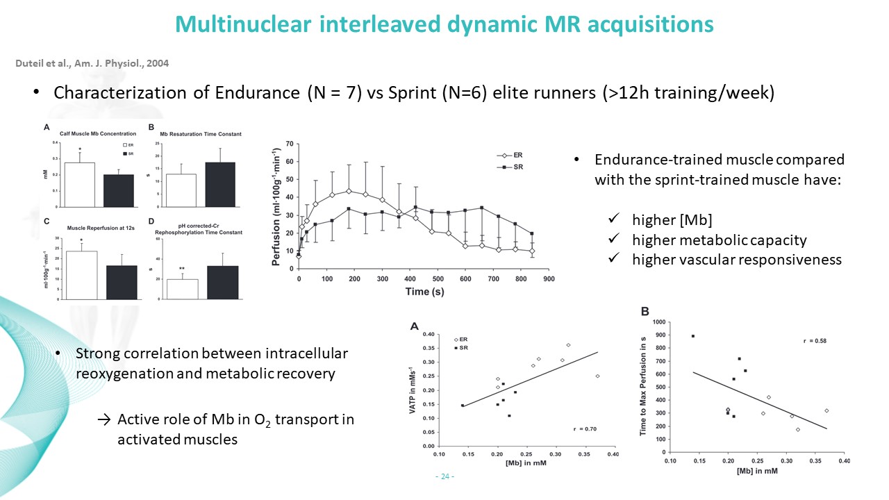 Slide #20
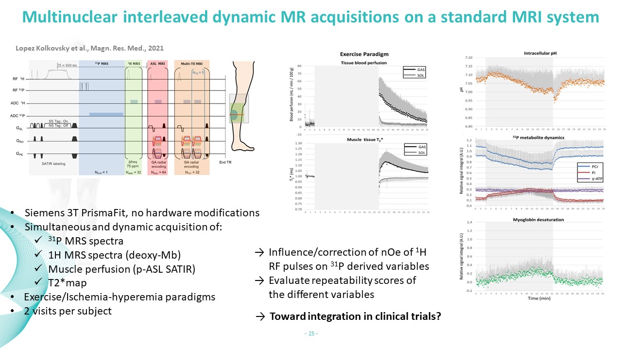 Slide #21
 Slide #22