Cardiovascular + Lung at Low Field
1National Institutes of Health, United States
Synopsis
Learn about how the physical properties at low field (<1T) can provide opportunities for novel cardiovascular and pulmonary imaging applications. In addition, we will discuss what imaging paradigms do and don’t work at low fields, and current research exploring low field cardiothoracic imaging.
Syllabus
Low field (<1T) MR imaging may enable accessible healthcare through reduced system costs, and this topic has been of recent research interest in the community [1-4].In this talk, attendees will learn about how the physical properties at low field can provide opportunities for novel cardiovascular and pulmonary imaging applications. In addition, attendees will learn about what structural and quantitative imaging paradigms do and don’t work at low fields, and current research exploring low field cardiothoracic imaging.
Acknowledgements
No acknowledgement found.References
[1] Wald, L.L., et al. Low-cost and portable MRI. J Magn Reson Imaging, 2020. 52(3): p. 686-696.
[2] Campbell-Washburn, A.E., et al. Opportunities in Interventional and Diagnostic Imaging by Using High-Performance Low-Field-Strength MRI. Radiology, 2019. 293(2): p. 384-393.
[3] Simonetti OP, Ahmad R. Low-Field Cardiac Magnetic Resonance Imaging: A Compelling Case for Cardiac Magnetic Resonance's Future. Circ Cardiovasc Imaging. 2017 Jun;10(6):e005446.
[4] Sarracanie, M., et al. Low-Cost High-Performance MRI. Sci Rep, 2015, 5, 15177.
Figures
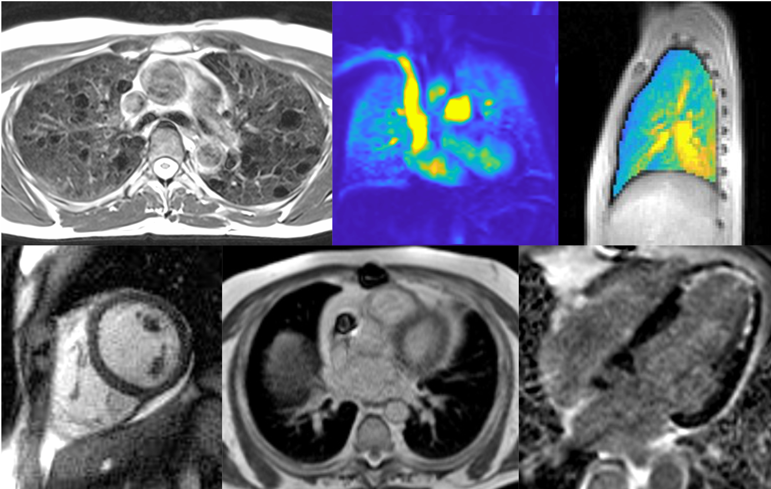
Example lung and cardiac imaging applications at 0.55T.
Top row, left-to-right: T2w lung imaging in a patient with lung cysts, pulmonary perfusion imaging, and lung water quantification.
Bottom row, left-to-right: Spiral bSSFP cine imaging, imaging a patient with metal implants, late-gadolinium enhancement imaging.