MRI of the Pancreas: Basics
Sikandar Shaikh1,2,3
1Department of Radiodiagnosis, Shadan Institute of Medical Sciences.. Hyderabad. India, Hyderabad, India, 2Department of Radiodiagnosis, Kasturba Medical College, Manipal, India, 3Department of Biomedical Engineering, Indian Institute of Technology, Hyderabad, India

Synopsis

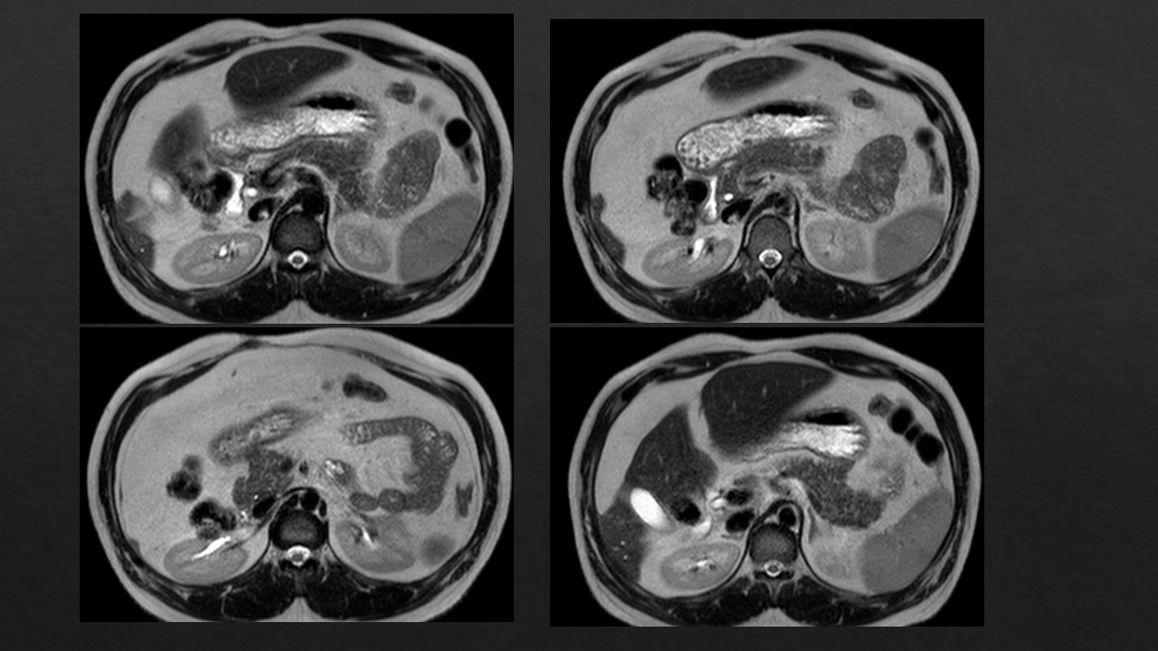
MRI is the important modality for the evaluation of the Pancreas. It has significant role in the evaluation of various pancreatic pathologies comprising of the congenital, infective, inflammation, neoplastic, traumatic, and miscellaneous pathologies. This is also important screening tool for differentiating the indeterminate pancreatic focal lesions like focal pancreatitis versus neoplastic. MRI using magnetic resonance cholangiopancreatography (MRCP) also evaluates associated hepatobiliary pancreatic pathologies. Due to technical advances in MRI, newer sequences as well as protocols like diffusion-weighted and T1-weighted dynamic contrast-enhanced MRI are used despite of various challenges of retroperitoneal location of the Pancreas.

Pancreas is one of the most important organs having both the exocrine and endocrine functions . MR imaging in pancreas includes the pancreatic and biliary ductal anatomy, detection and to characterize parenchymal disease, peripancreatic extension and also associated with the vascular anatomy. Due to the increased technological advances additional integrated coil elements and also the radiofrequency channels are applied . 1.5 T MR is being can do 3-dimensional T1-weighted and MR cholangiopancreatography (MRCP) sequences. The use of the Secretin-enhanced MRCP (S-MRCP) protocols are able to define the pancreatic ducts and the parenchymal glandular function also. Evaluation of the suspected biliopancreatic pain, in the staging of chronic pancreatitis, and diagnosis and follow-up of pancreatic tumours is more precise now. To evaluation of the pancreatic parenchyma and the pancreaticobiliary ductal system are dual-echo T1- weighted gradient-echo; T2-weighted axial and coronal sequences, turbo spin echo (TSE) or a variant of TSE; 2-dimensional and 3-dimensional MRCP; and fat-suppressed T1-weighted, 3- dimensional gradient-echo before and after gadolinium and the S-MRCP sequence. The basic T1-weighted sequences for haemorrhage, acute pancreatitis and identification of the pancreatic fat. T2-weighted sequences for the evaluation of the peripancreatic collections and cystic lesions. Fat suppression technique has two types for the suppression of the fat. Chemical shift fat suppression due to the difference of resonance frequency between fat and water gradient pulses affected by magnetic field inhomogeneity. Another fat suppression technique is the inversion-recovery (IR) fat suppression, such as short tau inversion recovery (STIR), having more T1 relaxation times between fat and water. MRCP is the commonly used technique for the acquisition by using the heavily T2- weighted images, with variants of TSE sequences. The sequences which use the TR and partial Fourier technique are called SSFSE or half Fourier acquisition single-shot turbo spin echo (HASTE). Two-dimensional MRCP has traditionally been used with coronal SSFSE slabs and by using 40-mm slabs in multiple coronal oblique planes which can image the pancreatic ductal system Three-dimensional MRCP is a 3-dimensional TSE sequence of very high spatial-resolution . The use of the thin sections without slice gap can identify the small stones, the small side branches of main pancreatic duct, and the intrahepatic biliary system. This three-dimensional MRCP can be acquired in the series of breath-holds or during free breathing. By using the 1- to 2- mm contiguous slices during free breathing and by using the navigator-echo techniques will be reducing motion artifacts. S-MRCP the concept here is that secretin will cause distention of the pancreatic ducts and due to this the evaluation of the pancreatic ductal anomalies, anomalous pancreaticobiliary junction and mild chronic pancreatitis . Contrast-Enhanced Sequences is 3-dimensional, fat suppressed, spoiled gradient echo known by various names like volume interpolated breath-hold (VIBE), fast gradient echo (F-GRE), liver acquisition with volume acceleration (LAVA), and T1-weighted, high-resolution, isotropic volume examination (THRIVE), these sequences are acquired by 2- to 5-mm contiguous slices within a 20-second breath-hold. PANCREATIC TRAUMA is more commonly presenting as blunt abdominal trauma ranging from 1% to 2%, pancreatic injuries .Mortality of pancreatic injuries from blunt trauma is 30%, . MR pancreatography can directly imaging of the pancreatic duct as well as pancreatic parenchyma and peripancreatic fat planes. Pancreatitis is the commonest of the pancreatic pathologies and having various causes, Cholelithiasis and alcoholism are the two most common causes , but other causes also are like autoimmune pancreatitis, various granulomatous causes are also there. MR can identify the peripancreatic fat planes , collections, pancreatic ducts and adjacent peritoneum and mesentery. The various other diffuse involvement of the pancreas is also seen in the various inflammatory, infiltrative, or neoplastic disorders. The inflammatory deposition disorders, including autoimmune pancreatitis, with the neoplastic processes include the hemochromatosis, fatty replacement, and amyloidosis. The infiltrative disorders comprises of the cystic fibrosis, lymphoma, and metastatic deposition. Fatty Replacement of the Pancreas Fat replacement, also termed lipomatosis, adipose atrophy, or fat infiltration of the exocrine pancreas, is commonly seen in obese and elderly patients. fatty replacement may be of 3 types: (1) diffuse fatty replacement, which reveals separation of pancreatic parenchyma with prominence of lobulations; (2) asymmetric fatty replacement, where a part of pancreas, such as the head region is spared (often mistaken as tumor); and (3) fatty pseudohypertrophy, where the pancreas is massively enlarged due to fatty replacement. Cystic Fibrosis Cystic fibrosis (CF) mainly manifests as chronic obstructive lung disease and pancreatic exocrine insufficiency, seen in 85% to 90% of patients MR imaging T1W MR imaging that shows fat replacement as high signal intensity and MR imaging directly proportional to the clinical compromise due to exocrine insufficiency. MRI and MRCP are routinely used clinically to assess the pancreas with pancreatic masses, or in those at risk for pancreatic malignancy. MRCP with higher resolution and breath-hold acquisitions, DWI with a greater number of b-values and IVIM modelling, and reduced FOV DWI can be assessed .

Acknowledgements

Dr. Bhuchandi

References

1. Ma J, Son JB, Zhou Y, et al. Fast spin-echo tripleecho Dixon (fTED) technique for efficient T2- weighted water and fat imaging. Magn Reson Med 2007;58:103–9.

2.Bayramoglu S, Kilickesmez O, Cimilli T, et al. T2- weighted MRI of the upper abdomen: comparison of four fat-suppressed T2-weighted sequences including PROPELLER (BLADE) technique. Acad Radiol 2010;17(3):368–74. [Epub 2009 Dec 30].

3.Nanko S, Oshima H, Watanabe T, et al. Usefulness of the application of the BLADE technique to reduce motion artifacts on navigation-triggered prospective acquisition correction (PACE) T2-weighted MRI (T2WI) of the liver. J Magn Reson Imaging 2009; 30:321–6.

4.Hirokawa Y, Isoda H, Maetani YS, et al. Evaluation of motion correction effect and image quality withthe periodically rotated overlapping parallel lines with enhanced reconstruction (PROPELLER) (BLADE) and parallel imaging acquisition technique in the upper abdomen. J Magn Reson Imaging 2008;28:957–62.

5.Purysko AS, Gandhi NS, Walsh RM, Obuchowski NA, Veniero JC. Does secretin stimulation add to magnetic resonance cholangiopancreatography in characterising pancreatic cystic lesions as side-branch intraductal papillary mucinous neoplasm? Eur Radiol 2014;24(12):3134-3141.

6.Canto MI, Harinck F, Hruban RH, et al. International cancer of the pancreas screening (CAPS) consortium summit on the management of patients with increased risk for familial pancreatic cancer. Gut 2013;62(3):339-347.

7.Jang KM, Kim SH, Kim YK, Song KD, Lee SJ, Choi D. Missed pancreatic ductal adenocarcinoma: Assessment of early imaging findings on prediagnostic magnetic resonance imaging. Eur J Radiol 2015;84(8):1473-1479.

8.Corrias G, Raeside MC, Agostini A, et al. Pilot study of rapid MR pancreas screening for patients with BRCA mutation. Eur Radiol 2019;29 (8):3976-3985.

9.Corral JE, Mareth KF, Riegert-Johnson DL, Das A, Wallace MB. Diagnostic yield from screening asymptomatic individuals at high risk for pancreatic cancer: A meta-analysis of cohort studies. Clin Gastroenterol Hepatol 2019;17(1):41-53.

10.Henrikson NB, Aiello Bowles EJ, Blasi PR, et al. Screening for pancreatic cancer: Updated evidence report and systematic review for the US preventive services task force. JAMA 2019;322(5):445-454.

11.Schawkat K, Eshmuminov D, Lenggenhager D, Endhardt K, Vrugt B, Boss A, et al. Preoperative Evaluation of Pancreatic Fibrosis and Lipomatosis: Correlation of Magnetic Resonance Findings With Histology Using Magnetization Transfer Imaging and Multigradient Echo Magnetic Resonance Imaging. Investigative radiology. 2018; 53(12):720–7.

Figures

Axial MR Images showing the normal appearance of the Pancreas in entire course

Proc. Intl. Soc. Mag. Reson. Med. 30 (2022)