T2, T2* & T1ρ Mapping
Graham Wright1
1Sunnybrook Research Institute, University of Toronto, Toronto, ON, Canada
1Sunnybrook Research Institute, University of Toronto, Toronto, ON, Canada
Synopsis
MRI measures of signal decay without refocusing, with intermittent refocusing and with continuous refocusing reflected by time constants T2*, T2, and T1ρ can yield important clinical information about cardiac tissue damage and response to injury. Reduced T2* reflects localized susceptibility effects as seen in hemorrhage and iron overload. T2 can depict changes in blood oxygenation reflecting ischemia and changes in water mobility reflecting inflammation. T1ρ can highlight chemical and spin exchange effects, increasing contrast between healthy and infarcted myocardium. Recently, higher dimensional and multi-parameter imaging methods have been developed for improved differentiation of myocardial pathophysiologies.
 Slide #1
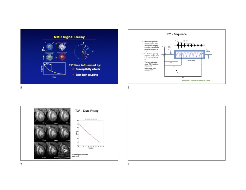 Slide #2
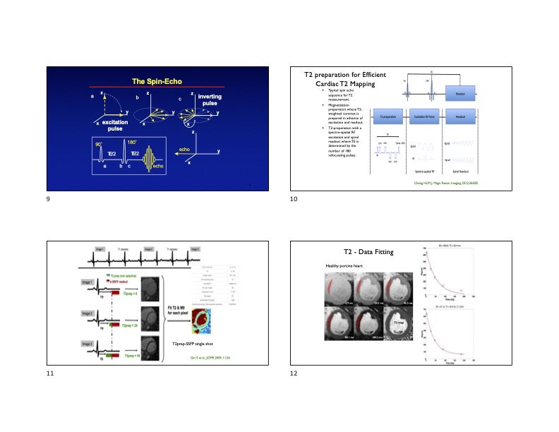 Slide #3
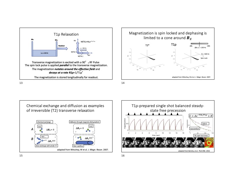 Slide #4
 Slide #5
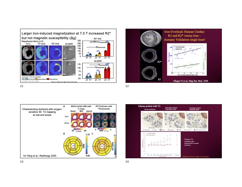 Slide #6
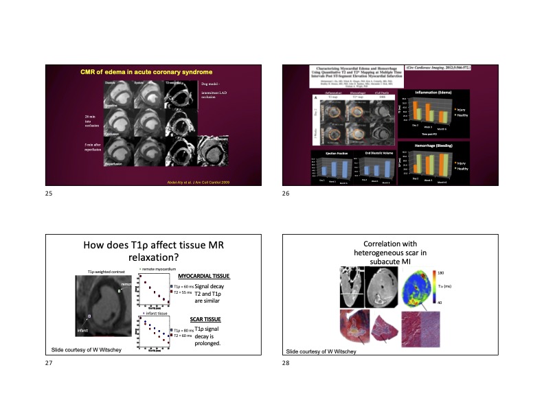 Slide #7
 Slide #8
 Slide #9
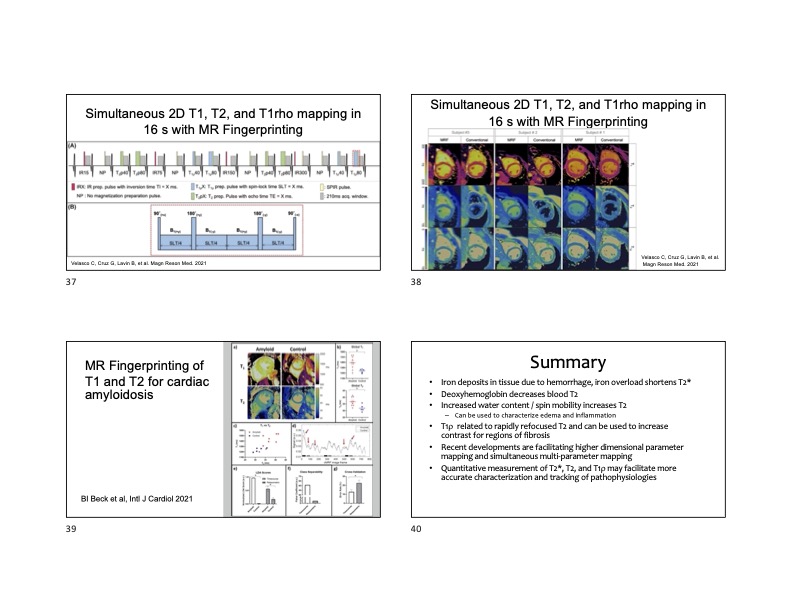 Slide #10