Advances in Carotid MR Vessel Wall Imaging Techniques
Chun Yuan1,2
1University of Utah, Salt Lake City, UT, United States, 2University of Washington, Seattle, WA, United States
1University of Utah, Salt Lake City, UT, United States, 2University of Washington, Seattle, WA, United States
Synopsis
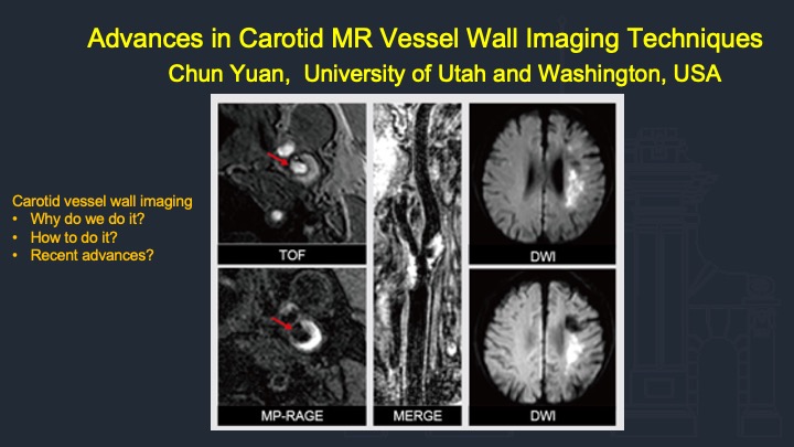
Carotid vessel wall imaging complements angiographic techniques, which are based on measurements of luminal stenosis, has the ability to identify atherosclerotic plaque features associated with increased risk for ischemic events. These features include luminal morphology, fibrous cap rupture, and ulcerations, intraplaque hemorrhage, lipid rich necrotic core, neovasculature and inflammation, and overall plaque burden. There are many imaging techniques developed for vessel wall imaging and quantitative analysis in recent years.This talk will summarize the current expert consensus on carotid vessel wall imaging and introduce recent advances in MRI based techniques.
Carotid vessel wall imaging, complements angiographic techniques which are based on measurements of luminal stenosis, has the ability to identify atherosclerotic plaque features associated with increased risk for ischemic events. These features include luminal morphology, fibrous cap rupture, and ulcerations, intraplaque hemorrhage, lipid rich necrotic core, neovasculature and inflammation, and overall plaque burden. There are many imaging techniques developed for vessel wall imaging and quantitative analysis in recent years.This talk will summarize the current expert consensus on clinical carotid vessel wall imaging. To be followed with introduction of established approach and technical challenges. A major part of the talk will be dedicated to the recent advances in MRI based techniques. This talk should provide the basics of current techniques in carotid vessel wall imaging.
Acknowledgements
No acknowledgement found.References
No reference found.Figures
Slide #1