DWI & DTI: Where, Why & How It Is & Is Not Used
Kei Yamada1
1Kyoto Prefectural Univ. of Medicine, Japan

Synopsis

Diffusion-weighted imaging (DWI)and diffusion-tensor imaging (DTI) has become one of the essential research/clinical tools in analyzing the brain in both normal and pathological states. In this presentation, I will first cover the brief history of DWI, and then explain how this tool has become an essential part of our daily practice.

Diffusion-weighted imaging (DWI)and diffusion-tensor imaging (DTI) has become one of the essential research/clinical tools in analyzing the brain in both normal and pathological states. In this presentation, I will first cover the brief history of DWI, and then explain how this tool has become an essential part of our daily practice. The breakthrough was the implementation of echo planer imaging (EPI) on clinical scanners. This led to motion-free image acquisition, which is crucial for DWI which is sensitive to motion in the order of microns. Following this introduction of this talk, I will explain about DTI and its related techniques, including tractography. These tools have also become vital elements of presurgical planning for brain tumors. Tractography has enabled visualization of three-dimensional fiber orientation in the living brain. The FACT algorithm was one of the most frequently used method [1]. Attempts to validate this technique have been made in the past, but are limited to comparisons of the images with known neuroanatomy. One has to interpret the results with cautions when applying this technique clinically [2,3].

Acknowledgements

No acknowledgement found.

References

1. Mori S, et al. Three-dimensional tracking of axonal projections in the brain by magnetic resonance imaging. Ann Neurol 1999; 45: 265-9

2. Yamada K. Diffusion tensor tractography should be used with caution. Proc Natl Acad Sci U S A. 2009 106:E14

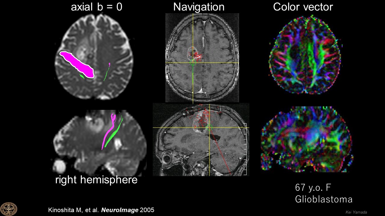
3. Kinoshita M, Yamada K, et al. Fiber-tracking does not accurately estimate size of fiber bundle in pathological condition. Neuroimage. 2005 1; 25: 424-429.

Figures

Figures from Kinoshita M, et al. NeuroImage 2005 [Reference 3]

Proc. Intl. Soc. Mag. Reson. Med. 30 (2022)