Dynamic Contrast: Basics
Ho-Ling Anthony Liu1
1Department of Imaging Physics, University of Texas MD Anderson Cancer Center, Houston, TX, United States
1Department of Imaging Physics, University of Texas MD Anderson Cancer Center, Houston, TX, United States
Synopsis
Dynamic susceptibility contrast (DSC) and dynamic contrast enhanced (DCE) MRI are two widely used methods in the clinic to evaluate perfusion and microvasculature of the tissue. They acquire dynamic T2*- and T1-weighted images, respectively, before and after a bolus injection of the Gd-based contrast agent (GBCA). The measured signal time curves can be converted to concentration time curves which are then used to quantify physiological parameters, such as blood flow, blood volume, transit times, vessel permeability, and volume fraction of the extravascular extracellular space. This lecture will cover basic principles, image acquisition, and quantitative analysis of the two MRI methods.
 Slide #1
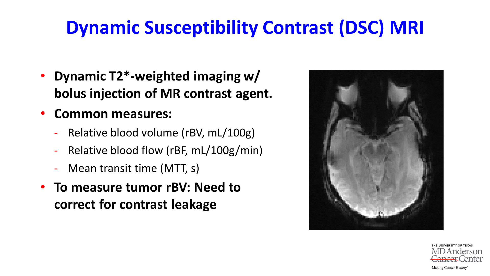 Slide #2
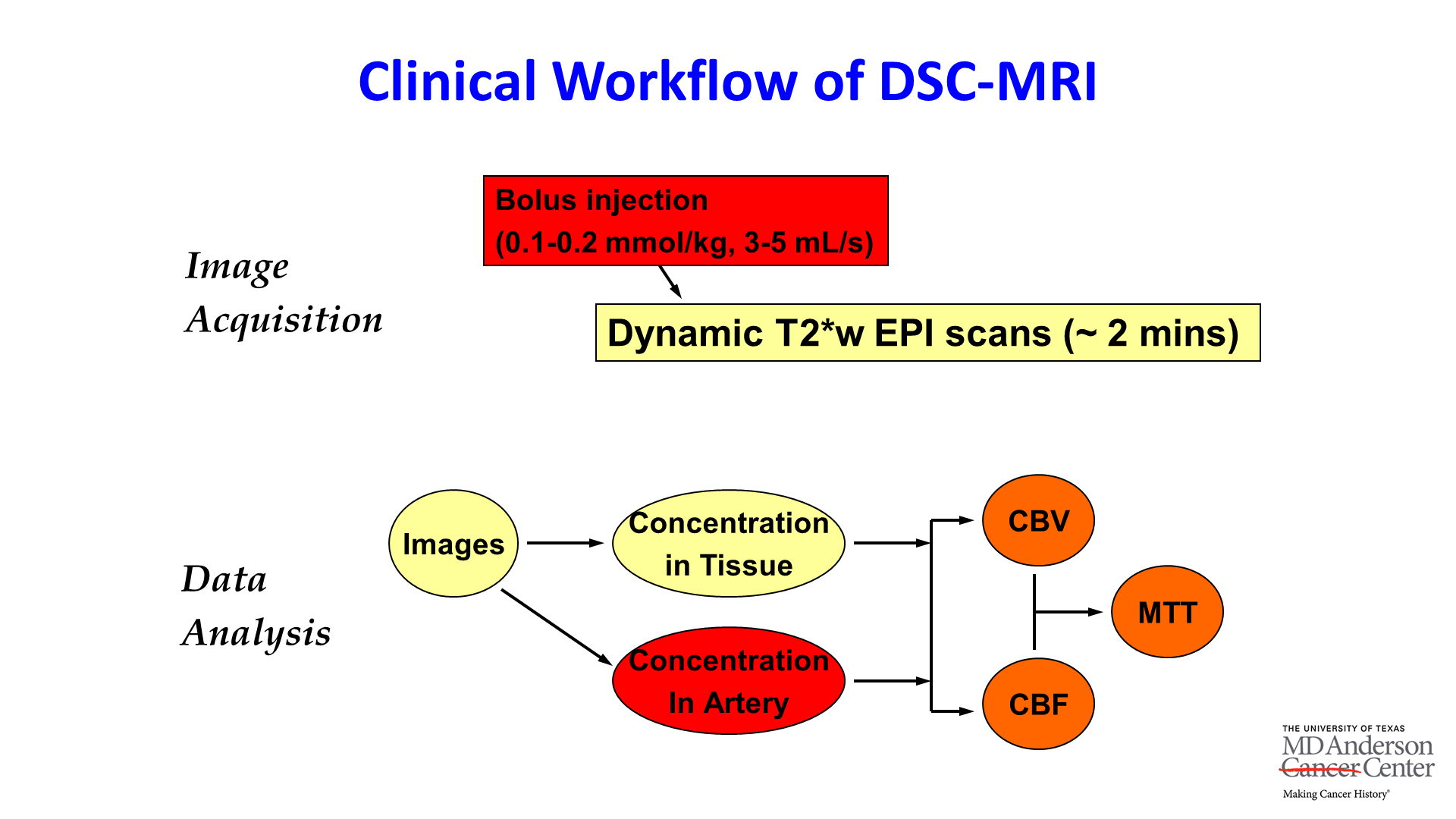 Slide #3
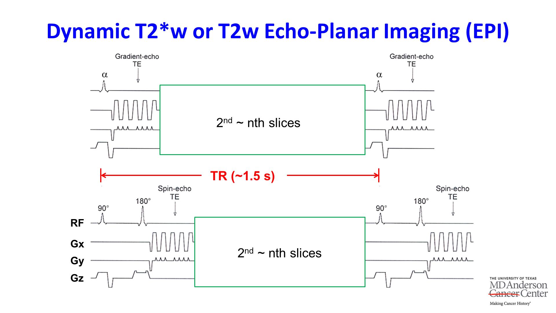 Slide #4
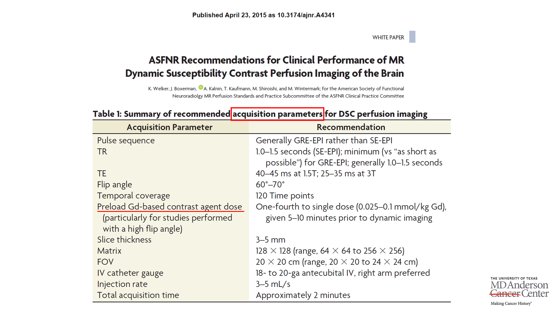 Slide #5
 Slide #6
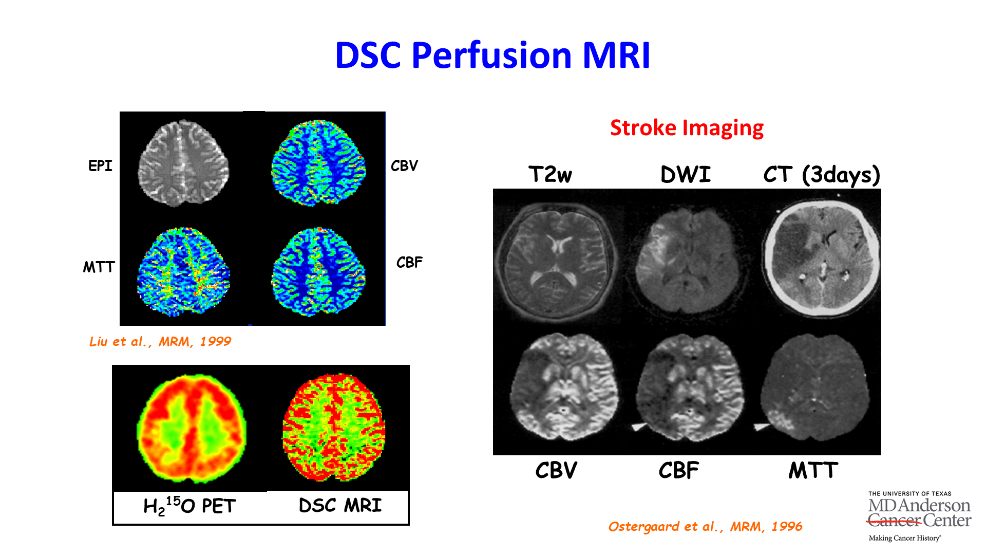 Slide #7
 Slide #8
 Slide #9
 Slide #10
 Slide #11
 Slide #12
 Slide #13
 Slide #14
 Slide #15
 Slide #16
 Slide #17
 Slide #18
 Slide #19
 Slide #20
 Slide #21
 Slide #22