5041
Impact of gradient scheme and shimming on out-of-voxel echo artefacts in edited MRS1Department of Radiology and Radiological Science, Johns Hopkins University School of Medicine, Baltimore, MD, United States, 2F.M. Kirby Research Center for Functional Brain Imaging, Kennedy Krieger Institute, Baltimore, MD, United States
Synopsis
Out-of-voxel (OOV) signal is a common spurious echo artifact, which interferes with spectral quantification. These signals arise from either incomplete suppression or accidental refocusing. Dephasing Optimization Through Coherence Order Pathway Selection (DOTCOPS) is an optimized gradient scheme that can be used to suppress unwanted signals. Here we explored the impact of gradient scheme and shimming-order on OOV artefacts in Hadamard-encoded edited MRS.
Introduction
Spectral quality in edited MR spectroscopy is important, especially when trying to measure low-concentration metabolites in the human brain1. One common source of artefacts seen in edited difference spectra is out-of-voxel (OOV) water echoes; broad OOV signals can occur at any chemical shift (arising from regions outside the shimmed voxel with uncontrolled local magnetic field). OOV echoes often appear in the spectrum with very strong first-order phase ‘ripple’, because they are not synchronized with the beginning of the acquisition window. These signals will affect quantification, by overlapping with targeted metabolite resonances. Dephasing Optimization Through Coherence Order Pathway Selection (DOTCOPS)2 gradient schemes are algorithmically optimized to suppress all potential alternative coherence transfer pathways (CTPs), and should suppress unwanted OOV echoes. Second-order shimming uses non-linear gradient fields to maximize field homogeneity inside the voxel, which unfortunately increases the diversity of local gradient fields outside of the voxel. Given that local spatial B0 gradients can refocus unintended CTPs, it is possible that OOVs are less prevalent when only first-order shimming is applied. Here we compare the size of unwanted OOV signals in Hadamard-edited (HERMES)3 data, acquired with local gradient scheme and DOTCOPS, and with first and second-order shimming.Methods
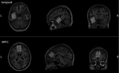
HERMES data were acquired in 15 healthy volunteers (4M/11F, average ages:23.8±2.8years old), using either the DOTCOPS or local gradient scheme (Philips-PRESS scheme with adjusted timing, denoted ‘Edden’) and first- or second-order shimming. Two 30x26x26mm3 voxels were sampled, medial pre-frontal (MPFC) and temporal (shown in Figure 1), two challenging brain regions, using the following parameters: Philips 3T Ingenia Elition X scanner; TR/TE = 2000/80 ms; 2 kHz spectral width; 1024 samples; 160 transients; acquiring 8 water-reference transients. All data were analyzed using Osprey4. We used the standard deviation of the linear-combination-modeling residual as a quantitative metric of OOV signal amplitude, since OOV echoes are largest contributor of unmodeled signal where present. As there are 3 categorical factors (region, gradient scheme, shimming order), ANOVA analysis and post-hoc Tukey and f tests were performed in R.Results
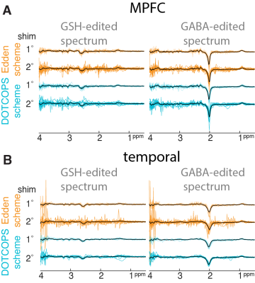
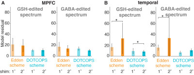
Edited difference spectra, overlaid for all subjects, are shown in Figure 2. Characteristic OOV echoes can be seen in both GABA- and GSH-edited spectra for all regions, gradient schemes and shimming approaches. Note that these spectra are presented without line-broadening which can dampen the amplitude of OOV echoes that occur late in the FID. The model residuals, which we interpret as a measure of OOV echo amplitude, are shown in Figure 3.In ANOVA, we found a statistically significant difference in average residual by the factor of gradient scheme in both GABA (f=5.416, p=0.0221) and GSH (f=12.088, p<0.001). A Tukey post-hoc test revealed that the DOTCOPS scheme gave smaller OOV signals on average than the ‘Edden’ scheme. Neither shimming scheme nor region were significant factors.
In the temporal region, a post-hoc f-test showed significantly greater variance in OOV amplitude for second-order shimming compared to first-order shimming in both edited spectra (GSH: p<0.001; GABA: p<0.0001) for the Edden gradient scheme. However, for the DOTCOPS scheme, only GSH-edited spectra showed this significant difference (GSH: p<0.05).
Discussion
OOV echoes are commonly seen for edited MRS across vendors, and are observed in both GABA- and GSH-edited data from two challenging brain regions in multiple subjects. Common strategies for minimizing them include changing the order and polarity of slice-selective gradients5. These unmodelled signal contributions can interfere with quantification both by obscuring the edited signals of interest and by masking the correct baseline level upon which signals are superimposed. Here we confirm that OOV echoes are reduced with the DOTCOPS gradient scheme compared to the widely-distributed local Edden scheme. Although the current sample size is modest, and parameters sampled not exhaustive, this strongly motivates switching to the DOTCOPS scheme for future experiments. Note that, while the DOTCOPS approach in general was supported by in vivo data in the original publication, the MEGA-PRESS scheme was not explicitly demonstrated and this abstract offers independent validation. OOV echo artifacts occur when signal from outside of the voxel - usually water signals, but also lipid and other smaller signals – are excited (either during water-suppression, saturation or PRESS sequence modules) and refocused by some combination of RF pulses. The gradient pulses used in the sequence to suppress signal outside of the voxel may not perform appropriately in regions of strong intrinsic field gradients, such as those hard-to-shim regions in the frontal and temporal lobes, close to tissue-bone-air interfaces.The hypothesis that shimming to the second order might exacerbate out-of-voxel artifacts was not unequivocally supported, although there is some supporting evidence in the temporal data. Shim order might be considered as an additional factor to adjust in optimizing protocols for challenging brain regions.Conclusion
The DOTCOPS gradient scheme for J-difference-edited PRESS acquisitions gives spectra with smaller OOV echo artifacts than the gradient scheme used in a widely disseminated editing sequence.Acknowledgements
This work has been supported by NIH grants P41EB015909, R01EB016089, R01EB023963, R21AG060245, and R00AG062230.References
1.Choi IY, Andronesi OC, Barker P, et al. Spectral editing in 1H magnetic resonance spectroscopy: Experts’ consensus recommendations. NMR Biomed. 2021;34(5):e4411. doi:10.1002/nbm.4411
2.Landheer K, Juchem C. Dephasing optimization through coherence order pathway selection (DOTCOPS) for improved crusher schemes in MR spectroscopy. Magn Reson Med. 2019;81(4):2209-2222. doi:10.1002/mrm.27587
3.Chan KL, Saleh MG, Oeltzschner G, Barker PB, Edden RAE. Simultaneous measurement of Aspartate, NAA, and NAAG using HERMES spectral editing at 3 Tesla. NeuroImage. 2017;155:587-593. doi:10.1016/j.neuroimage.2017.04.043
4.Oeltzschner G, Zöllner HJ, Hui SCN, et al. Osprey: Open-source processing, reconstruction & estimation of magnetic resonance spectroscopy data. J Neurosci Methods. 2020;343:108827. doi:10.1016/j.jneumeth.2020.108827
5.Carlsson Å, Ljungberg M, Starck G, Forssell-Aronsson E. Degraded water suppression in small volume 1H MRS due to localised shimming. Magn Reson Mater Phys Biol Med. 2011;24(2):97-107. doi:10.1007/s10334-010-0239-2
Figures


