5016
Advanced Signal Combination for Improved Image Quality in the Presence of Motion1Canon Medical Research USA, Inc., Mayfield Village, OH, United States
Synopsis
Motion is a major challenge in MRI. Averaging multiple acquisitions of the same target can help suppress motion artifacts. However, combining multiple acquisitions with equal weighting can produce non-diagnostic image quality if one acquisition suffers from significantly more motion artifacts than the other acquisition. For this study, a framework for suppressing motion artifacts was developed by assigning higher weighting to the acquisition with less motion. The performance of the proposed strategy, called advanced signal combination (ASC), was evaluated in sagittal T2-weighted C-spine MRI obtained with number of acquisitions=2. ASC was found to suppress motion artifact while maintaining SNR.
Introduction
Patient motion is a major challenge in MRI because of long acquisition times. A multitude of solutions have been proposed for motion correction. These include PROPELLER1, COCOA2, navigator-based motion correction3,4, shot rejection3,5, iterative methods with entropy-related criterion6, aligned SENSE7, and prospective motion correction8. One relatively simple strategy for suppressing motion artifacts and increasing signal-to-noise ratio (SNR) is to average multiple acquisitions of the same target. However, combining multiple acquisitions with equal weighting (default combination) can produce non-diagnostic image quality if one acquisition suffers from significantly more motion artifacts than the other acquisition. The aim of this study was to develop a framework for suppressing motion artifacts by assigning higher weighting to the acquisition with less motion. We evaluated the performance of our proposed strategy, which we call advanced signal combination (ASC), in sagittal T2-weighted C-spine MRI obtained with number of acquisitions (NAQ)=2.Methods
(Theory) ASC arose from the following two observations: 1) If one acquisition in a NAQ=2 scan has significantly more motion than the other acquisition, then that acquisition contributes the majority of motion artifacts to the final combined image (Figure 1). 2) During combination, more heavily weighting the acquisition which has less motion suppresses motion artifacts while maintaining SNR (Figure 1). (Implementation) The proposed solution was implemented as follows: 1) An Energy of Edge Difference (EED) metric was used to determine if one acquisition had significantly more motion than the other acquisition. 2) If significantly more motion was detected, then the phase encodings from the central 20% of the combined image were replaced with the equivalent encodings from the acquisition with lesser motion. The remaining 80% of the phase encodings were generated with equal weighting for both acquisitions. (EED Calculation) As shown in Figure 2, EED was calculated by first combining the two acquisitions using the default (simple average) combination. Two variants were then generated by replacing the central 20% of PE lines in k-space with either the first acquisition (AQ1 Comb) or second acquisition (AQ2 Comb). Edge content for both variants and the default combination were calculated using Sobel edge detection kernels and normalized with the 95th percentile pixel intensity. EED was calculated using the following formula:$$EED = \frac{\sum\mid AQ1\ Comb\ Edges -AQ2\ Comb\ Edges\mid^{2}}{\sum\mid Default \ Combination \ Edges\mid^{2}} \times100$$
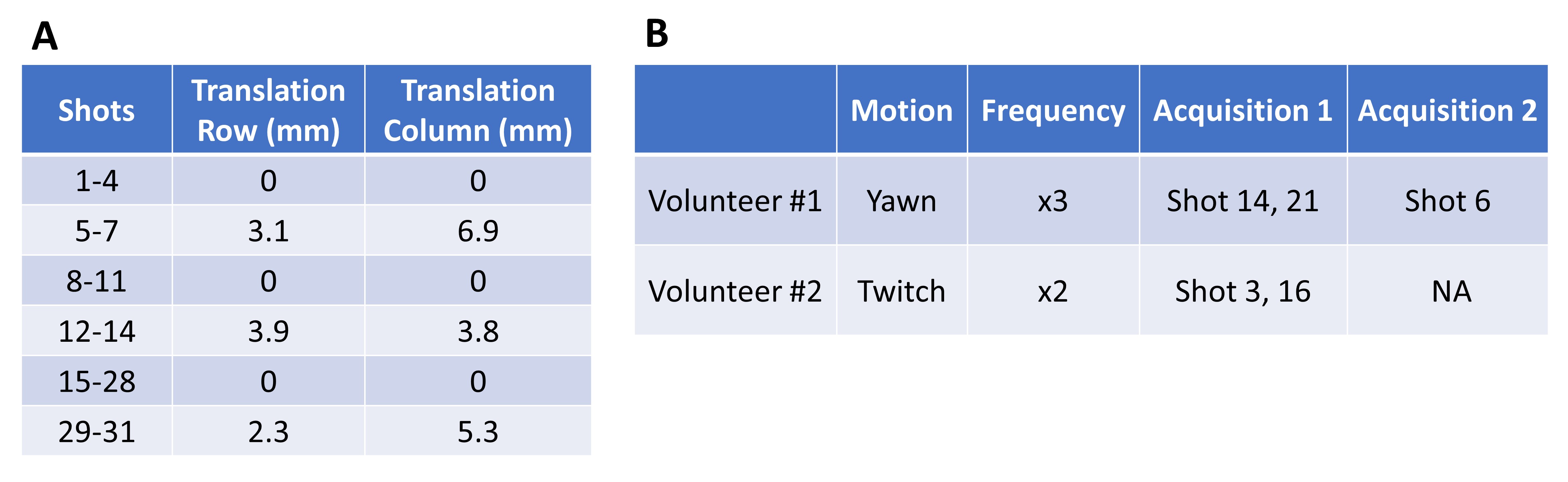
Empirical studies with training data showed that an EED>19.0 could discriminate whether or not significantly more motion existed in one of the two acquisitions. (Simulation Study): One volunteer (male) was scanned using a Vantage Galan 3T MR (Canon Medical Systems Corporation, Tochigi, Japan) and 16-ch Atlas SPEEDER Head/Neck coil under institutional IRB-approved protocol. Data from a 2D FSE pulse sequence were acquired while the volunteer remained still. Sequence parameters were as follows: spatial resolution=0.76 mm x 0.76 mm, slice thickness=3 mm, slice gap=0.6 mm, echo train length=17, number of shots=31, NAQ=2, slices=13. In the first acquisition, rigid-body motion was simulated for 9 of 31 shots. Motion parameters used in the simulation study are presented in Table 1A. For the simulation study, SNR was calculated by drawing regions of interest (ROIs) on the background and spinal cord. (Volunteer studies) Two volunteers (1 male and 1 female) were scanned under institutional IRB-approved protocol with the same system and pulse sequence used for the simulation study. Both volunteers were scanned under different motion conditions for each acquisition (Table 1B), and non-rigid motion correction2 was applied to each acquisition. EED was calculated for each volunteer scanned, and the variant with lower total spatial variation was selected as optimal for cases where EED>19.0.
Results
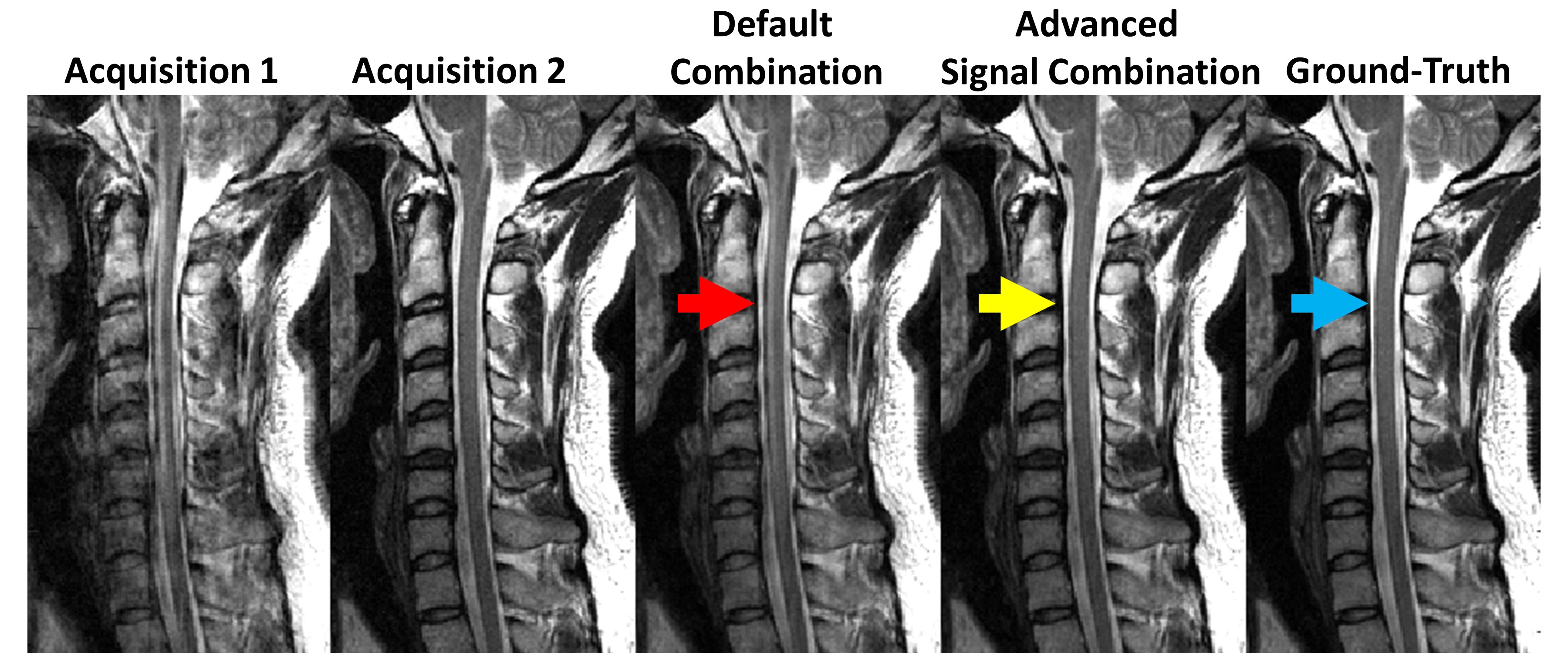
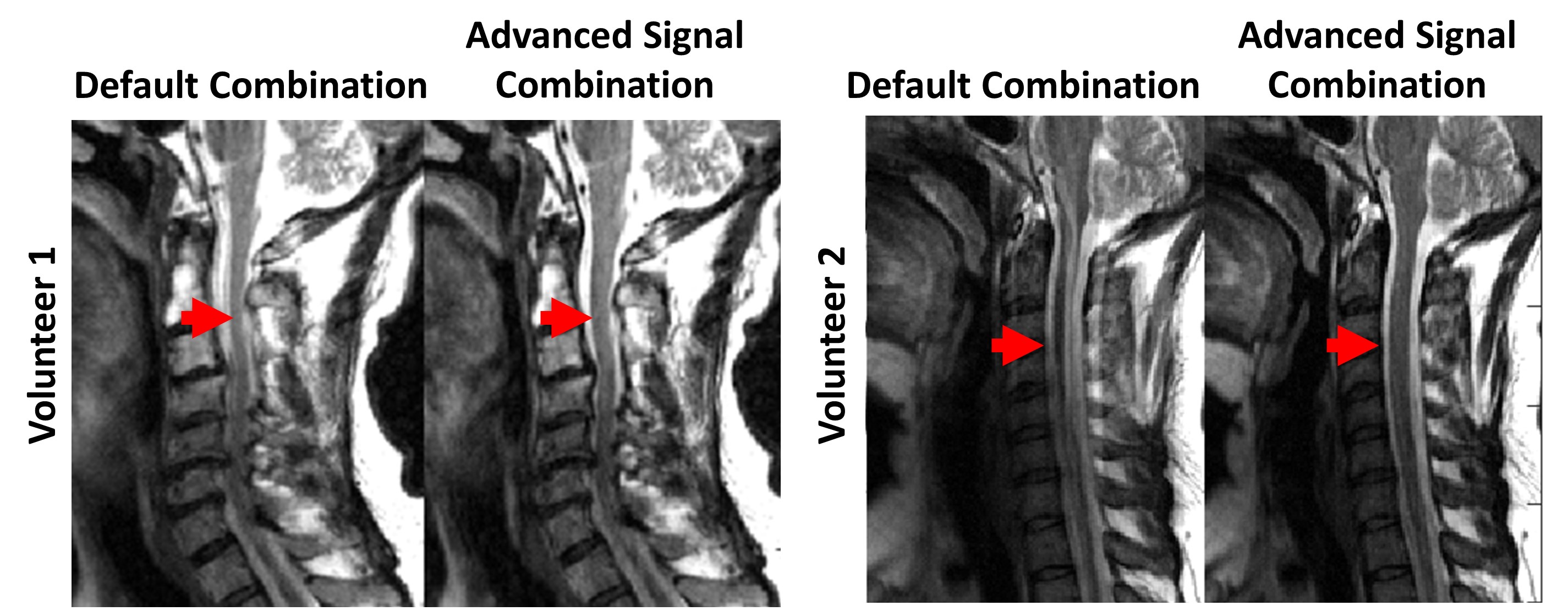
A simulation study (Figure 3) showed that ASC produced better image quality (IQ) and SNR than the second acquisition alone (i.e. the acquisition without simulated motion), but worse SNR than default combination without simulated motion (ground-truth) (SNR: Acquisition 2=24.2, ASC=30.5, ground-truth=35.0). SNR was not calculated for acquisition 1 and default combination with simulated motion because of low IQ. EED>19.0 was found for both volunteer scans, and ASC produced better IQ than default combination (Figure 4).Discussion
During volunteer studies, ASC suppressed motion artifacts in sagittal T2-weighted C-spine MRI with non-rigid motion correction. A simulation study showed that ASC could suppress motion artifacts while generating better SNR than a single acquisition. (Limitations) (1) During simulation study, ASC resulted in lower SNR than default combination with simulated motion and ground-truth. This limitation is due to the fact that ASC rejected 10% of total k-space lines acquired during NAQ=2 scanning. (2) ASC could fail to improve IQ when both acquisitions exhibit high motion artifact. (3) ASC may lead to reduced IQ if the wrong acquisition (i.e. the acquisition with higher motion) is selected, or if ASC is applied when no significant difference in motion exists between acquisitions. (Future Direction) Additional studies are needed to extend ASC to NAQ>2, additional anatomies, different imaging planes, and alternate contrasts. Additionally, studies with large patient cohorts are needed to evaluate ASC’s performance in a clinical setting.Conclusion
This preliminary study showed that ASC could suppress motion artifact in sagittal T2-weighted C-spine MRI with NAQ=2 while maintaining SNR.Acknowledgements
No acknowledgement found.References
1. Pipe JG. Motion correction with PROPELLER MRI: Application to head motion and free-breathing cardiac imaging. Magn Reson Med. 1999;42(5):963-969.
2.Huang F, Lin W, Börnert P, Li Y, Reykowski A. Data convolution and combination operation (COCOA) for motion ghost artifacts reduction. Magn Reson Med. 2010;64(1):157-166.
3. Lin W, Huang F, Börnert P, Li Y, Reykowski A. Motion correction using an enhanced floating navigator and GRAPPA operations. Magn Reson Med. 2010;63(2):339-348.
4.Wallace TE, Afacan O, Waszak M, Kober T, Warfield SK. Head motion measurement and correction using FID navigators. Magn Reson Med. 2019;81(1):258-274.
5. Kober T, Marques JP, Gruetter R, Krueger G. Head motion detection using FID navigators. Magn Reson Med. 2011;66(1):135-143.
6.Atkinson D, Hill DLG, Stoyle PNR, et al. Automatic compensation of motion artifacts in MRI. Magn Reson Med. 1999;41(1):163-170.
7.Cordero-Grande L, Teixeira RPAG, Hughes EJ, Hutter J, Price AN, Hajnal JV. Sensitivity Encoding for Aligned Multishot Magnetic Resonance Reconstruction. IEEE Trans Comput Imaging. 2016;2(3):266-280.
8. Lange T, Taghizadeh E, Knowles BR, et al. Quantification of patellofemoral cartilage deformation and contact area changes in response to static loading via high-resolution MRI with prospective motion correction. J. Magn. Reson. 2019;50(5):1561-1570.
Figures
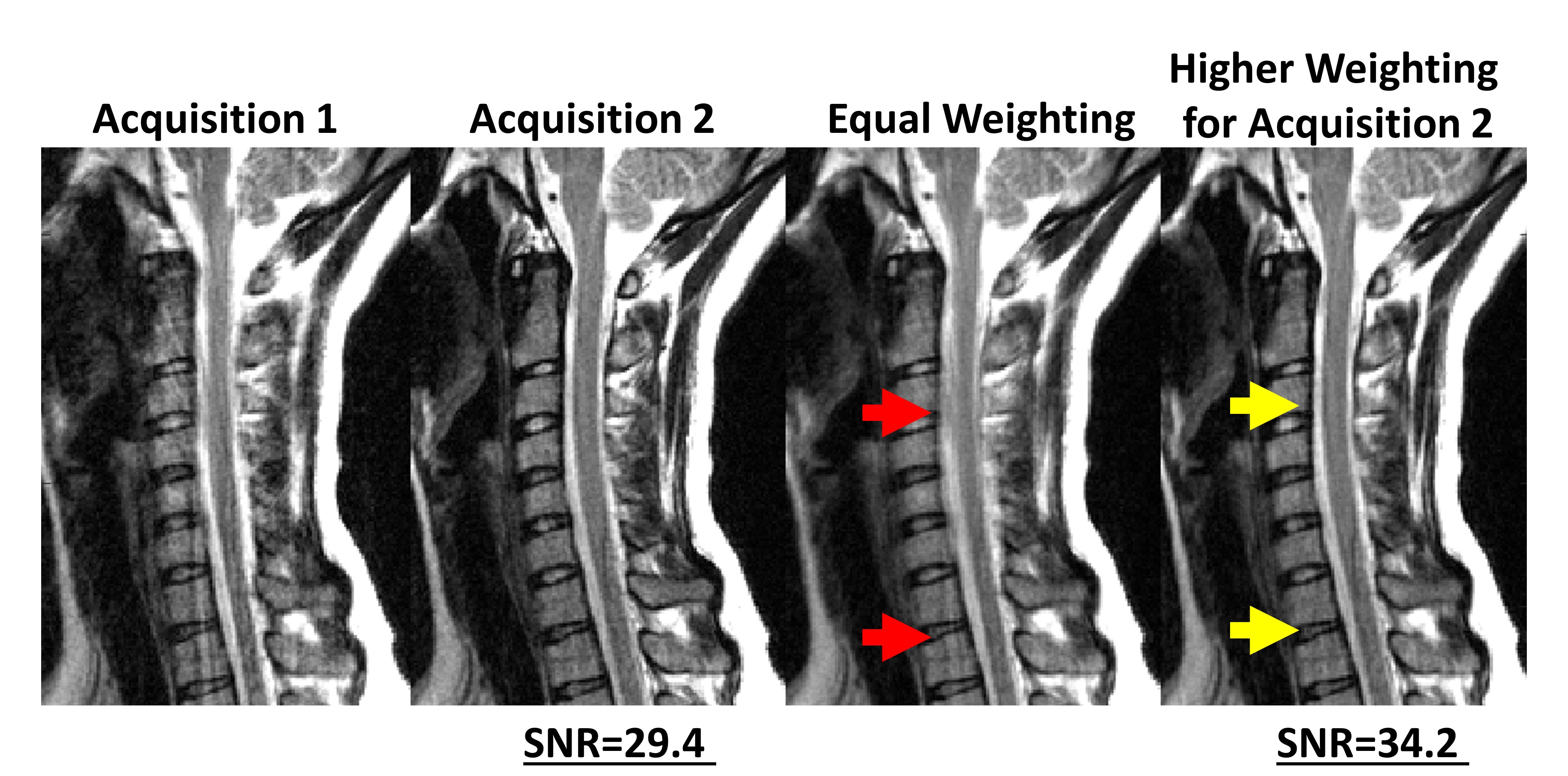
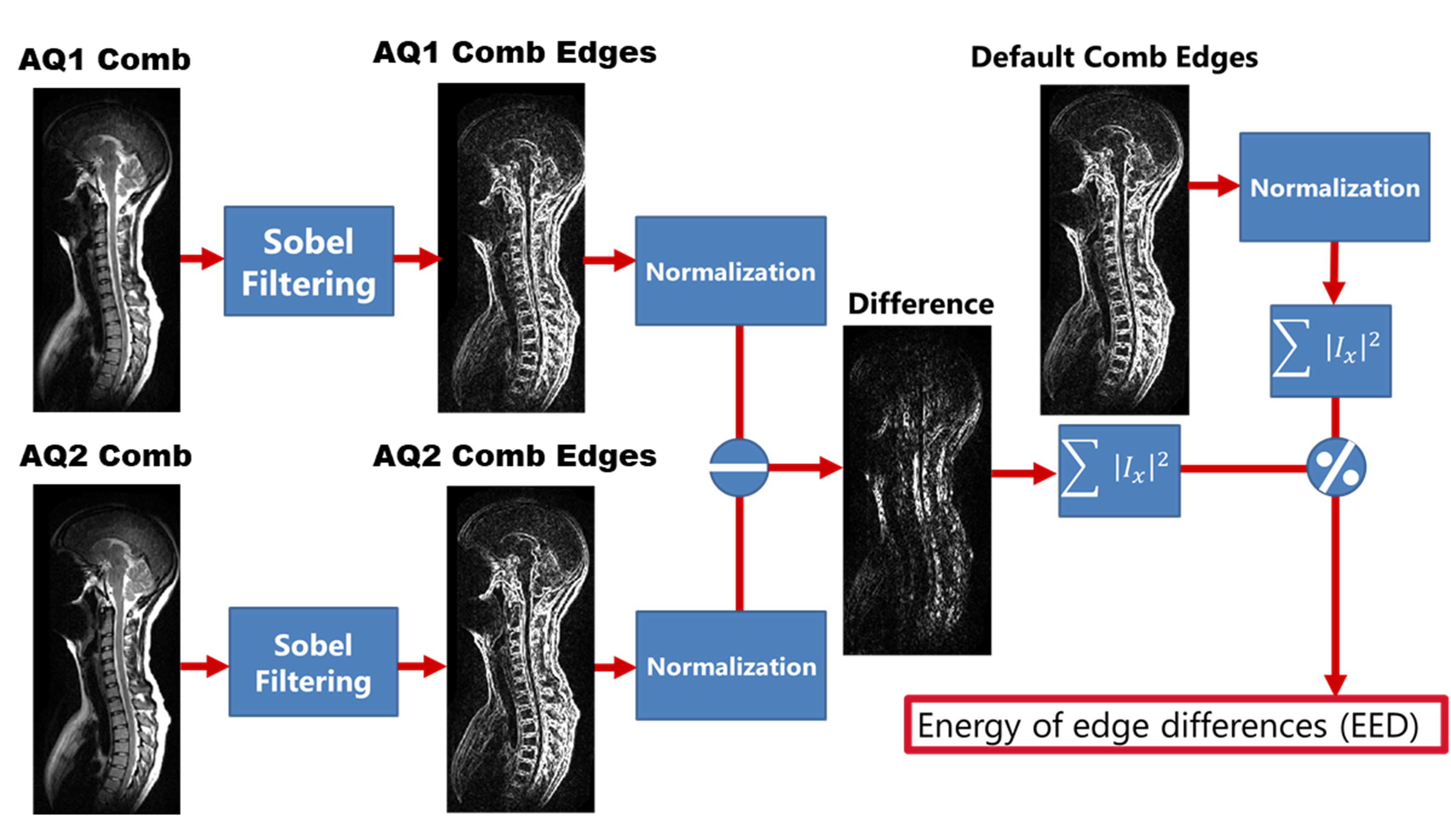
Figure 2: The procedure used to calculate energy of edge difference (EED) is shown. Initially, two acquisitions are combined with equal weighting (Default Comb). Afterwards, the central 20% of PE lines in k-space were replaced with either the first acquisition (AQ1 Comb) or second acquisition (AQ2 Comb). Image edge content for both variants and default comb were calculated using Sobel edge detecting kernels and normalized with the 95th percentile pixel intensity. EED was then calculated using the following formula:
EED=Σ |AQ1 Comb Edges - AQ2 Comb Edges|2/Σ| Default Comb Edges|2 x100