4979
Highly Accelerated Multiple Parametric MR Imaging with Wave-CAIPI and MULTIPLEX1Shenzhen Institute of Advanced Technology, Chinese Academy of Sciences, Shenzhen, China, 2UIH America, Inc., Houston, TX, United States
Synopsis
A novel single-scan hybrid 3D imaging method using Wave-CAIPI and MULTIPLEX technologies, named as WAMP, is proposed for 3D high-resolution rapid imaging. One single scan of the proposed rapid imaging method not only generates simultaneous B1t and T1 maps, but also qualitative images of T1W, PDW, T2*W, augmented T1W (aT1W), SWI and MRA, as well as parametric maps of T2* (R2*), PD and QSM. The in vivo experiments have showed that the proposed method could rapidly achieve high image quality and high quantification accuracy at the high acceleration factors in clinical applications.
Introduction
Recently, many multi-parametric MRI techniques have been developed and explored for quantifying one or more relaxometric parameters and for generating multiple contrast-weighted images[1-20]. Such as, MRF[4], STAGE[5-7], MPME[8-9], MAGiC[10], EPIMix[11-13], QRAPMASTER[14], EPTI[15], ME-MP2RAGE[16], Multitasking[17-18], MULTIPLEX[20], and so on. However, the current challenges of multi-parametric imaging still lie in 3D high-resolution (e.g. voxel size <1mm3) imaging capacity, high data acquisition efficiency, well-formulated signal model, compatibility with various sequence design, acceleration techniques, etc[20]. In this study, we proposed a hybrid rapid imaging method of single scan, incorporating Wave-CAIPI[21-24] and MULTIPLEX[20] technologies, for 3D high-resolution multi-parametric imaging of single scan which was named as WAMP. One single WAMP scan could not only generate simultaneous B1t and T1 maps, but also qualitative images of T1W, PDW, T2*W, augmented T1W (aT1W), SWI and MRA, as well as parametric maps of T2* (R2*), PD and QSM. The in vivo experiments demonstrated the proposed method can achieve high image quality and high quantification accuracy with higher imaging speed than before.Methods
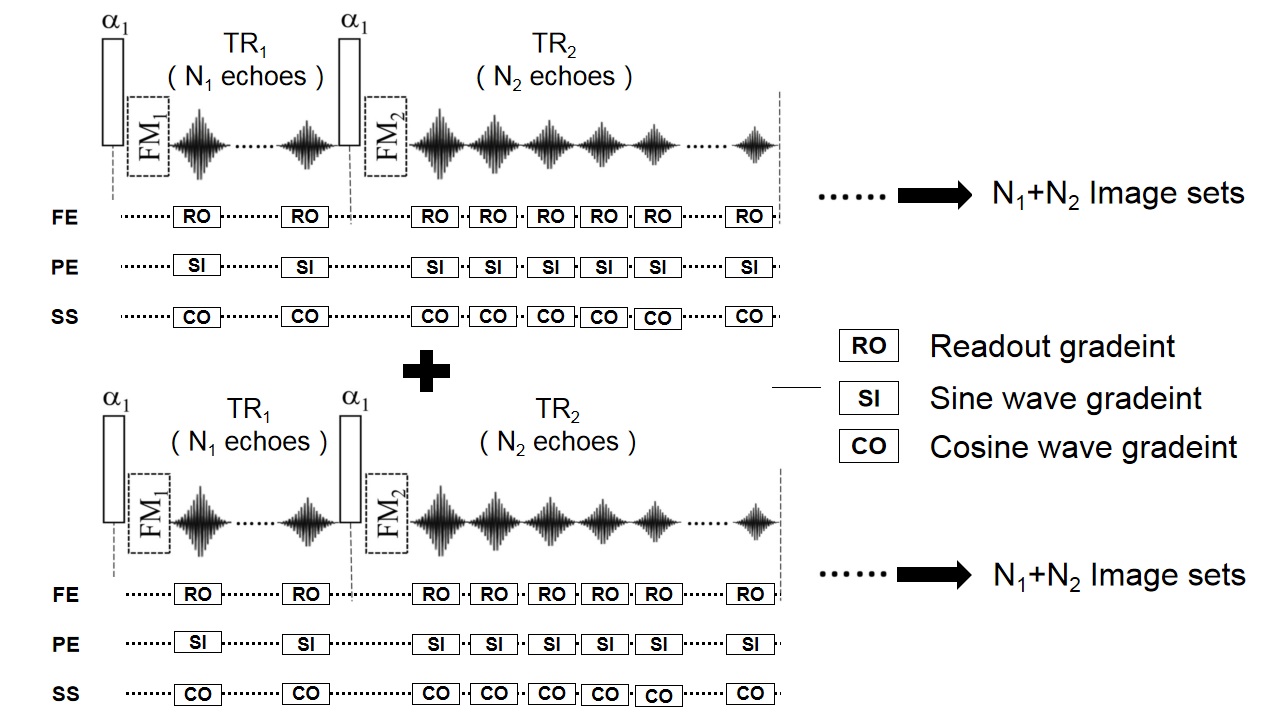
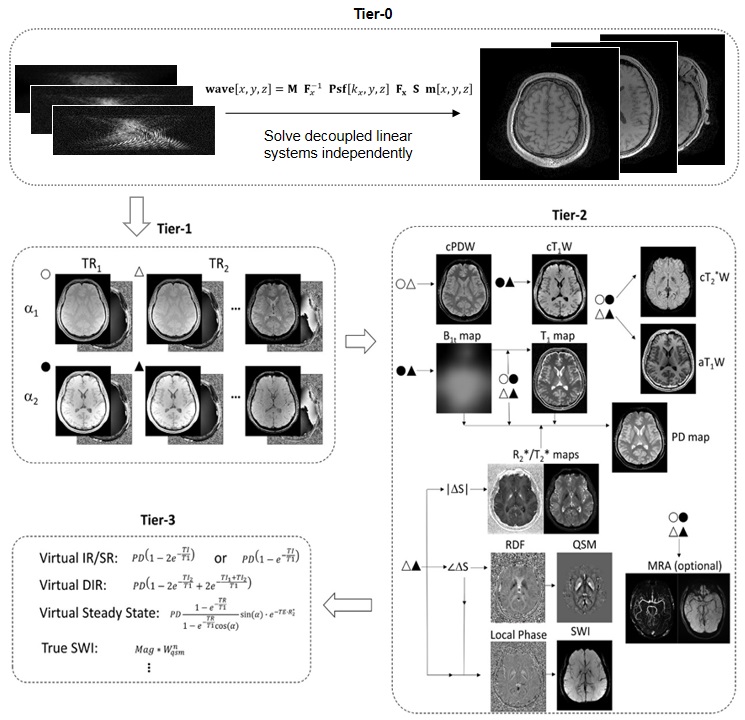
Pulse Sequence: The proposed sequence features in a dual-TR, dual-FA (flip angle) and multi-echo design, as illustrated in Figure 1. The dual-TR feature is derived from the actual flip angle imaging (AFI) technique[25] for B1t mapping. Within each TR (i.e. TR1 and TR2), a number (i.e. N1 and N2) of gradient echoes can be acquired, providing different levels of susceptibility weighting effects[26]. Simultaneously with the readout gradients of each gradient echoes, two wave gradients of sinusoidal waveforms with phase shift of π/2 are applied in the phase encoding direction and the slice encoding direction, respectively. The WAMP sequence is composed of two repetitions of the dual-TR acquisition, each time using a different FA to introduce different levels of T1 weightings. In addition to the wave gradients, the 2D-CAIPI sampling scheme[27] employs for imaging accelerations in the phase encoding direction and the slice encoding direction. In sum, one WAMP scan generates 2(N1+N2) sets of echo images. Furthermore, flow modulation (FM) blocks can be optionally inserted after each excitation pulses.Image Reconstruction: Image reconstructions and processing were implemented by in-house C++ programs on the ADEPT (UIH, Shanghai, China) platform. As shown in Figure 1, 2(N1 + N2) echo image sets can be generated in a WAMP sequence scan. In addition, flow modulation (FM) blocks can be selectively inserted after each excitation pulse. The processing pipeline of image data scanned by the proposed WAMP sequence is shown in Figure 2. For simplicity, WAMP data images are divided into four layers (Tier-0, Tier-1, Tier-2, and Tier-3), depending on their computational order and relationships in the MDI[28] processing pipeline for complex image processing. The Tier-0 layer is to reconstruct images from the "original" echo image of 2(N1+N2) groups through the Wave-CAIPI model[21]. The Tier-1 layer is to model images using the 2(N1+N2) group echo image from the Tier-0 layer[20]. The Tier-2 layer is to calculate the composite PDW/T1W/T2*W (cPDW/cT1W/cT2W), B1t, T1, aT1W, T2*/R2*, PD, SWI, QSM, MRA images; The Tier-2 layer is to calculate cPDW, cT1W, and QSM images, using the Bloch simulation. The proposed method can provide multiple sets of qualitative images and multiple contrast parameter maps with high SNR, quantitative accuracy and acquisition/reconstruction efficiency.
In Vivo Experiment: All MRI scans were performed on a 3T system (uMR790, UIH, Shanghai, China). The protocols were approved by the Institutional Reviews Board (IRB) of Shenzhen Institutes of Advanced Technology, Chinese Academy of Science. A healthy volunteer was scanned using a 24-channel head coil. The scan parameters of MULTIPLEX and WAMP were: α1/α2 = 4°/16°, TR1/TR2 =8/39.3ms, BW=275Hz/px, matrix size=224×224×60, voxel size=1×1×3 mm3. The wave amplitudes of the WAMP were chosen as 12 mT/m with wave cycles of 7.
Maximum Acceleration Experiment: The whole brain of another volunteer were also scanned on the same 3T MR system (uMR790, UIH, Shanghai, China). The 32-channel head coil were used and IRB-approved healthy subjects were enrolled in the experiments. The point spread functions were estimated using two-dimension projection data[22]. The scan parameters of MULTIPLEX and WAMP were as similar as above with the maximum acceleration factors.
Results
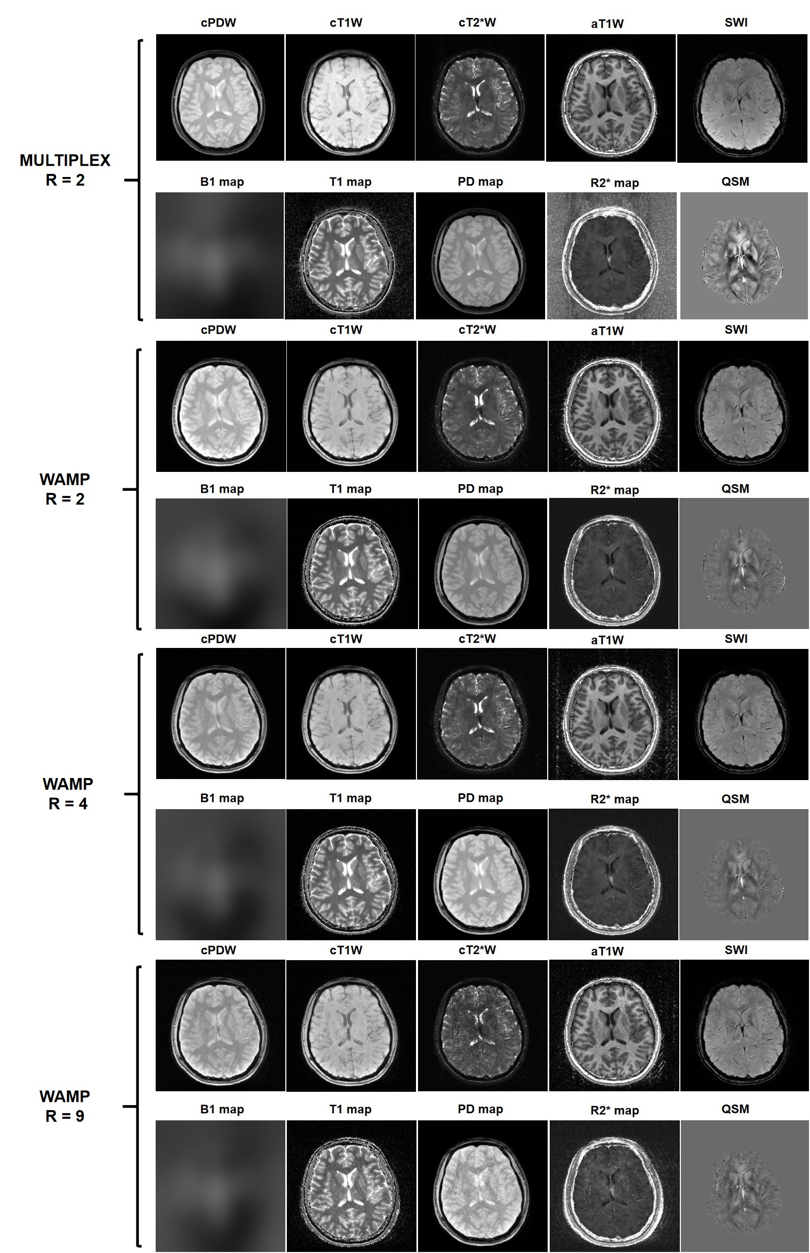
Figure 3 showed some axial slices of in vivo human whole-brain. The imaging acceleration factor of MULTIPLEX was 2; the imaging acceleration factors of WAMP were 2, 4, and 9. The scan time of MULTIPLEX was 11:02 min; the scan time of WAMP were 11:13 min, 5:34 min, and 2:39 min. The proposed method can provide multiple contrast images with higher acquisition efficiency than before.Figure 4 presented some slices of in vivo human brain. The maximum acceleration factor of MULTIPLEX was 3, but the acceleration factors of WAMP were 3 and 9. And the MULTIPLEX and WAMP comparisons of cT1W, cPDW, cT2*W, T1 map, PD map and R2* map, the proposed method can also provide high-quality images at the high accelerations.
Discussion and Conclusion
In this paper, the proposed method has implemented with the dual-TR, dual-FA, multi-echo GRE-based sequence design, wave gradients, 2D-CAIPI sampling scheme, the MDI algorithm, etc. All of the in vivo experiments have demonstrated the proposed single-scan method can yield high good-quality images under high resolutions and high acceleration factors for diagnose applications.Acknowledgements
Some of the work was partially supported by the National Natural Science Foundation of China (61871373, 81729003, and 81901736), the Strategic Priority Research Program of Chinese Academy of Sciences (No. XDB25000000), the Chinese Academy of Sciences Engineering Laboratory for Medical Imaging Technology and Equipment (No. KFJ-PTXM-012), the Pearl River Talent Recruitment Program of Guangdong Province (2019QN01Y986), the Shenzhen Science and Technology Program (JCYJ20210324115810030), the Shenzhen Peacock Plan Team Program (No. KQTD20180413181834876), and the Shenzhen Key Laboratory of Ultrasound Imaging and Therapy (No. ZDSYS201802061806314).References
[1]. Schmitt P, Griswold MA, Jakob PM, et al. Inversion recovery TrueFISP: Quantification of T1, T2, and spin density. Magn Reson Med. 2004; 51(4): 661-667.
[2]. Gulani V, Schmitt P, Griswold MA, Webb AG, Jakob PMI Jr. Towards a single-sequence neurologic magnetic resonance imaging examination: Multiple-contrast images from an IR TrueFISP experiment. Invest Radiol. 2004; 39(12): 767-774.
[3]. Warntjes JB, Leinhard OD, West J, Lundberg P. Rapid magnetic resonance quantification on the brain: Optimization for clinical usage. Magn Reson Med. 2008; 60(2): 320-329.
[4]. Ma D, Gulani V, Seiberlich N, et al. Magnetic resonance fingerprinting. Nature. 2013;495(7440):187-192.
[5]. Chen Y, Liu S, Wang Y, Kang Y, Haacke EM. Strategically acquired gradient echo (STAGE) imaging, part I: Creating enhanced T1 contrast and standardized susceptibility weighted imaging and quantitative susceptibility mapping. Magn Reson Imaging. 2018; 46: 130-139.
[6]. Wang Y, Chen Y, Wu D, et al. Strategically acquired gradient echo (STAGE) imaging, part II: Correcting for RF inhomogeneities in estimating T1 and proton density. Magn Reson Imaging. 2018; 46: 140-150.
[7]. Haacke EM, Chen Y, Utriainen D, et al. Strategically acquired gradient echo (STAGE) imaging, part III: Technical advances and clinical applications of a rapid multi-contrast multi-parametric brain imaging method. Magn Reson Imaging. 2020; 65: 15-26.
[8]. Cheng CC, Preiswerk F, Hoge WS, Kuo TH, Madore BJM. Multipathway multi-echo (MPME) imaging: All main MR parameters mapped based on a single 3D scan. Magn Reson Med. 2019; 81(3): 1699-1713.
[9]. Cheng CC, Preiswerk F, Madore B. Multi-pathway multi-echo acquisition and neural contrast translation to generate a variety of quantitative and qualitative image contrasts. Magn Reson Med. 2020; 83(6): 2310-2321.
[10]. Tanenbaum LN, Tsiouris AJ, Johnson AN, et al. Synthetic MRI for clinical neuroimaging: Results of the Magnetic Resonance Image Compilation (MAGiC) prospective, multicenter, multireader trial. AJNR Am J Neuroradiol. 2017; 38(6): 1103-1110.
[11]. Skare S, Sprenger T, Norbeck O, et al. A 1-minute full brain MR exam using a multicontrast EPI sequence. Magn Reson Med. 2018;79:3045-54.
[12]. Delgado AF, Kits A, Bystam J, et al. Diagnostic performance of a new multicontrast one-minute full brain exam (EPIMix) in neuroradiology: a prospective study. J Magn Reson Imaging. 2019;50:1824-33.
[13]. Ryu KH, Baek HJ, Skare S, Moon JI, Choi BH, Park SE, Ha JY, Kim TB, Hwang MJ, Sprenger T. Clinical Experience of 1-Minute Brain MRI Using a Multicontrast EPI Sequence in a Different Scan Environment. AJNR Am J Neuroradiol. 2020 Mar;41(3):424-429.
[14]. Warntjes JBM, Dahlqvist O, Lundberg P. A novel method for rapid, simultaneous T1, T2* and proton density quantification. Magn Reson Med. 2007;57:528-537.
[15]. Wang F, Dong Z, Reese TG, et al. Echo planar time-resolved imaging (EPTI). Magn Reson Med. 2019;81(6):3599-3615.
[16]. Metere R, Kober T, Moller HE, Schafer A. Simultaneous Quantitative MRI Mapping of T1, T2* and Magnetic Susceptibility with Multi-Echo MP2RAGE. PLoS One. 2017;12(1):e0169265.
[17]. Christodoulou AG, Shaw JL, Nguyen C, et al. Magnetic resonance multitasking for motion-resolved quantitative cardiovascular imaging. Nat Biomed Eng. 2018;2(4):215-226.
[18]. Ma S, Nguyen CT, Han F, et al. Three-dimensional simultaneous brain T1 , T2 , and ADC mapping with MR Multitasking. Magn Reson Med. 2020;84(1):72-88.
[19]. Ji S, Yang D, Lee J, Choi SH, Kim H, Kang KM. Synthetic MRI: Technologies and Applications in Neuroradiology. J Magn Reson Imaging. 2020 Nov 13. doi: 10.1002/jmri.27440.
[20]. Ye Y, Lyu J, Hu Y, Zhang Z, Xu J, Zhang W. MULTI-parametric MR imaging with fLEXible design (MULTIPLEX). Magn Reson Med. 2021 Aug 31. doi: 10.1002/mrm.28999.
[21]. Bilgic B, Gagoski BA, Cauley SF, et al. Wave-CAIPI for highly accelerated 3D imaging. Magn Reson Med. 2015;73(6):2152-2162.
[22]. Wang H, Qiu Z, Su S, Jia Z, Li Y, Liu X, Zheng H, Liang D. Parameter optimization framework on wave gradients of Wave-CAIPI imaging. Magn Reson Med. 2020 May;83(5):1659-1672.
[23]. Su S, Qiu Z, Luo C, Shi C, Wan L, Zhu Y, Li Y, Liu X, Zheng H, Liang D, Wang H. Accelerated 3D bSSFP Using a Modified Wave-CAIPI Technique With Truncated Wave Gradients. IEEE Trans Med Imaging. 2021 Jan;40(1):48-58.
[24]. Qiu Z, Jia S, Su S, Zhu Y, Liu X, Zheng H, Liang D, Wang H. Highly accelerated parallel MRI using wave encoding and virtual conjugate coils. Magn Reson Med. 2021 Sep;86(3):1345-1359.
[25]. Yarnykh VL. Actual flip-angle imaging in the pulsed steady state: a method for rapid three-dimensional mapping of the transmitted radiofrequency field. Magn Reson Med 2007;57(1):192-200.
[26]. Denk C, Rauscher A. Susceptibility weighted imaging with multiple echoes. J Magn Reson Imaging. 2010;31(1):185-191.
[27]. Breuer F, Blaimer M, Mueller MF, Seiberlich N, Heidemann RM, Griswold MA, Jakob PM. Controlled aliasing in volumetric parallel imaging (2D CAIPIRINHA). Magn. Reson. Med. 2006;55.3:549-556.
[28]. Ye Y, Lyu J, Sun W, et al. A multi-dimensional integration (MDI) strategy for MR T2 * mapping. NMR Biomed. 2021:e4529.
Figures