4963
Analysis of white matter tissue integrity of COVID-19 recovered patients using diffusion weighted imaging1School of Engineering and Science, Tecnologico de Monterrey, Mexico City, Mexico, 2School of Medicine and Health Sciences, Tecnologico de Monterrey, Monterrey, Mexico, 3School of Engineering and Science, Tecnologico de Monterrey, Monterrey, Mexico, 4Neuroimaging, Instituto Nacional de Neurologia y Neurocirugía, Mexico City, Mexico, 5Instituto Nacional de Enfermedades Respiratorias, Mexico City, Mexico
Synopsis
Evidence supports that the virus SARS-CoV-2 has a potential neuroinvasion causing neurological symptoms, even after recovery. Diffusion Weighted Imaging (DWI) allows us to detect the microstructural changes in white matter tissue integrity. In this work, we studied the neurological manifestations of COVID-19 in apparent diffusion coeficient (ADC), fractional anisotropy (FA), and metrics derived from fixel-based analysis (FBA) such as fiber density (FD), fiber cross-section (FC), and fiber density and cross-section (FDC). We found statistically significant differences in 34/43 fiber bundle regions from the IIT Human Brain Atlas in the five different metrics studied.
Introduction
Evidence supports that the virus SARS-CoV-2 has a potential neuroinvasion causing neurological symptoms, even after recovery1,2. Previous studies report neurological findings on clinical manifestations associated to the virus. Nervous system symptoms such as dizziness, headaches, impaired consciousness, acute cerebrovascular disease, ataxia, seizures, nerve pain, and impairment of taste and smell had been reported since the beginning of the pandemic1. This has been denominated as neuro-covid2 and these reported abnormalities might cause a long-term burden in these patients after recovery. Previous work using diffusion tensor imaging (DTI) compared COVID-19 recovered patients with non-COVID controls found significant changes in MD and FA3. However, it is difficult to attribute differences to specific structural changes at the microscopic level. Fixel based analysis has proven to provide better insight into white matter (WM) tissue integrity when compared to DTI. Based on such framework, the purpose of this preliminary study was to evaluate WM tissue integrity of COVID-19 recovered patients using both DTI and FBA analysis.Methods
Participants were recruited at the National Institute of Neurology and Neurosurgery in Mexico City. A total of 27 COVID-19 recovered patients (9 female, 64 ±4.7 years) and 25 Non-COVID healthy controls (12 female, 65±4.5 years) were studied. No statistical difference was found in the groups ages.MR data were acquired using a Siemens 3 Tesla Magnetom Skyra (Siemens Healthenieers, Germany). DWI was performed using the following parameters: 72 axial slices, 64 directions, b-values= 0, 1000 and 2000 s/mm2, TR/TE=5000/102 ms, 2.2 mm isotropic resolution, obtained in anterior-posterior (AP) phase encoding direction. Also one b0 volume was acquired in posterior-anterior (PA) phase encoding (PE) direction for eddie currents and EPI distortion correction.
Diffusion data analysis was performed using MRtrix 3.04. Preprocessing of DWI data included denoising, Gibbs unringing, motion and eddy current-induced distortion correction, and bias field correction. EPI distortion correction was performed using a pair of b0s in PE and reversed PE directions for the patient’s data, however for the control group the reversed PE direction b0 volume was performed using the Synthesized b0 for diffusion distortion correction (Synb0-DisCo)5 . Then we computed FA and ADC maps for the DTI analysis and FD, FC and FDC for the Fixel Based Analysis6.
Data was then registered to the Illinois Institute of Technology (IIT) Human Brain Atlas (v.5.0)6 to compute mean values of the DTI and FBA metrics for the 42 white matter fiber bundles and the sum of all tracts (TDIsum).
Statistical analysis was performed in python using the stats package (The SciPy community, 2021). First, we proved for normality and homoscedasticity to then perform either a t-test for equal means when the requirements were met, or a Mann-Whitney test when not.
Results
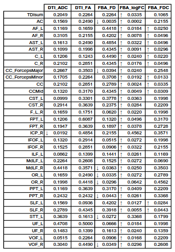
When comparing COVID-19 recovered patients to the control group, we found statistically significant differences in a total of 34 out of 43 regions of interest studied. For the DTI analysis, we only found a reduction in the ADC for the right inferior cerebellar peduncle (ICP_R) (p = 0.0192). No significant difference for FA. On the other hand, for FBA metrics we found significant reductions in 8, and increment in 31 and 11 white matter bundles for FD, FC and FDC, respectively (TABLE 1) when comparing COVID-19 recovered patients to healthy controls.Discussion
In this preliminary study we used both DTI and FBA derived metrics for the assessment of brain white matter alterations in COVID-19 recovered patients when compared to age matched healthy controls. Our preliminary results confirm the superiority of FBA over DTI as a more sensitive method for the analysis of DWI6. Overall we found a generalized increase in fiber crossection (reported as log_FC) and decrease in fiber density for some regions. These results suggest white matter alterations derived from COVID-19 infection, however a more in-depth analysis is required in order to correlate them with the reported symptoms. Also, limitations of our study include a relatively low sample size and the use of Synb0-DisCo for the preprocessing of the control group. In conclusion, we showed WM alterations in COVID-19 recovered patients when compared to healthy controls using FBA analysis.Acknowledgements
We would likw to acknowledge the medical staff fighting the pandemic in Mexico City at the Instituto Nacional de Enfermedades Respiratorias and Instituto Nacional de Neurología y NeurocirugíaReferences
1. Mao L, e. a. (2020). JAMA Neurology, 683-690.
2. Ferrarese, C, et al. Neurological Sciences 41 (2020): 1355-1359.
3. Lu M, e. a. (2020). EClinicalMedicine, 12.
4. Tournier, J‐D et al. International journal of imaging systems and technology 22.1 (2012): 53-66.
5. Schilling, Kurt G. et al. Magnetic resonance imaging 64 (2019): 62-70.
6. Raffelt, David A., et al. Neuroimage 144 (2017): 58-73.
7. Farquharson S., e. a. (2013). Journal of Neurosurgery, 1367-1377.
Figures