4952
Bone-marrow fatty-acids profiles from women calcaneus by 1H-MR Spectroscopy to search early osteoporotic biomarkers1Physics Dpt., Sapienza University of Rome, Rome, Italy, 2Center for Prevention, Diagnosis and Treatment of Osteoporosis, IRCCS Santa Lucia Foundation, Rome, Italy, 3Institute for Complex Systems, National Research Council CNR-ISC, Rome, Italy, 4Department of Orthopaedics and Traumatology, Tor Vergata University of Rome, Rome, Italy, 5Institute for Complex Systems, National Research Council CNR-ISC, Roma, Italy
Synopsis
Changes occurring in the bone-marrow fatty-acids (BMFA) could be precursors of the bone mineral loss due to the development of osteoporosis. In this view, early biomarkers of osteoporosis among BMFA could be highlighted by quantifying fatty-acids with 1H-MRS in young, premenopausal, postmenopausal women with normal t-score and in osteoporotic women. In this study, L13, L09, fUFA, and fPUFA together with TL quantification obtained in women calcanei could be early osteoporosis markers. The present work confirms the potential of MRS to investigate BMFA metabolic variations due to postmenopause and to the development of osteoporosis.
Introduction
Aging is associated with loss of bone density and increased fracture risk (i.e., osteoporosis). Moreover, in women, menopause status is an additional factor risk for osteoporosis. During the last years1,2 there is increasing evidence that age-associated changes in bone marrow fatty acids (BMFA) may severely affect musculoskeletal system and adipose distribution. In particular, the changes occurring in the BMFA could be precursors of the bone mineral loss due to the development of osteoporosis. In this view, early biomarkers of osteoporosis among BMFA could be highlighted by quantifying fatty-acids with 1H-MRS in young, premenopausal, postmenopausal women with normal t-score and in osteoporotic women. Aim of this study was to identify early relevant changes in the metabolic profile of BMFA of women during healthy aging to osteoporosis status by 1H-MR Spectroscopy. A better understanding of metabolism change via the alterations of selected fatty acids may pave the way for novel medical interventions in musculoskeletal diseases such as osteoporosis.Methods
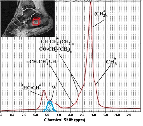
Thirty-five Caucasian subjects (age range: 22-59y) were recruited to investigate calcaneous bone-marrow. Subjects were excluded in the case of clinical evidence for metabolic bone disease or metastasis, previous history of irradiation and current use of steroids or hormone replacement therapy. From each volunteer clinical data (age, menopausal age, T-score) were collected. The study was approved by the Local Ethics Committee. Written informed consent was obtained from each subject before MRS examination. The following group were analysed: young group, with age less than 35y (mean age =26.5±3.8y), pre-menopausal group (age range 37-47, mean age =42.0±3.6y), postmenopausal group ( age range 50-59y, mean age =55.4±2.9y), and osteoporotic subjects (age range 50-58, mean age =53±2.8y). Experiments: Single-voxel MRS was performed by using point resolved spectroscopy (PRESS) sequence. Axial and coronal T2-weighted images of foot site were acquired for positioning a 15×15×15mm3 voxel in the center of the calcaneus of each subject. Before all acquisitions, the static field homogeneity was optimized in the volume of interest using a second-order shimming routine. Each subject underwent a MRS examination of the right foot, using an 3T MR scanner(Allegra; Siemens Medical System, Erlangen, Germany), equipped with a circularly polarized volume head coil for radiofrequency transmission and reception. The acquisition parameters were: TR=4000ms, TE=22ms, NSA=32 without water suppression, spectral bandwidth (BW) 2000Hz and 1024 data points. A homemade positioning device was used to immobilize foot during MR acquisition.Processing and statistical analysis: Row-data of spectra acquired from each subject were analyzed using LcModel software (SPTYPE 6). Zero and first order phase corrections were performed. Nine proton resonances were quantified in bone-marrow spectra: CH3 (labeled L09, at 0.90ppm), bulk CH2 (L13 at 1.3ppm), the CH2α- (L23 at 2.3ppm) and β- (L16 at 1.59ppm) to the carbonyl, allylic CH2 α to a double-bound (L21 at 2.03ppm), water (W at 4.7ppm ), glycerol CH2, glycerol CH (L41+L43 at 4.10-4.35ppm) and olefinic double-bond protons (L52+L53 at 5.20ppm) (Fig.1). Data with a %SD equal or higher than 10% were discarded. The following parameters were evaluated after fatty-acid quantification3,4: Total lipids (TL), unsaturated index (UI), unsaturated fatty-acids fraction, fUFA, polyunsaturated fatty-acids fraction, fPUFA3. Homogeneity of variances between groups was tested by using Levene’s. Pairwise comparisons between groups were made using a Welch ANOVA. Pearson correlation was studied between fatty-acids parameters, age, menopausal age, T-score.
Results
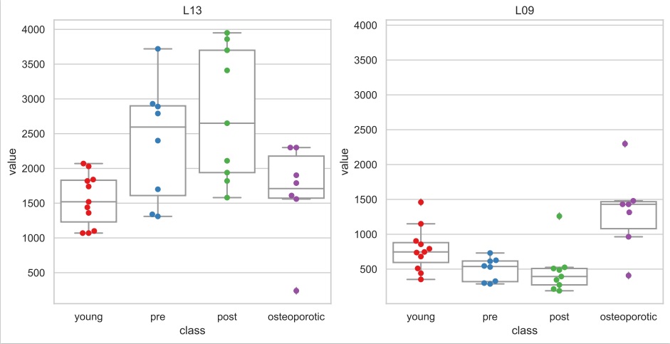
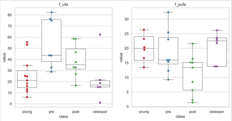
A reproducibility study of BMFA quantification was performed as reported in a previous work2. The reproducibility was in agreement with that reported in Di Pietro et al. paper 2.As expected 5, a strong linear correlation was found between the age at menopause and T-score (r = 0.737, p < 0.01). A significant correlation was found between TL and age (r=0.919, p<0.001) in young and premenopausal subjects (Fig.2) and between TL and time elapsed from menopause (r=0.520, p<0.05) in over 47y subjects. L13 and L09 significantly discriminate between postmenopausal and osteoporotic women (p=0.03 and p=0.006, respectively), (Fig.3). L52+L53 was significantly lower in young group compared to all the other groups. L21 and fPUFA significantly discriminates between pre- and postmenopausal women (p=0.02 and p=0.04, respectively). Moreover, fPUFA and fUFA discriminate between postmenopausal and osteoporotic group (Fig.4). No significant discrimination in UI was found between groups.
Discussion
Compared to the other skeletal sites, the calcaneus is already very rich in fat and probably it becomes fat-saturated with aging (Fig.2). This determines a different metabolism/quantification of BMFA in the calcaneus compared to that obtained in the vertebrae/femur1,2,6,7. According to literature7,8, fUFA decreases from normal (pre+post-menopausal) to osteoporotic women (Fig.4). In this work, we also highlight that fPUFA is significantly lower in postmenopausal compared to osteoporotic subjects. This data suggests that osteoporosis is related to an unbalance between saturated and unsaturated fatty acid proportions. On the other hand, L13 and L09, which are related to saturated fat, are candidate markers to detect osteoporosis (Fig.3).Conclusion
L13, L09, fUFA, and fPUFA together with TL quantification obtained in women calcaneus could be early osteoporosis markers. The present work confirms the potential diagnostic utility of MRS to investigate BMFA metabolic variations due to postmenopause and to the development of osteoporosis.Acknowledgements
This work is a part of the project DETERMINER Founded by: Lazioinnova – Regione Lazio
A0375-2020-36705 (Italy)
References
1. Patsch, Janina M., et al. "Bone marrow fat composition as a novel imaging biomarker in postmenopausal women with prevalent fragility fractures." Journal of bone and mineral research 28.8 (2013): 1721-1728.
2. Di Pietro, Giulia, et al. "Bone marrow lipid profiles from peripheral skeleton as potential biomarkers for osteoporosis: a 1H-MR spectroscopy study." Academic radiology 23.3 (2016): 273-283.
3. Peterson P, Trinh L and Månsson S. Quantitative H MRI and MRS of fatty acid composition. Magn Reson Med. 00 (2020):1–19.
4. Corbin, Ian R., et al. "In vivo assessment of hepatic triglycerides in murine non-alcoholic fatty liver disease using magnetic resonance spectroscopy." Biochimica et Biophysica Acta (BBA)-Molecular and Cell Biology of Lipids 1791.8 (2009): 757-763.
5. Ibrahim, Nihal A., Nessrin Nabil, and Sana Ghaleb. "Pathophysiology of the risk factors associated with osteoporosis and their correlation to the T-score value in patients with osteopenia and osteoporosis in the United Arab Emirates." Journal of pharmacy & bioallied sciences 11.4 (2019): 364.
6. Griffith, James F., et al. "Vertebral marrow fat content and diffusion and perfusion indexes in women with varying bone density: MR evaluation." Radiology 241.3 (2006): 831-838.
7. Yeung, David KW, et al. "Osteoporosis is associated with increased marrow fat content and decreased marrow fat unsaturation: a proton MR spectroscopy study." Journal of Magnetic Resonance Imaging: An Official Journal of the International Society for Magnetic Resonance in Medicine 22.2 (2005): 279-285.
8. Martel, Dimitri, et al. "Chemical shift-encoded MRI for assessment of bone marrow adipose tissue fat composition: Pilot study in premenopausal versus postmenopausal women." Magnetic resonance imaging 53 (2018): 148-155.
Figures