4944
Correlation of 4D MRI and Motion Capture during Dynamic Wrist Movements1Radiology, Medical College of Wisconsin, Milwaukee, WI, United States, 2Department of Rehabilitation Sciences & Technology, University of Wisconsin-Milwaukee, Milwaukee, WI, United States
Synopsis
4D MRI and external motion capture systems were utilized to track unconstrained movement of wrist carpal bones. Using slab-to-volume registration of dynamic MRI to static MRI reference images, wrist kinematic profiles for 19 healthy subjects were computed and compared to gold-standard motion analysis metrics during ulnar/radial deviation and flexion/extension motions. The agreement of kinematic measures derived using the intrinsic (i.e. carpal bone volume) MRI-based approach and external (sensor-based) motion capture methods provided validation of the deployed MRI-based kinematic profiling methodology.
INTRODUCTION
Compared to single-frame static diagnostic images, images of moving joints (kinematic imaging) can provide crucial differentiating information in orthopedic assessments of connective tissue injuries, structural deformities, and structural integrity [1-4]. In the present study, a new approach for kinematic MRI was deployed to capture high resolution static images and dynamic images of the moving joints during wrist radial/ulnar deviation and flexion/extension tasks in 19 healthy subjects. MRI kinematic profiles of angular displacement were obtained by registering the acquired dynamic imaging volumetric frames to a high-resolution static volume. Each imaged subject also underwent motion capture evaluations of the same movements. Results from the motion analysis were used to validate the new MRI-based kinematic profiling technology.METHODS
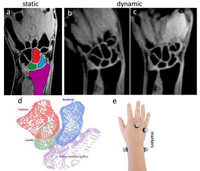
MRI acquisition: After providing written consent to participate in a locally approved IRB protocol, 19 healthy subjects with no prior reported wrist pathology were evaluated. Subjects were positioned in a prone "superman" position for both static and dynamic imaging. MRI data were collected on a GE HealthCare Signa Premier 3T MRI scanner using a 16-channel large flex coil and a 3D LAVA Flex sequence (sample static and dynamic images are shown in Figs. 1a, 1b, and 1c). Static images were acquired with 0.9×0.9×1 mm3 voxel size and an acquisition matrix size of 224×224×60. During dynamic wrist tasks, forty sub-volumes with a temporal resolution of 2.57s were acquired using multi-phase 3D LAVA Flex series with 1.6×1.6×2.5 mm3 voxel size with 128×128×12 acquisition matrix size. Dynamic imaging parameters were chosen to minimize motion artifacts and was a fast enough rate for subjects to complete comfortably. For dynamic imaging, no motion-restriction constraints were utilized. Instead, visual feedback ensured a constant angular velocity during a 103 second acquisition.Segmentation: Individual scaphoid, capitate, and lunate bones were initially auto-segmented from the static and dynamic images using a 3D V-Net neural network [5] that was trained on manually segmented data from a previous study cohort. The neural net segmentations were then screened and modified (if needed) by a locally developed semi-automated seed-based connected-component region growing algorithm. The articular surface of the radius was also segmented using the semi-automated algorithm (Example segmented bones are colored distinctively on Fig. 1a).
Registration: Dynamic sub-volumes of scaphoid, capitate and lunate were registered to their corresponding high-resolution complete static volumes using a point-cloud-based registration method [6] (sample registered point clouds are shown in Fig. 1d). For comparison with motion capture, the angular displacement of the combined center-of-mass of the tracked scaphoid, lunate, and capitate bones relative to the center-of-mass of the distal radial surface was computed from the segmented volumes.
Motion Capture: 3D trajectories of retro-reflective markers placed on the subjects’ wrists and hands (Fig. 1e) were obtained using a 15-camera Vicon TS motion capture system during 5 trials of maximal radial/ulnar and flexion/extension tasks. The trajectories were processed and filtered using Vicon Nexus software. An inverse kinematics model was then applied via MATLAB to calculate the 3D wrist joint angles over the duration of each task [7].
RESULTS AND DISCUSSION
Representative kinematic profiles of the ulnar/radial deviation and flexion/extension wrist joint angles derived from the MR images were compared to those derived from the 3D motion capture data for 5 sample subjects (Fig. 2). The maximum angular displacement (range of motion), which occurs at extreme positions relative to the neutral position, is narrower for the MRI than the motion capture experiments. This range of motion restriction relative to the motion capture analysis was manually measured from the MR images at their visually identified extreme angular displacements, allowing the MR results to be rescaled accordingly. After applying a linear transformation, the MR data showed excellent agreement with the motion capture results thus validating the MR kinematic profiles in both motions.Figure 3 quantifies the agreement between the MR and motion capture kinematic profiles through Bland-Altman and correlation plots (insets) of the rate (i.e., slope of the profiles shown in Fig. 2), which is effectively a measure of the matched curvature between the two modality profiles. Across the 19 studied subjects no consistent bias was found between the two modality measurements. Correlation parameters of R2=0.8, R2=0.9 were obtained for the flexion/extension and radial/ulnar deviation motions, respectively.
CONCLUSIONS
In this study, kinematic profiles of carpal bones relative to the distal radius derived from 4D MRI of unconstrained wrist motion were validated using motion analysis methods. Beyond the simple composite angular displacement measures analyzed in this validation study, many more complex metrics such as gaps between the carpal bones, rotational angles of individual carpal bones, and translational shifts of individual carpal bones can be computed and analyzed using the presented kinematic MRI approach. The MRI acquisition methods demonstrated in this work do not use any prototype pulse-sequences and can easily be implemented using most imaging systems. Ongoing work is utilizing this validated MRI approach to develop normative kinematic profiling templates and assessing variations in pathological subjects with known wrist pathology.Acknowledgements
This research was supported by National Institute of Health (NIH) grant R21AR075327. The authors wish to thank Tugan Muftuler, Andrew Nencka, Robin Ausman, and Brad Swearingen for valuable technical support.References
[1] C. Muhle, et al., Kinematic CT and MR imaging of the patellofemoral joint. European Radiology, 9(3):508–518, 1999.
[2] J.E. Johnson, et al., Scapholunate ligament injury adversely alters in vivo wrist joint mechanics: An MRI-based modeling study. Journal of Orthopedic Research, 31(9):1455–1460, 2013.
[3] G. Li, et al., Feasibility of using orthogonal fluoroscopic images to measure in vivo joint kinematics. J. of biomechanical engineering, 126(2):313, 2004.
[4] B.H. Foster, et al., A principal component analysis-based framework for statistical modeling of bone displacement during wrist maneuvers. J. of Biomechanics, 85:173-181, 2019.
[5] F. Milletari, N. Navab, S-A. Ahmadi, V-Net: fully convolutional neural networks for volumetric medical image segmentation. In: IEEE International Conference on 3D Vision. IEEE Computer Society; 2016:565-571.
[6] M. Zarenia, et al, Unconstrained Kinematic MRI Tracking of Wrist Carpal Bones, arXiv, 2021, 2111.04582.
[7] A.J. Schnorenberg, et al., Biomechanical model for evaluation of pediatric upper extremity joint dynamics during wheelchair mobility. Journal of Biomechanics, 47(1):269, 2014.
Figures


