4935
Characterization of a novel rodent model of arteriovenous malformations (AVMs) with time-of-flight magnetic resonance angiography
Chul Han1, Gregory Harrison Turner2, Candice Nguyen1, Michael Lawton1, and S. Paul Oh1
1Barrow Aneurysm and AVM Research Center, Barrow Neurological Institute, Phoenix, AZ, United States, 2Neuroimaging Research, Barrow Neurological Institute, Phoenix, AZ, United States
1Barrow Aneurysm and AVM Research Center, Barrow Neurological Institute, Phoenix, AZ, United States, 2Neuroimaging Research, Barrow Neurological Institute, Phoenix, AZ, United States
Synopsis
A mouse model of arteriovenous malformations was developed with a genetics-based approach that conditionally deleted the causative activin receptor-like kinase 1 (ACVRL1 or ALK1) gene. This model was characterized in vivo using time-of-flight angiography (TOF_MRA). We correlated radiographic and histopathologic findings and determined that the 55 Tagln-Cre (+);Alk12f/2f mouse was a promising experimental brain AVM model for future studies of AVM pathophysiology, growth, rupture, and therapeutic regression.
Introduction
An arteriovenous malformation (AVM) is an abnormal connection of arteries and veins that forms a low-resistance, high-flow tangle of vessels that is prone to life-threatening hemorrhage and seizure [1]. Recently a genetic mouse model that spontaneously generate AVMs has been developed. This new model has been found to replicate human AVMs better than any previous mouse model of the disease [2]. Further evaluation was needed, however, to determine if this model allowed for extended examination of the disease over time. For this study the Tagln-Cre (+);Alk12f/2f mouse model was characterized in vivo using time of flight angiography (TOF-MRA).Methods
Magnetic resonance imaging was performed using a 7T small-animal, 30-cm horizontal-bore magnet and BioSpec Avance III spectrometer (Bruker, Billerica, MA) with a 116-mm high-power gradient set (600 mT/m) and a 30-mm whole-body mouse quadrature coil. Coronal T2-weighted rapid acquisition with relaxation enhancement images spanning the volume of brain were acquired to identify AVM lesions (TR=5000 ms, TE=12 ms, TEeff=60 ms, Rare Factor=8, 180×180 matrix, 0.1×0.1×0.5-mm voxels, 30 slices, NEX=4). The 3D time of flight MRA images were acquired using a flow-compensated gradient echo method (TR=30 ms, TE=3.5 ms, α= 30°, 180×180×200 matrix, 0.1×0.1×0.1 voxels, NEX=4). Magnetization transfer pulses were added to the MRA acquisition to provide additional background tissue suppression (offset frequency= 1500 Hz, r.f. amplitude= 0.5 μT).Results
Brains of control mice appeared normal on T2 MRI and showed no evidence of AVMs or hemorrhage while MRA showed normal arterial anatomy. The brains of 55 Tagln-Cre (+);Alk12f/2f mutant mice were studied with MRI and MRA and categorized into three groups. Group 1 had no detectable vascular lesions (23 of 55, 42%); group 2 had AV fistulas with no nidus and a dilated draining vein (10 of 55, 18%); and group 3 had AVMs with true nidus consisting of a stereotypical tangle of arteries and dilated veins (22 of 55, 40%). Histologic analysis also validated findings on MRI. AVMs observed in MR images corresponded AVMs and dilated, tortuous vessels in histology. Microhemorrhage was detected adjacent to AVMs in MRI corresponded to iron deposition detected by Prussian blue staining.Conclusion
This study confirms that the novel Tagln-Cre (+);Alk12f/2f mouse model of brain AVM has characteristics similar to human AVMs that can be observed in vivo through MRA. The use of TOF MRI, with its emphasis on vessels with high flow, was particularly adept at detecting the abnormal flow due to the abnormal arterial-venous connection that is a hallmark of AVMs.Acknowledgements
The authors would like to acknowledge the BNI-ASU Center for Preclinical Imaging This work was supported by grants from Barrow Neurological Foundation, Leducq Foundation, the US Department of Defense, and the National Institutes of Health.References
[1] Pan, P, et al., JCBFM, June 2021
[2] Han, C, et al., JNS, in press
Figures
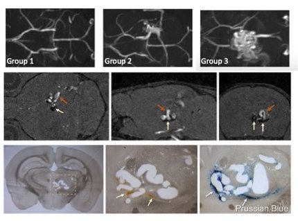
Top Row: MRA images corresponding to three AVM groups. Group 1 (left) contains no discernible lesion. Group 2 (middle) contains a fistula with no nidus. Group 3 (right) contains both a fistula along with a nidus with a large tangle of arteries and veins. Middle Row: 2D MRA sections of an AVM in corresponding axial, sagittal, and coronal views. White arrows indicate the lesion of microhemorrhage adjacent to the nidus. Bottom Row: Coronal section of the brain showing the AVM in the thalamus. White arrows indicate hemosiderin and Prussian blue staining indicating microhemorrhage.
DOI: https://doi.org/10.58530/2022/4935