4934
Vascular map assessment - the dual use of an analysis tool to evaluate imaging technique reproducibility and AI technique’s performance1Vascular Imaging Lab and BioMolecular Imaging Center, Department of Radiology, University of Washington, Seattle, WA, United States, 2Carle Illinois College of Medicine, University of Illinois at Urbana-Champaign, Urbana-Champaign, IL, United States, 3Institute of Science and Technology for Brain-Inspired Intelligence, Fudan University, Shanghai, China
Synopsis
Intracranial artery feature extraction (iCafe), a custom-made semi-automatic vascular map construction software, can quantitatively measure intracranial vascular features on time of flight (TOF) MRA with good scan-rescan and inter/intra operator reproducibility. To fully extend the use of iCafe to a novel MRA technique named simultaneous non-contrast angiography and intraplaque hemorrhage (SNAP), we will first evaluate its vascular feature measurements reproducibility and then compare these measurements with those acquired with a newly developed AICafe (AI + iCafe) technique on the same dataset. Good reproducibility was obtained on SNAP MRA using iCafe, as well as the AICafe technique.
Introduction:
Intracranial artery feature extraction (iCafe), a custom-made semi-automatic vascular map construction software, can measure intracranial vascular features, e.g. artery length, number of branches, and artery volume1 using time of flight (TOF) or simultaneous non-contrast angiography and intraplaque hemorrhage (SNAP) MRA2. iCafe measures on TOF have shown good reproducibility3 and have been used in many studies4,5. More interestingly, compared to TOF, SNAP can provide additional information in distal arteries because of the sensitivity to slow blood flow6 However, the reproducibility of iCafe measurement on SNAP has not yet been reported. Moreover, with the help of deep learning-based open snake tracking algorithm7, artificial intelligence intracranial artery feature extraction (AICafe) was developed to automatically trace arteries on TOF MRA similar to iCafe. Use of AICafe on SNAP MRA has not yet been reported either.Aims:
In this study, we aim to:1) Determine the scan-rescan reproducibility of SNAP MRA using iCafe.
2) Compare the performance of AICafe with iCafe, if SNAP MRA demonstrates good reproducibility.
Methods:
Fifteen subjects were scanned on a Philips Ingenia 3T scanner. The local Institutional Review Board approved this prospective study and all participants gave written informed consent. All subjects were scanned twice within two weeks using the same SNAP imaging protocol: voxel size 0.6mm isotropic, FOV 180×180×70mm3, TR/TE 11ms/6ms, flip angle 11 degrees, with spoiled gradient echo readout. Registration was performed to obtain the common coverage of the first and the second scan. Intracranial artery centerlines were drawn manually by iCafe (A. G., 3-year experience in SNAP MRA review). Independently, the same SNAP images were automatically traced by AICafe. Manual checking of the AICafe traces were carried out to ensure quality of results and remove extracranial arteries if they were traced. Then, artery length, branch number, and artery volume (considering artery as a cylinder with changing radius) were calculated. The coefficient of variation (CV) and intraclass correlation coefficient (ICC) were calculated separately for iCafe and AICafe to measure the scan-rescan reproducibility.Results:
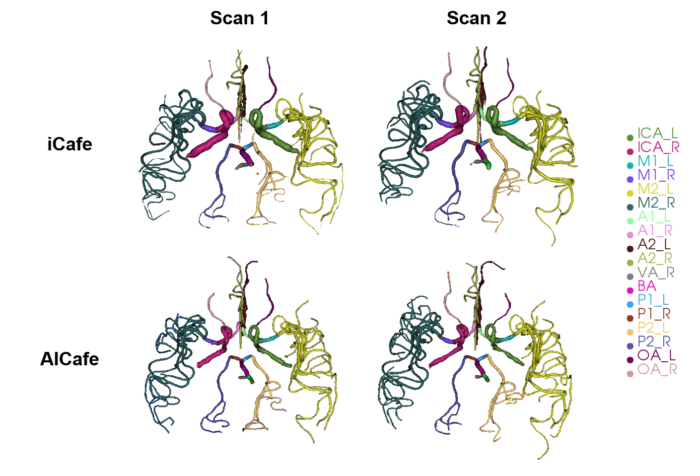
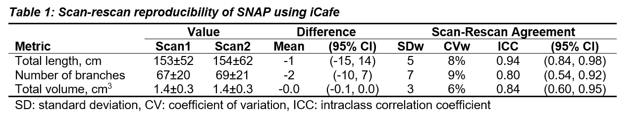
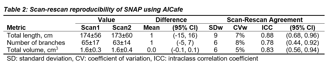
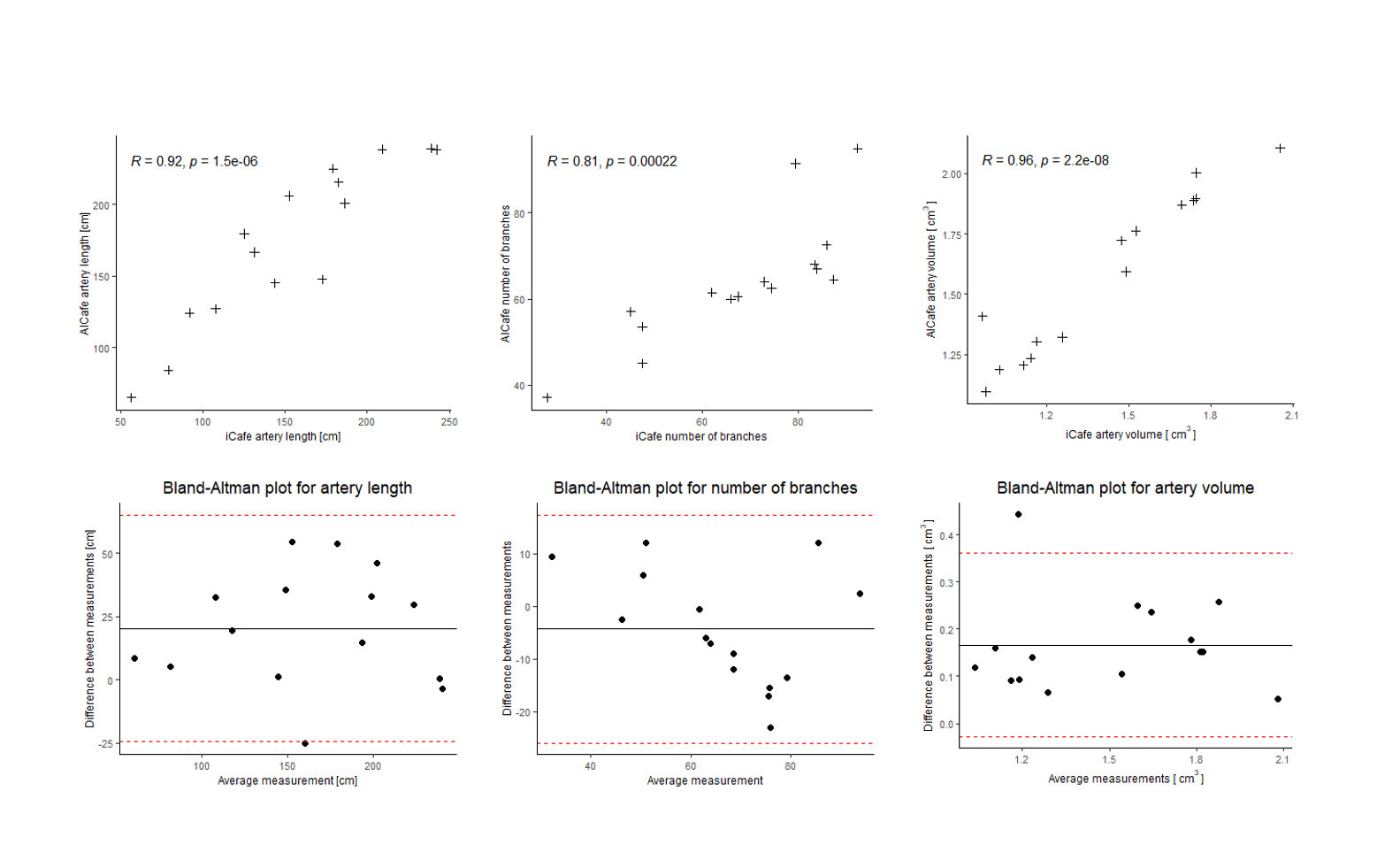
Intracranial artery centerline tracing using iCafe and AICafe for the first and second scan are shown in Figure 1, which highlights the consistency between the two scans as well as the two vascular map construction methods. Table 1 summarizes the scan-rescan quantitative measurements of intracranial arteries from iCafe, while Table 2 summarizes the results from AICafe. The artery length has a high ICC value (0.94) and low CV value (8%) by iCafe and ICC value (0.88) and CV value (7%) by AICafe. Other vascular features including branch number and volume also show similar good CV values (<10%) and lower ICC values (0.78~0.84). All CVs were lower for AICafe than for iCafe. Figure 2 shows good correlations in artery length, number of branches, and artery volume between iCafe and AICafe. Figure 2 also includes Bland-Altman plots for artery length, number of branches, and artery volume.Conclusion:
The vascular maps generated by both iCafe and AICafe on SNAP imaging are highly reproducible. Artery length has the highest ICC value among all the vascular features both in iCafe and AICafe, while other features also have good reproducibility (0.75~0.9). These findings suggest that SNAP, as well as iCafe and AICafe, can be used together to track changes of intracranial arteries in longitudinal studies. Moreover, the lower CV values of AICafe compared to the values of iCafe suggests that AICafe may render more consistent measurements than the manual measurements using iCafe. Although iCafe and AICafe showed good correlations, there are still differences in the artery length and volume measured by iCafe and AICafe, which might be caused by systemic errors. In summary, SNAP is a robust imaging technique that provides consistent intracranial vascular features measured by iCafe and AICafe on scan-rescan testing.Acknowledgements
This work was supported in part by NIH grant R01NS092207.References
1. Chen, Li, et al. "Development of a quantitative intracranial vascular features extraction tool on 3D MRA using semiautomated open‐curve active contour vessel tracing." Magnetic resonance in medicine 79.6 (2018): 3229-3238.
2. Wang, Jinnan, et al. "Simultaneous noncontrast angiography and intraplaque hemorrhage (SNAP) imaging for carotid atherosclerotic disease evaluation." Magnetic resonance in medicine 69.2 (2013): 337-345.
3. Chen, Li, et al. "Quantification of morphometry and intensity features of intracranial arteries from 3D TOF MRA using the intracranial artery feature extraction (iCafe): a reproducibility study." Magnetic resonance imaging 57 (2019): 293-302.
4. Liu, Wenjin, et al. "Uncontrolled hypertension associates with subclinical cerebrovascular health globally: a multimodal imaging study." European Radiology 31.4 (2021): 2233-2241.
5. Chen, Li, et al. "Quantitative assessment of the intracranial vasculature in an older adult population using iCafe." Neurobiology of aging 79 (2019): 59-65.
6. Xiong, Yuhui, et al. "Intracranial simultaneous noncontrast angiography and intraplaque hemorrhage (SNAP) MRA: Analyzation, optimization, and extension for dynamic MRA." Magnetic resonance in medicine 82.5 (2019): 1646-1659.
7. Chen, Li, et al. "Deep Open Snake Tracker for Vessel Tracing." International Conference on Medical Image Computing and Computer-Assisted Intervention. Springer, Cham, 2021.
Figures