4905
Resting-state based Cerebrovascular Reactivity (CVR) mapping at high spatial resolutions1Department of Diagnostic Radiology & Nuclear Medicine, University of Maryland School of Medicine, Baltimore, MD, United States, 2Department of Radiology, Johns Hopkins University School of Medicine, Baltimore, MD, United States
Synopsis
Cerebrovascular reactivity (CVR) is typically assessed with a carbon dioxide (CO2) stimulus combined with BOLD fMRI. Recently, resting-state BOLD fMRI has been shown capable of generating CVR maps, providing a potential for broader CVR applications in neuroimaging studies. However, prior RS-CVR studies have primarily been performed at a spatial resolution of 3-4mm3 voxel sizes. It remains unknown whether RS-CVR can also be obtained at high-resolution without major degradation in image quality. In this study, we investigated the feasibility of high-resolution CVR mapping based on resting-state fMRI data. Our results showed good CVR quality can be obtained at 2-3mm3 voxel sizes.
INTRODUCTION
Cerebrovascular reactivity (CVR), an index of the dilatory function of cerebral blood vessels, is typically assessed with a carbon dioxide (CO2) stimulus combined with BOLD fMRI1. Recently, resting-state BOLD fMRI has been shown capable of generating CVR maps without the need for a gas (CO2 inhalation) or drug (acetazolamide administration) challenge2,3, indicating a potential for broader CVR applications in neuroimaging studies. However, compared with CO2-CVR, resting-state CVR (RS-CVR) is known to suffer from lower SNR and prior RS-CVR studies have primarily been performed at a spatial resolution of 3-4mm3 voxel sizes. As the fMRI field marches towards higher spatial resolutions, it remains unknown whether RS-CVR can also be obtained at high resolution without a major degradation in image quality. Therefore, the goal of the present study is to investigate the effect of spatial resolution of the resting-state BOLD sequence on CVR mapping. To achieve this goal, resting-state BOLD scans were collected with three resolutions at decreasing voxel size, and the resulting CVR maps were compared against maps obtained with the conventional CO2-inhalation method.METHODS
Image Acquisition: Data were collected from eight participants (3F/5M, 26.5±4.9 years) using a 3 Tesla Prisma Siemens scanner with a 32-channel head coil. Four BOLD scans were acquired in each subject: three ten-minute resting-state scans with different resolutions (2mm, 2.4mm, and 3mm isotropic resolution), and one five-minute scan (2mm isotropic resolution) with CO2 challenge (two 50s periods of 5% CO2 inhalation interleaved with room air inhalation). Other BOLD imaging parameters were: TR/TE/FA=720ms/37ms/52º, multiband factor=8; for the 2mm, 2.4mm and 3mm resolutions, FOV=208*208*144 mm3, 210*210*155 mm3 and 210*210*168 mm3, respectively. A high-resolution T1-MPRAGE sequence (1x1x1 mm3) was also acquired.Image Analysis: The resting-state and CO2-inhalation BOLD data were analyzed following previous literature4. For resting-state data, after motion correction and detrending, the BOLD images were temporally filtered to [0, 0.1164 Hz], and a whole-brain-averaged time course was calculated3. Then, using the whole-brain BOLD time course as the independent variable, motion vectors and linear drift as covariates, voxel-wise regression analysis was performed to calculate a CVR index map in %BOLD signal change, which was then normalized to the whole-brain average to yield a relative CVR map. For the CO2-inhalation data, after motion correction, the BOLD images were smoothed by 0mm, 1.33mm and 2.24mm Gaussian kernels to match the intrinsic resolution of the 2mm, 2.4mm and 3mm resting-state scans, respectively. CVR maps were then generated for the three resolutions following the conventional method5. Briefly, the EtCO2 time course was shifted to account for the lung-to-brain delay, and then this shifted EtCO2 was used as the independent variable in a similar voxel-wise regression analysis, generating an absolute CVR map in %∆BOLD/mmHg CO2 which was then normalized to the whole-brain average to yield a relative CVR map. Accuracy of RS-CVR mapping was measured using Pearson’s R2, intraclass correlation (ICC) A1 (absolute agreement), and ICC-C1 (consistency) to compare the RS-CVR maps to the standard CO2-inhalation CVR maps. Sensitivity was explored using Z-score maps from the voxel-wise analysis. Mean Z-scores of each resting-state scan of each subject were calculated and compared across difference resolutions.
Statistical analysis was performed using Kruskal-Wallis Non-parametric ANOVA with Dunn’s test and Holm correction as a post-hoc analysis.
RESULTS
Figure 1 shows the CVR maps obtained from each scan of each subject. Visual inspection suggested that good map quality is seen in all scans, with clear gray/white matter contrast. Lower resolution yielded images that are somewhat blurry but appear more stable. Figure 2 demonstrates the accuracy comparisons among the three resolutions. The 2mm resolution showed slightly lower correlation with the CO2-inhalation CVR maps, whereas the 2.4mm resolution showed slightly higher correlation. Nonetheless, no significant differences between the resolutions in Pearson’s R2 or ICC were found (p>0.089), suggesting comparable accuracy among different resolutions. We conducted the same accuracy analysis using BOLD data pre-smoothed with 4mm and 8mm kernels, and found similar results, with no significant differences between resolutions with different image smoothing.Histograms of the mean Z-score distributions across all subjects for the three resolutions are shown in Figure 3a. Boxplots of the mean Z-scores for each subject are shown in Figure 3b. A significant difference between the resolutions was found, and post-hoc analysis showed a pattern of increasing mean Z-score with increasing voxel size, with the 2x2x2 mm3 and 2.4x2.4x2.4 mm3 resolutions being significantly lower than the 3x3x3 mm3 resolution (p=0.0007, p=0.03569 respectively). This is expected as larger voxel size should have higher SNR, and thus, yield higher sensitivity6.
DISCUSSION
In this study, we have investigated the feasibility of high-resolution CVR mapping based on resting-state fMRI data. Our results show that good CVR quality can be obtained at 2-3 mm3 voxel sizes. Within this resolution range, the higher resolutions gave more resolved information but the lower resolution yielded more robust maps. These findings suggest that the resting-state CVR mapping method can be applied at a similar resolution as functional connectivity mapping, which should broaden the use of CVR mapping in basic science and clinical applications.Acknowledgements
No acknowledgement found.References
1. Sleight E, Stringer MS, Marshall I, Wardlaw JM, Thrippleton MJ. Cerebrovascular Reactivity Measurement Using Magnetic Resonance Imaging: A Systematic Review. Front Physiol. 2021 Feb 25;12:643468.
2. Liu P, Li Y, Pinho M, Park DC, Welch BG, Lu H. Cerebrovascular reactivity mapping without gas challenges. Neuroimage. 2017 Feb 1;146:320-326.
3. Golestani AM, Wei LL, Chen JJ. Quantitative mapping of cerebrovascular reactivity using resting-state BOLD fMRI: Validation in healthy adults. Neuroimage 2016;138:147–163.
4. Liu P, Liu G, Pinho MC, Lin Z, Thomas BP, Rundle M, Park DC, Huang J, Welch BG, Lu H. Cerebrovascular Reactivity Mapping Using Resting-State BOLD Functional MRI in Healthy Adults and Patients with Moyamoya Disease. Radiology. 2021 May;299(2):419-425.
5. Lu H, Liu P, Yezhuvath U, Cheng Y, Marshall O, Ge Y. MRI mapping of cerebrovascular reactivity via gas inhalation challenges. J Vis Exp. 2014 Dec 17;(94):52306.
6. Huettel SA, Song AW, McCarthy G. Functional magnetic resonance imaging. Third edition. pp, 259-262. 2014. Sunderland, Massachusetts, U.S.A.: Sinauer Associates, Inc., Publishers.
Figures
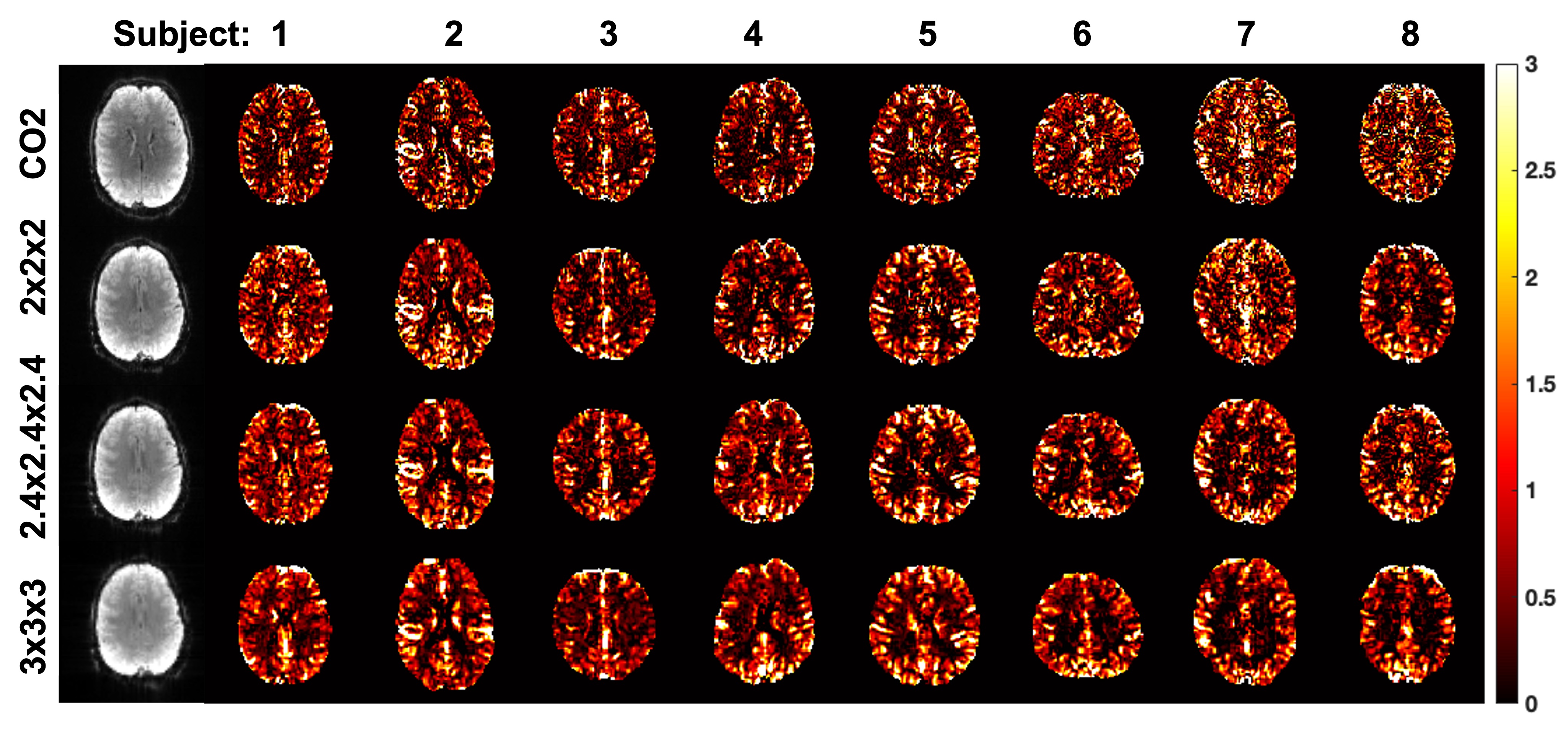
Figure 1: Relative CVR maps for all subjects and resolutions. The first column shows the raw BOLD images from Subject 1.
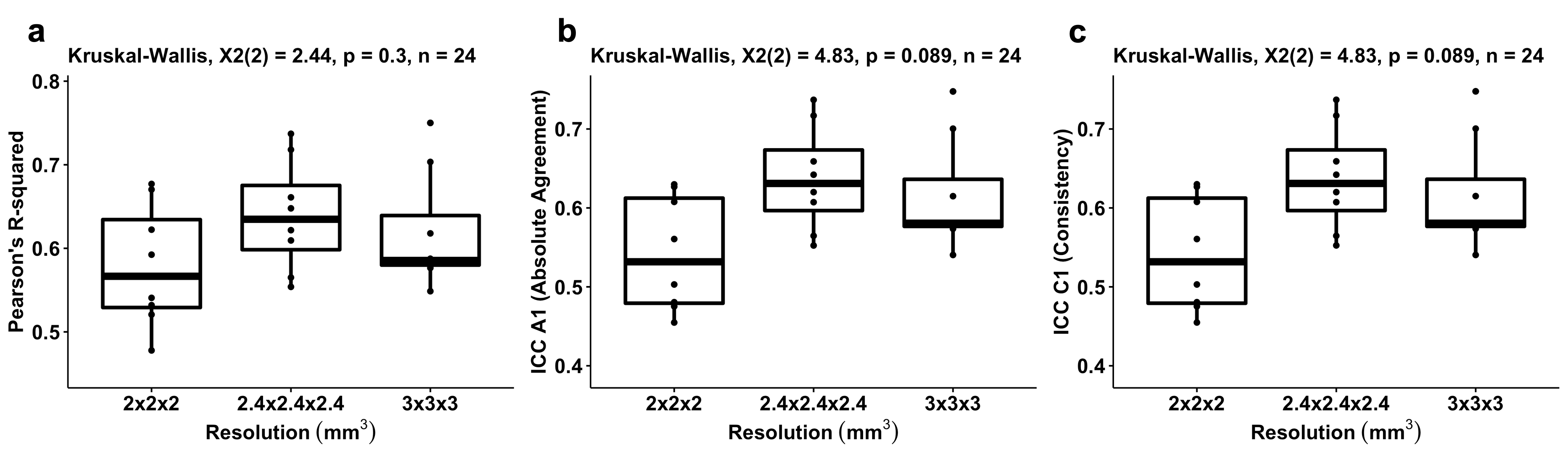
Figure 2: Results of accuracy analysis at the intrinsic RS-CVR resolutions. (a) Comparisons of Pearson’s R2. (b) Comparisons of ICC-A1 (absolute agreement). (c) Comparisons of ICC-C1 (consistency).
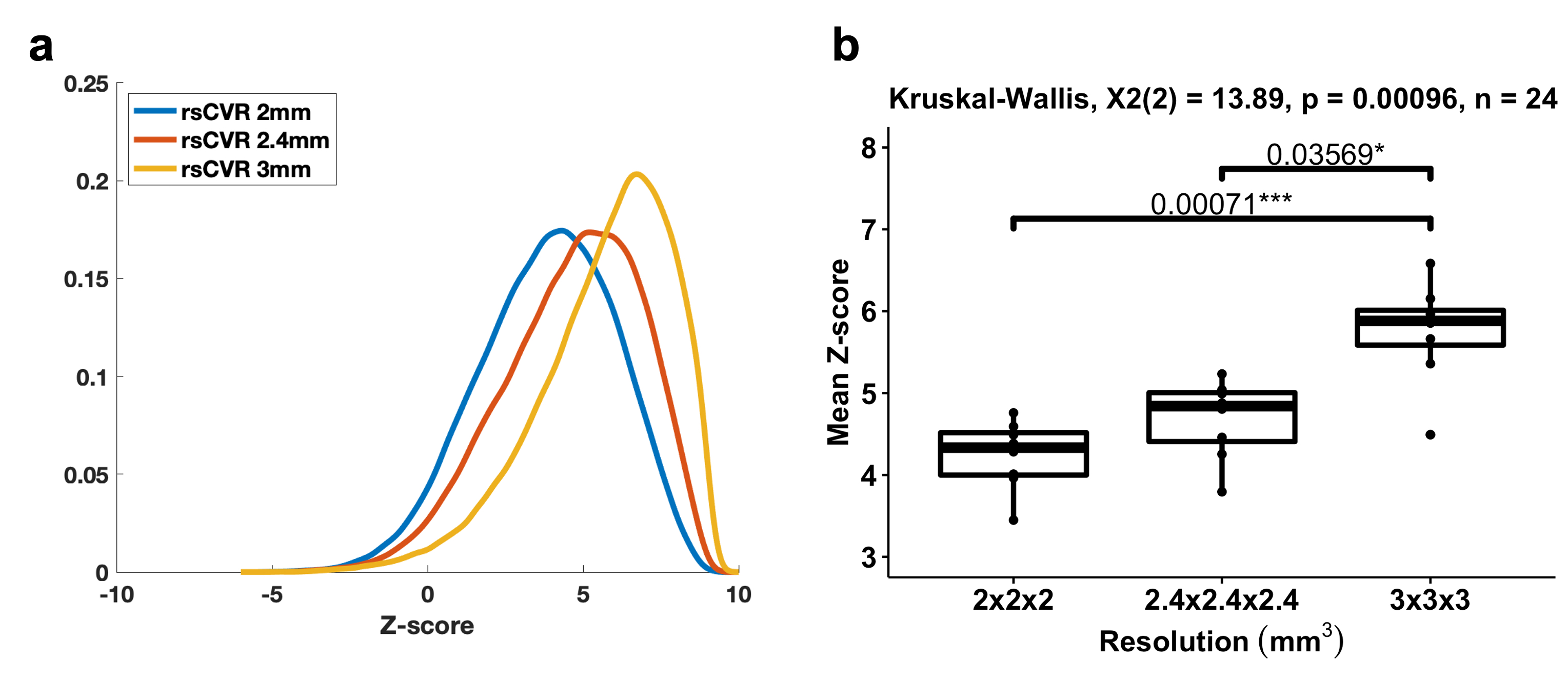
Figure 3: Results of the sensitivity comparisons. (a) Distributions of average Z-scores of RS-CVR maps for each resolution. (b) Comparisons of mean Z-scores of RS-CVR maps across different resolutions.